Comparative Study
. 2006 Dec;35(6):681-8.
doi: 10.1165/rcmb.2006-0169OC. Epub 2006 Jul 13.

Cigarette smoke induces cellular senescence

Affiliations
Comparative Study

Cigarette smoke induces cellular senescence

Toru Nyunoya et al. Am J Respir Cell Mol Biol. 2006 Dec.

Abstract

Chronic obstructive pulmonary disease (COPD) is the fourth leading cause of death in the United States, and cigarette smoking is the major risk factor for COPD. Fibroblasts play an important role in repair and lung homeostasis. Recent studies have demonstrated a reduced growth rate for lung fibroblasts in patients with COPD. In this study we examined the effect of cigarette smoke extract (CSE) on fibroblast proliferative capacity. We found that cigarette smoke stopped proliferation of lung fibroblasts and upregulated two pathways linked to cell senescence (a biological process associated with cell longevity and an inability to replicate), p53 and p16-retinoblastoma protein pathways. We compared a single exposure of CSE to multiple exposures over an extended time course. A single exposure to CSE led to cell growth inhibition at multiple phases of the cell cycle without killing the cells. The decrease in proliferation was accompanied by increased ATM, p53, and p21 activity. However, several important senescent markers were not present in the cells at an earlier time point. When we examined multiple exposures to CSE, we found that the cells had profound growth arrest, a flat and enlarged morphology, upregulated p16, and senescence-associated beta-galactosidase activity, which is consistent with a classic senescent phenotype. These observations suggest that while a single exposure to cigarette smoke inhibits normal fibroblast proliferation (required for lung repair), multiple exposures to cigarette smoke move cells into an irreversible state of senescence. This inability to repair lung injury may be an essential feature of emphysema.


Figures
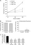
<b>Figure 1.</b>
Figure 1.
A short-term exposure to CSE induces growth inhibition, but not all of the classic features of cellular senescence. (A) HFL-1 cells were cultured in regular tissue culture plates with or without CSE 2.5 and 5% solution for 48 h. The cell counts were measured at various time points (0, 24, and 48 h). Data are expressed as mean ± SEM for three independent experiments (*P < 0.01). (B) HFL-1 cells were treated as in A for 48 h. The cell viability was measured by the Guava ViaCount Assay using a propidium iodide stain. Data are expressed as mean ± SEM for three independent experiments (*P < 0.05). SA β-gal staining was also performed at 48 h. The percentage of SA β-gal–positive cells/total cell number is shown. Data are expressed as mean ± SEM for three independent experiments (*P < 0.05). (C) HFL-1 cells were cultured at the starting cell density of 0.15 × 10⁶/ml in regular tissue culture plates with or without CSE 5% solution. CSE was prepared with or without filtering (0.22 μm) for the immediate use, and filtered CSE solution was kept at 4°C or −70°C for 24 h before use. The cell counts were measured at 48 h. Data are expressed as mean ± SEM for three independent experiments (*P < 0.05).
<b>Figure 2.</b>
Figure 2.
A single exposure to CSE induces cell cycle arrest at multiple phases of the cell cycle. (A) Cell cycle synchronization was induced with serum deprivation. Then, HFL-1 cells were cultured with or without CSE 5% solution in the presence of serum in regular tissue culture plates for 24 h. The harvested cells were stained with propidium iodide and were evaluated with a FACScan flow cytometer. The percentage of cells at each cell cycle phase/total cells was shown. Data are expressed as mean ± SEM for three independent experiments (*P < 0.05). (B) HFL-1 cells were treated as in A, except that they were not synchronized by prior exposure to serum deprivation. The percentage of cells at each cell cycle phase/total cells is shown. Data are expressed as mean ± SEM for three independent experiments (*P < 0.01; **P < 0.05). (C) HFL-1 cells were cultured with or without CSE 5% solution for 24 h. Western blot analysis was performed for phosphorylation of Histone 3 (Ser 10) at 24 h. Equal loading was determined by stripping the blot and reprobing with antibodies to β-actin. Western blotting data are representative of three experiments.
<b>Figure 3.</b>
Figure 3.
A single exposure to CSE causes an early activation of the ATM-p53-p21-pRb pathway. (A) HFL-1 cells were cultured with or without CSE 5% solution for 24 h. Western blot analysis was performed for phosphorylation of ATM (serine 1981), p53 (serine 15), and pRb (serine 807/811) and the amount of p53, p21, and p16 at 24 h. Equal loading was determined by stripping the blot and reprobing with antibodies to β-actin. Western blotting data are representative of three experiments. (B) HFL-1 cells were cultured with or without CSE 5% solution for 48 h, and were washed out, then were cultured with a serum media for up to 2 wk. Western blot analysis was performed for phosphorylation of pRb (inactive state) and the amount of p53 and p16. Equal loading was determined by stripping the blot and reprobing with antibodies to β-actin. Western blotting data are representative of three experiments. HFL-1 cells were treated as in B. SA β-gal staining was performed at 7 d and at 14 d. The percentage of SA β-gal–positive cells/total cell number is shown. Data are expressed as mean ± SEM for three independent experiments (*P < 0.01).
<b>Figure 4.</b>
Figure 4.
Multiple exposures to CSE induce cellular senescence. (A) HFL-1 cells were cultured on regular tissue culture plates with multiple exposures to CSE 3.5% solution or a single exposure to CSE 5% solution (for 48 h followed by no exposure to CSE for an additional 12 d) or without CSE for up to 14 d. The cell counts were measured at various time points (Days 0, 2, 4, 7 and 14). Data are expressed as mean ± SEM for three independent experiments (*P < 0.01). (B) HFL-1 cells were treated as in A. SA β-gal staining was performed for the control and multiple CSE-exposed cells on Days 7 and 14. The percentage of SA β-gal–positive cells/total cell number was shown. Data are expressed as mean ± SEM for three independent experiments (*P < 0.01). The digital photographs were obtained on Day 14, and represent one of three identical experiments.
<b>Figure 5.</b>
Figure 5.
CSE-induced cellular senescence is linked to activation of both the p16 and p53 pathways. HFL-1 cells were cultured on regular tissue culture plates with multiple exposures to CSE 3.5% solution for up to 14 d. Western blot analysis was performed for accumulation of p53 and p16 and phosphorylation of pRb. Equal loading was determined by stripping the blot and reprobing with antibodies to β-actin. Western blotting data are representative of three experiments.
<b>Figure 6.</b>
Figure 6.
A proposed pathway for CSE-induced cellular senescence. Repeated exposure to CSE activates both the p53 and p16 pathways. Both pathways likely contribute to a senescent phenotype, including cell cycle arrest, an increase in SA β-gal activity, and a flat and enlarged cellular morphology.

Similar articles

Cited by

References

    1. Pauwels RA, Buist AS, Calverley PM, Jenkins CR, Hurd SS. Global strategy for the diagnosis, management, and prevention of chronic obstructive pulmonary disease. NHLBI/WHO Global Initiative for Chronic Obstructive Lung Disease (GOLD) Workshop summary. Am J Respir Crit Care Med 2001;163:1256–1276. - PubMed
    1. Murphy SL. Deaths: final data for 1998. Natl Vital Stat Rep 2000;48:1–105. - PubMed
    1. Mannino DM, Homa DM, Akinbami LJ, Ford ES, Redd SC. Chronic obstructive pulmonary disease surveillance--United States, 1971–2000. MMWR Surveill Summ 2002;51:1–16. - PubMed
    1. Fishman AP. One hundred years of COPD. Am J Respir Crit Care Med 2005;171:941–948. - PubMed
    1. Absher M. Fibroblasts. In: Massaro D, editor. Lung biology in health and disease. New York: Marcel Dekker Inc.; 1995. pp. 401–439.

Publication types

MeSH terms