. 2006 Aug;116(8):2142-2151.
doi: 10.1172/JCI27821.

Neonatal Fc receptor for IgG regulates mucosal immune responses to luminal bacteria

Affiliations

Neonatal Fc receptor for IgG regulates mucosal immune responses to luminal bacteria

Masaru Yoshida et al. J Clin Invest. 2006 Aug.

Abstract

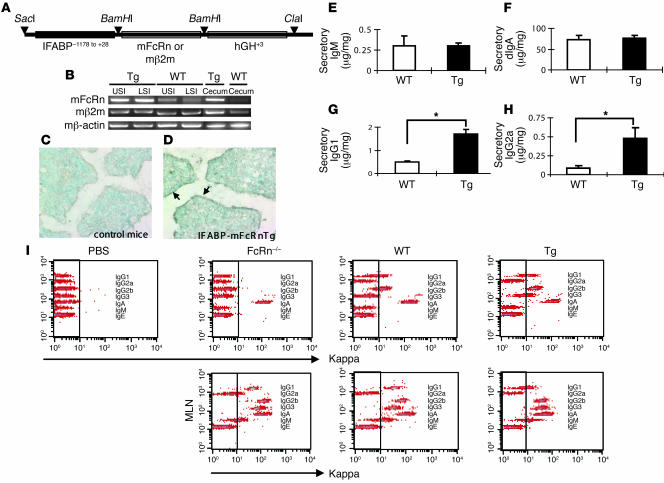
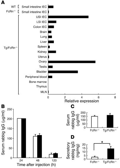
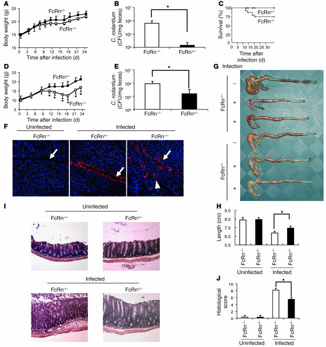
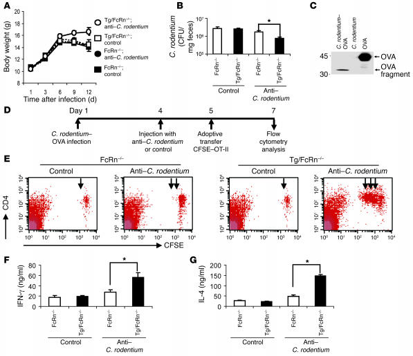
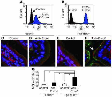
The neonatal Fc receptor for IgG (FcRn) plays a major role in regulating host IgG levels and transporting IgG and associated antigens across polarized epithelial barriers. Selective expression of FcRn in the epithelium is shown here to be associated with secretion of IgG into the lumen that allows for defense against an epithelium-associated pathogen (Citrobacter rodentium). This pathway of host resistance to a bacterial pathogen as mediated by FcRn involves retrieval of bacterial antigens from the lumen and initiation of adaptive immune responses in regional lymphoid structures. Epithelial-associated FcRn, through its ability to secrete and absorb IgG, may thus integrate luminal antigen encounters with systemic immune compartments and as such provide essential host defense and immunoregulatory functions at the mucosal surfaces.


Figures

Figure 1
Figure 1. Absence of intestinal luminal IgG in FcRn–/– mice.
(A) The construct for the Tg IFABP-mFcRnTg/mβ2mTg mouse, designed to express mFcRn and mβ2m under the control of the IFABP. (B) Increased mFcRn expression in epithelial cells of IFABP-mFcRnTg/mβ2mTg (Tg) mouse. RNA was extracted from epithelial cells of upper and lower small intestines (USI and LSI, respectively) and cecum in 6-week-old IFABP-mFcRnTg/mβ2mTg founder BALB/c mice and littermate WT BALB/c mice and subjected to RT-PCR. (C and D) Immunohistochemical analysis of lower small intestine in WT (C) and IFABP-mFcRnTg/mβ2mTg mice (D). Arrows indicate staining of FcRn. (EH) The levels of Igs secreted into the intestinal lumen. Secretory IgM (E), dimeric IgA (F), IgG1 (G), and IgG2a (H) were measured by ELISA. The mean ± SD are shown for each group (n = 8). *P < 0.05. (I) The levels of Igs (IgG1, IgG2a, IgG2b, IgG3, IgA, IgM, and IgE) secreted into the lumen of the indicated mouse strains on a C57BL/6 background were measured by a cytometric bead array.
Figure 2
Figure 2. IgG transport into the lumen of IFABP-mFcRnTg/mβ2mTg/FcRn–/– mice.
(A) Quantitative PCR of cDNA using PCR primers originating in exon 2 in variety of tissues in IFABP-mFcRnTg/mβ2mTg/FcRn–/– (Tg/FcRn–/–) mice. IEC, intestinal epithelial cells. (B) Serum rabbit IgG levels in IFABP-mFcRnTg/mβ2mTg/FcRn–/– (black bars) and littermate control FcRn–/– mice (white bars) at 24, 48, and 120 hours after injection of rabbit IgG. (C) Serum rabbit IgG levels in IFABP-mFcRnTg/mβ2mTg/FcRn–/– mice measured at 12 hours after injection of rabbit IgG (n = 4). (D) Secretory rabbit IgG levels in feces of IFABP-mFcRnTg/mβ2mTg/FcRn–/– mice measured at 12 hours after injection of rabbit IgG as ng per mg of feces (n = 4). *P < 0.05.
Figure 3
Figure 3. Susceptibility toC. rodentium infection in the presence of FcRn.
(A and B) Susceptibility to infection with 1 × 10⁹ CFU of C. rodentium in FcRn–/– BALB/c mice. (A) Body weight changes in FcRn–/– and FcRn+/– mice with C. rodentium infection. (B) CFU of C. rodentium in feces of FcRn–/– and FcRn+/– mice 21 days after infection. Mean ± SD are shown for each group (n = 6). (CE) Susceptibility to infection with C. rodentium in FcRn–/– C57BL/6 mice. Survival rate (C) and body weight changes (D) in FcRn–/– and FcRn+/– mice with C. rodentium infection. (E) CFU of C. rodentium in feces of FcRn–/– and FcRn–/+ mice 21 days after infection. Mean ± SD are shown for each group (n = 8). (F) Immunohistochemical analysis of the colon to detect intimin in mice with C. rodentium infection. Colonic tissues were collected at day 7 from selected mice on a C57BL/6 background. Sections were stained for intimin using a polyclonal rabbit anti–C. rodentium intimin antibody (red) and nuclei (blue) and were examined by confocal microscopy. Magnification, ×400. Macroscopic findings (G) and the length of colon (H) in FcRn–/– and FcRn+/– C57BL/6 mice, uninfected or infected with C. rodentium, at 21 days after infection. (I) Histological findings of colon in FcRn–/– and littermate FcRn+/– C57BL/6 mice with or without C. rodentium infection (21 days after infection). Magnification, ×100. (J) Histological score of colonic tissue in the mice with or without C. rodentium infection at day 21. *P < 0.05.
Figure 4
Figure 4. Induction of immune response toC. rodentium –derived antigen in the presence of circulating IgG and FcRn in intestinal epithelial cells.
(A and B) The effect of FcRn-mediated IgG transport into the intestinal lumen in C. rodentium infection. Body weight changes (A) and CFU of C. rodentium in feces 21 days after infection (B) in IFABP-mFcRnTg/mβ2mTg/FcRn–/– and FcRn–/– C57BL/6 mice with i.v. injection of anti–C. rodentium IgG or control IgG. Mean ± SD are shown for each group (n = 6). (C) Establishment of a genetically engineered C. rodentium strain that constitutively produces an OVA fragment. The immunoblot confirms the expression of the OVA fragment by C. rodentium. (D) Summary of the experimental protocol, with the inoculation of C. rodentium–OVA or control bacteria, the injection of anti–C. rodentium IgG or control IgG, and the adoptive transfer of CD45.1+CD4+OVA-specific T cells from CD45.1+OT-II mice. (E) The number of OVA-specific CD4+ T cells in the MLNs in IFABP-mFcRnTg/mβ2mTg/FcRn–/– and FcRn–/– mice increased in the presence of anti–C. rodentium IgG or control IgG (n = 3). Arrows indicate increasing rounds of cell division. (F and G) Cytokine production in OVA-specific CD4+ T cells purified from the MLNs and cultured with OVA for 48 hours in vitro. Cytokine production of IFN-γ (F) and IL-4 (G) was measured by ELISA. Mean ± SD are shown for each group (n = 4). *P < 0.05.
Figure 5
Figure 5. Luminal bacterial antigens transported as an immune complex across intestinal epithelial cells via FcRn in vivo.
(A and B) Flow cytometry of rabbit anti–E. coli IgG and control IgG followed by PE-conjugated goat anti-rabbit IgG (A) and FITC-conjugated E. coli (B). (CF) Confocal microscopy analysis of transport of bacterial antigens across intestinal epithelial cells. Sections were stained for actin (phalloidin; red) and nuclei (blue). Magnification, ×400. Arrow in F illustrates transported FITC-conjugated E. coli in intestinal epithelial cells in the presence of rabbit anti–E. coli IgG. (G) Presence of FITC-conjugated E. coli in CD11c+ cells of the MLNs of IFABP-mFcRnTg/mβ2mTg/FcRn–/– mice 5 hours after FITC-conjugated E. coli administration in the presence of rabbit anti–E. coli IgG. Mean fluorescence intensity (MFI) on gated CD11c+ cells is shown. The mean ± SD was shown for each group (n = 4). *P < 0.05.

References

    1. Robert-Guroff M. IgG surfaces as an important component in mucosal protection. Nat. Med. 2000;6:129–130. - PubMed
    1. Yoshida M., et al. Human neonatal Fc receptor mediates transport of IgG into luminal secretions for delivery of antigens to mucosal dendritic cells. Immunity. 2004;20:769–783. - PubMed
    1. Woof J.M., Mestecky J. Mucosal immunoglobulins. Immunol. Rev. 2005;206:64–82. - PubMed
    1. Hanson, L.A., and Brandzaeg, P. 1980. The mucosal defense system. InImmunologic disorders in infants and children. Stiehm, E.R., editor. W.B. Saunders. Philadelphia, Pennsylvania, USA. 137–164.
    1. Kozlowski P.A., Cu-Uvin S., Neutra M.R., Flanigan T.P. Comparison of the oral, rectal, and vaginal immunization routes for induction of antibodies in rectal and genital tract secretions of women. Infect. Immun. 1997;65:1387–1394. - PMC - PubMed

Publication types