. 2006 Jul 15;78(14):4820-9.
doi: 10.1021/ac060132w.

Mapping of protein disulfide bonds using negative ion fragmentation with a broadband precursor selection

Affiliations

Mapping of protein disulfide bonds using negative ion fragmentation with a broadband precursor selection

Mingxuan Zhang et al. Anal Chem. .

Abstract

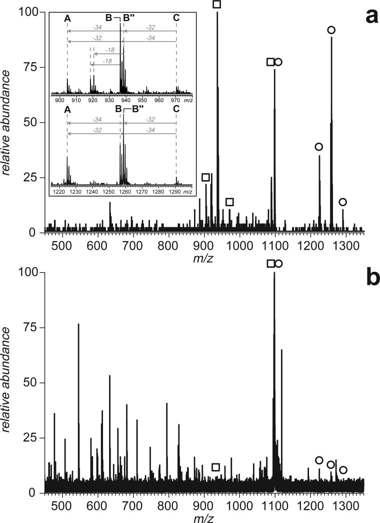
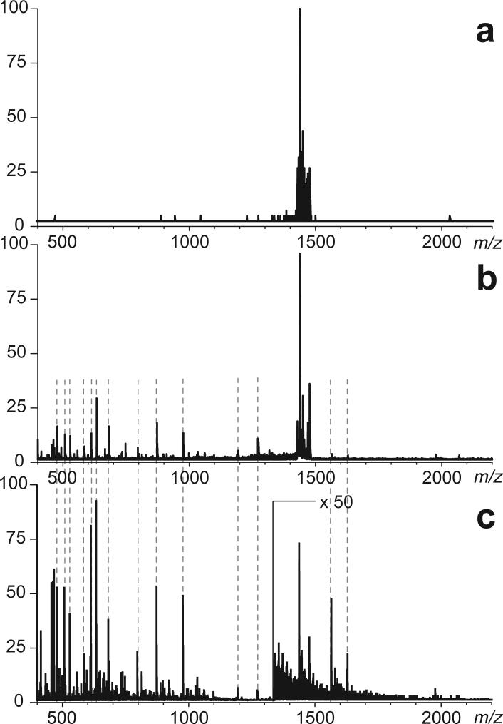
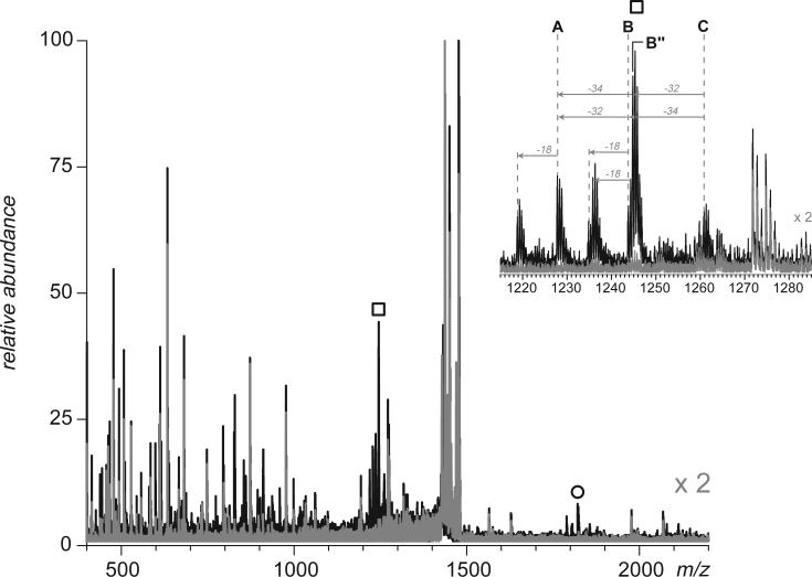
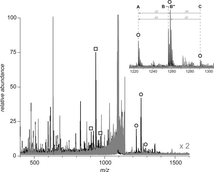
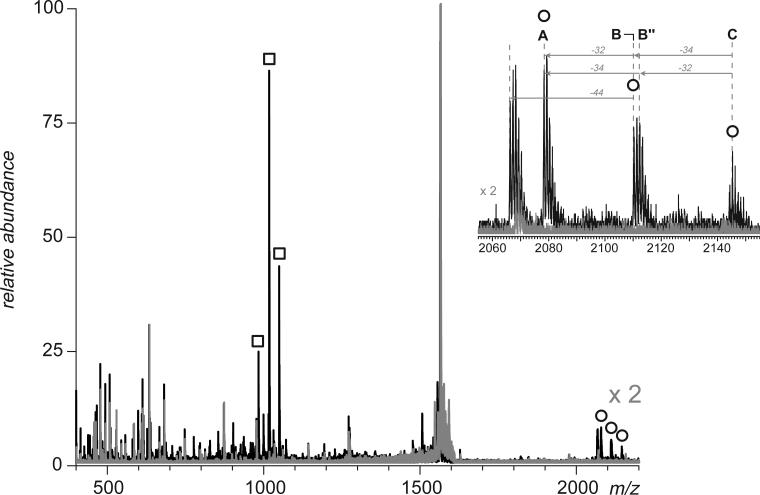
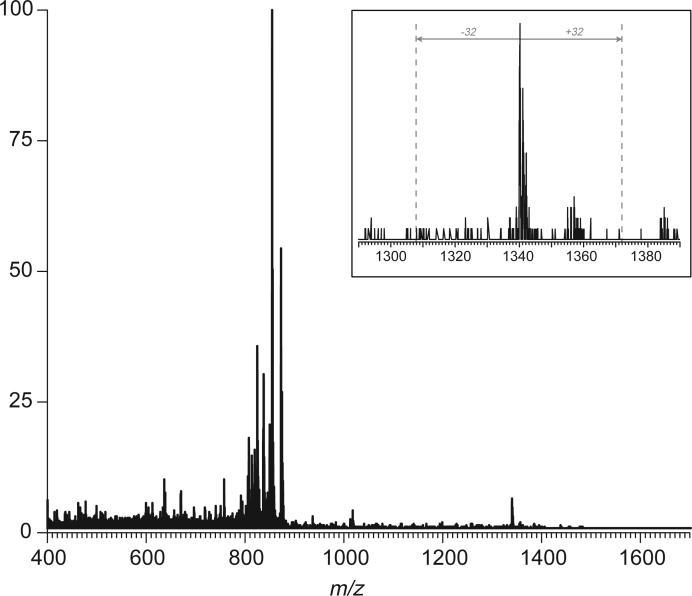
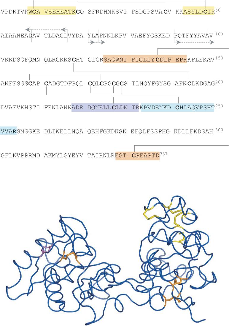
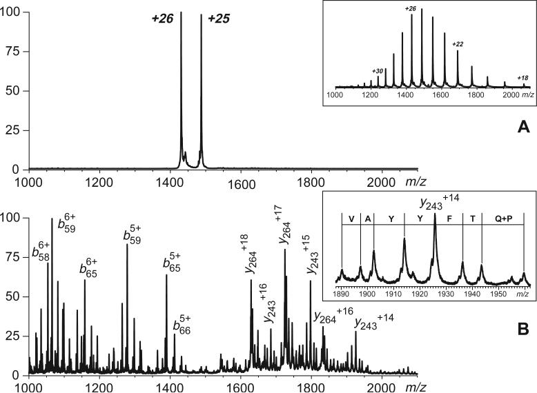
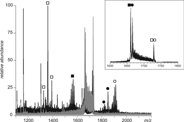
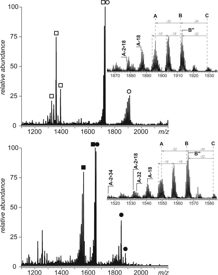
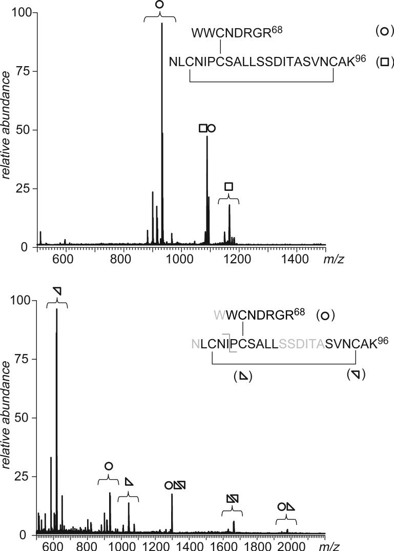
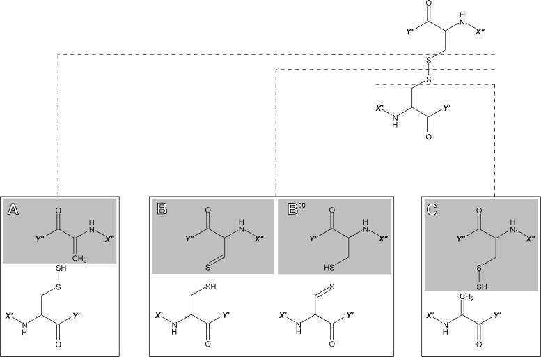
Fast mapping of disulfide bonds in proteins containing multiple cysteine residues is often required in order to assess the integrity of the tertiary structure of proteins prone to degradation and misfolding or to detect distinct intermediate states generated in the course of oxidative folding. A new method of rapid detection and identification of disulfide-linked peptides in complex proteolytic mixtures utilizes the tendency of collision-activated peptide ions to lose preferentially side chains of select amino acids in the negative ion mode. Cleavages of cysteine side chains result in efficient dissociation of disulfide bonds and produce characteristic signatures in the fragment ion mass spectra. While cleavages of other side chains result in insignificant loss of mass and full retention of the peptide ion charge, dissociation of external disulfide bonds results in physical separation of two peptides and, therefore, significant changes of both mass and charge of fragment ions relative to the precursor ion. This feature allows the fragment ions generated by dissociation of external disulfide bonds to be easily detected and identified even if multiple precursor ions are activated simultaneously. Such broadband selection of precursor ions for consecutive activation is achieved by lowering the dc/rf amplitude ratio in the first quadrupole filter of a hybrid quadrupole time-of-flight mass spectrometer. The feasibility of the new method is demonstrated by partial mapping of disulfide bridges within a 37-kDa protein containing 16 cysteine residues and complete disulfide mapping within a lysozyme (14.5 kDa) containing 8 cysteine residues. In addition to detecting peptide pairs connected by a single external disulfide, the new method is also shown to be capable of identifying peptides containing both external and internal disulfide bonds. The two major factors determining the efficiency of disulfide mapping using the new methodology are the effectiveness of proteolysis and the ability of the resulting proteolytic fragments to form multiply charged negative ions.


Figures

Figure 1
Figure 1
Mass spectra of fragment anions generated by CAD of a disulfide-linked peptide dimer [8−18]-[43−50] derived from digestion of hTf/2N with trypsin. The spectrum shown on panel (a) was obtained with a hybrid QqTOF mass spectrometer (CAD carried out in an RF-only quadrupole), and the spectrum shown on panel (b) was acquired with an FT ICR mass spectrometer (SORI CAD). Open squares and circles indicate ions corresponding to intact peptide monomers produced by dissociation of the disulfide bond in the peptide dimer ion without any backbone cleavages (zoomed views are shown in insets).
Figure 2
Figure 2
The effect of ion kinetic energy on transparency of the first quadrupole filter (Q) of a hybrid QqTOF mass spectrometer. Top panel shows a broadband selection of a range of low-intensity precursor ions (m/z 1430−1480) derived from digestion of hTf/2N with trypsin. The middle panel shows the broadband MS/MS spectrum acquired below the fragmentation threshold. Origin of all ions outside of the precursor window can be traced to abundant peptide anions in MS1 spectrum (bottom trace).
Figure 3
Figure 3
Detection and identification of a 227−241 disulfide bridge in hTf/2N by broadband CAD of anions (m/z 1430−1480) derived from digestion of this protein with trypsin. The broadband spectra acquired below and above the fragmentation threshold are shown in gray and black, respectively. The inset shows a group of peaks corresponding to peptide [233−254]. Anionic species representing another peptide from the dimer, [218−232], are marked with circles.
Figure 4
Figure 4
Detection and identification of a 9−48 disulfide bridge in hTf/2N by broadband CAD of anions (m/z 1090−1150) derived from digestion of this protein with trypsin. The broadband spectra acquired below and above the fragmentation threshold are shown in gray and black, respectively. The inset shows a group of peaks corresponding to peptide [8−18]. Anionic species representing another peptide from the dimer, [43−50], are marked with squares.
Figure 5
Figure 5
Detection and identification of a 137−331 disulfide bridge in hTf/2N by broadband CAD of anions (m/z 1550−1620) derived from digestion of this protein with trypsin. The broadband spectra acquired below and above the fragmentation threshold are shown in gray and black, respectively. The inset shows a group of peaks corresponding to peptide [125−143]. Anionic species representing another peptide from the dimer, [328−337], are marked with squares.
Figure 6
Figure 6
An example of broadband CAD of monomeric peptide anions derived from digestion of hTf/2N with trypsin. The only ion outside of the precursor ion window (m/z 810−860), whose origin cannot be traced to abundant peptide ions in MS1 able to pass the quadrupole filter at elevated kinetic energy, is the one at m/z 1340. The appearance of this ion peak (zoomed view, inset) is inconsistent with a notion of this ion being a product of disulfide dissociation.
Figure 7
Figure 7
Amino acid sequence (top) and three-dimensional structure of hTf/2N (bottom) showing locations of disulfide bonds. Disulfide bonds whose existence has been determined by broadband CAD in the negative ion mode (tryptic digest) are colored in orange (disulfide-linked tryptic peptide dimers are highlighted in the top diagram). The disulfide bond whose existence is inferred from the results of positive ion CAD of intact protein is colored in purple (positions of amide bond cleavages leading to abundant b- and y-ions are shown on the top diagram).
Figure 8
Figure 8
Broadband selection (top) and fragmentation (bottom) of intact multiply charged hTf/2N cations carried out with a hybrid QqTOF mass spectrometer. The inset in the top panel shows positive ion ESI mass spectrum of hTf/2N prior to precursor ion selection. The inset in the bottom panel shows a series of y-ions carrying 14 charges produced by dissociation of amide bonds in the [90−99] region of the protein (ladder sequencing).
Figure 9
Figure 9
Detection of four tryptic peptides of BSA involved in formation of external disulfide bridges by broadband CAD in the negative ion mode (m/z 1650−1740). The broadband spectra acquired below and above the fragmentation threshold are shown in gray and black, respectively. The two precursor ions that were identified as disulfide-linked dimers are labeled in the inset.
Figure 10
Figure 10
Narrow-band negative ion CAD of the two dimeric tryptic peptides of BSA whose dissociation reveals the presence of external disulfide bonds. Labeling of fragment ions is the same as in Figure 9. The insets show fragment ions peaks corresponding to peptides with three cysteine residues each, [106−138] (top) and [569−597] (bottom).
Figure 11
Figure 11
Top: detection of a lysozyme tryptic peptide dimer containing two disulfide bonds by broadband CAD in the negative ion mode (the peptide dimer structure is shown in the inset). Bottom: additional processing of the peptide mixture with pepsin generates a peptide trimer connected by two disulfide bonds (amino acid residues cut by pepsin are typed in gray). CAD of the corresponding anionic species gives rise to five groups of fragment ions produced by dissociation of one or both disulfide bonds in the gas phase. Individual peptide monomers are marked with open circles and triangles, as shown on the inset.
Scheme 1
Scheme 1

References

    1. Standing KG. Peptide and protein de novo sequencing by mass spectrometry. Curr. Opin. Struct. Biol. 2003;13:595–601. - PubMed
    1. Coon JJ, Syka JE, Shabanowitz J, Hunt DF. Tandem mass spectrometry for peptide and protein sequence analysis. Biotechniques. 2005;38:519–523. - PubMed
    1. Kaltashov IA, Eyles SJ. Studies of biomolecular conformations and conformational dynamics by mass spectrometry. Mass Spectrom. Rev. 2002;21:37–71. - PubMed
    1. Welker E, Wedemeyer WJ, Narayan M, Scheraga HA. Coupling of conformational folding and disulfide-bond reactions in oxidative folding of proteins. Biochemistry. 2001;40:9059–9064. - PubMed
    1. Yu J. Intentionally degrading protein pharmaceuticals to validate stability-indicating analytical methods. BioPharm. Appl. 2000;13:46–50.

Publication types