

Computational models of the hippocampal region: implications for prediction of risk for Alzheimer's disease in non-demented elderly
- PMID: 16842102
- PMCID: PMC1626443
- DOI: 10.2174/156720506777632826
Computational models of the hippocampal region: implications for prediction of risk for Alzheimer's disease in non-demented elderly
Abstract
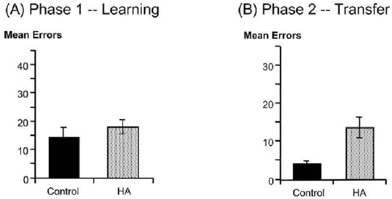
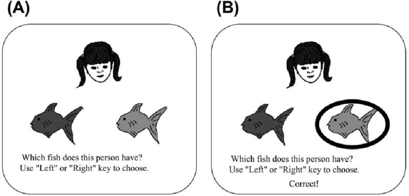
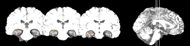
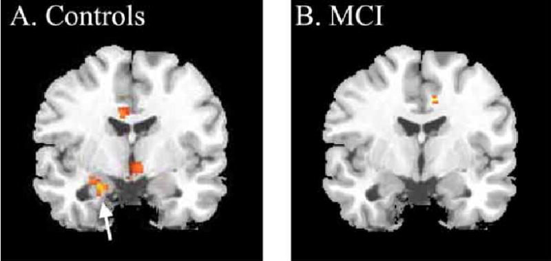
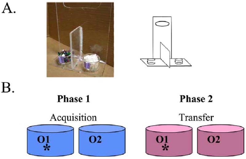
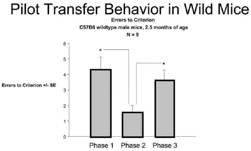
We have pursued an interdisciplinary research program to develop novel behavioral assessment tools for evaluating specific memory impairments following damage to the medial temporal lobe, including the hippocampus and associated structures that show pathology early in the course of Alzheimer's disease (AD). Our approach uses computational models to identify the functional consequences of hippocampal-region damage, leading to testable predictions in both rodents and humans. Our modeling argues that hippocampal-region dysfunction may selectively impair the ability to generalize when familiar information is presented in novel recombinations. Previous research has shown that specific reductions in hippocampal volume in non-demented elderly individuals correlate with future development of AD. In two previous studies, we tested non-demented elderly with and without mild hippocampal atrophy (HA) on stimulus-response learning tasks. Individuals with and without HA could learn the initial information, but the HA group was selectively impaired on transfer tests where familiar features and objects were recombined. This suggests that such generalization deficits may be behavioral markers of HA, and an early indicator of risk for subsequent cognitive decline. Converging support for the relevance of these tasks to aging and Alzheimer's disease comes from our recent fMRI studies of individuals with mild cognitive impairment (MCI). Activity in the hippocampus declines with progressive training on these tasks, suggesting that the hippocampus is important for learning new stimulus representations that support subsequent transfer. Individuals with HA may be able to learn, but in a more hippocampal-independent fashion that does not support later transfer. Ultimately, this line of research could lead to a novel battery of behavioral tests sensitive to very mild hippocampal atrophy and risk for decline to AD, allowing early diagnosis and also allowing researchers to test new Alzheimer's drugs that target individuals in the earliest stages of the disease - before significant cognitive decline. A new mouse version of one of our tasks shows promise for translating these paradigms into rodents, allowing for future studies of therapeutic interventions in transgenic mouse models of AD.
Figures


Similar articles
-
Hippocampal atrophy disrupts transfer generalization in nondemented elderly.J Geriatr Psychiatry Neurol. 2002 Summer;15(2):82-90. doi: 10.1177/089198870201500206. J Geriatr Psychiatry Neurol. 2002. PMID: 12083598
-
Automated 3D mapping of hippocampal atrophy and its clinical correlates in 400 subjects with Alzheimer's disease, mild cognitive impairment, and elderly controls.Hum Brain Mapp. 2009 Sep;30(9):2766-88. doi: 10.1002/hbm.20708. Hum Brain Mapp. 2009. PMID: 19172649 Free PMC article.
-
Early diagnosis of Alzheimer's disease: contribution of structural neuroimaging.Neuroimage. 2003 Feb;18(2):525-41. doi: 10.1016/s1053-8119(02)00026-5. Neuroimage. 2003. PMID: 12595205
-
[Mild Cognitive Impairment or pre-demential Alzheimer's disease?].Rev Neurol (Paris). 2002;158(10 Suppl):S30-4. Rev Neurol (Paris). 2002. PMID: 12529583 Review. French.
-
Neuroimaging of hippocampal atrophy in early recognition of Alzheimer's disease--a critical appraisal after two decades of research.Psychiatry Res Neuroimaging. 2016 Jan 30;247:71-8. doi: 10.1016/j.pscychresns.2015.08.014. Psychiatry Res Neuroimaging. 2016. PMID: 26774855 Review.
Cited by
-
Promising developments in neuropsychological approaches for the detection of preclinical Alzheimer's disease: a selective review.Alzheimers Res Ther. 2013 Nov 21;5(6):58. doi: 10.1186/alzrt222. eCollection 2013. Alzheimers Res Ther. 2013. PMID: 24257331 Free PMC article. Review.
-
A neural model of hippocampal-striatal interactions in associative learning and transfer generalization in various neurological and psychiatric patients.Brain Cogn. 2010 Nov;74(2):132-44. doi: 10.1016/j.bandc.2010.07.013. Epub 2010 Aug 21. Brain Cogn. 2010. PMID: 20728258 Free PMC article.
-
Beneficial effects of multisensory and cognitive stimulation on age-related cognitive decline in long-term-care institutions.Clin Interv Aging. 2014 Feb 14;9:309-20. doi: 10.2147/CIA.S54383. eCollection 2014. Clin Interv Aging. 2014. PMID: 24600211 Free PMC article. Clinical Trial.
-
Novel age-dependent learning deficits in a mouse model of Alzheimer's disease: implications for translational research.Neurobiol Aging. 2011 Jul;32(7):1273-85. doi: 10.1016/j.neurobiolaging.2009.08.003. Epub 2009 Aug 31. Neurobiol Aging. 2011. PMID: 19720431 Free PMC article.
-
Longitudinal MRI and cognitive change in healthy elderly.Neuropsychology. 2007 Jul;21(4):412-8. doi: 10.1037/0894-4105.21.4.412. Neuropsychology. 2007. PMID: 17605574 Free PMC article.
References
-
- de Leon M, Golomb J, George A, et al. Hippocampal formation atrophy: Prognostic significance for Alzheimer’s Disease. In: Corain B, Iqbal K, Nicolini M, et al., editors. Alzheimer’s Disease: Advances in Clinical and Brain Research’. New York: John Wiley and Sons; 1993. pp. 35–46.
-
- de Leon M, George A, Golomb J, Tarshish C, Convit A, Kluger A, et al. Frequency of hippocampal formation atrophy in normal aging and Alzheimer’s disease. Neurobiol Aging. 1997;18:1–11. - PubMed
-
- de Toledo-Morrell L, Goncharova I, Dickerson B, Wilson R, Bennett D. From healthy aging to early Alzheimer’s disease: In vivo detection of entorhinal cortex atrophy. Ann N Y Acad Sci. 2000;911:240–253. - PubMed
-
- Grundman M, Sencakova D, Jack C, et al. Brain MRI hippocampal volume and prediction of clinical status in a mild cognitive impairment trial. J Mol Neurosci. 2002;19(1–2):23–28. - PubMed
-
- de Leon M, George A, Stylopoulos L, et al. Early marker for Alzheimer’s disease: The atrophic hippocampus. Lancet. 1989:672–673. - PubMed
Publication types
MeSH terms
Grants and funding
LinkOut - more resources
Full Text Sources
Medical