. 2006 Jul 16:2:22.
doi: 10.1186/1744-8069-2-22.

The mu opioid agonist morphine modulates potentiation of capsaicin-evoked TRPV1 responses through a cyclic AMP-dependent protein kinase A pathway

Affiliations

The mu opioid agonist morphine modulates potentiation of capsaicin-evoked TRPV1 responses through a cyclic AMP-dependent protein kinase A pathway

Irina Vetter et al. Mol Pain. .

Abstract

Background: The vanilloid receptor 1 (TRPV1) is critical in the development of inflammatory hyperalgesia. Several receptors including G-protein coupled prostaglandin receptors have been reported to functionally interact with the TRPV1 through a cAMP-dependent protein kinase A (PKA) pathway to potentiate TRPV1-mediated capsaicin responses. Such regulation may have significance in inflammatory pain. However, few functional receptor interactions that inhibit PKA-mediated potentiation of TRPV1 responses have been described.

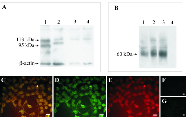
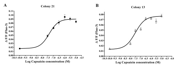
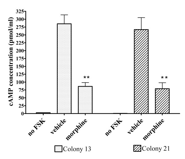
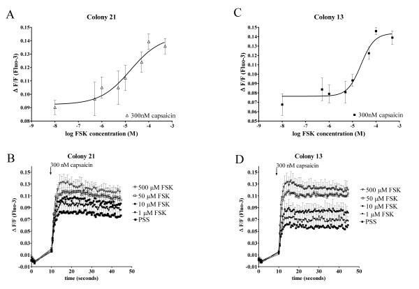
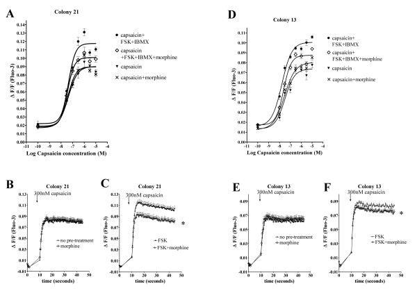
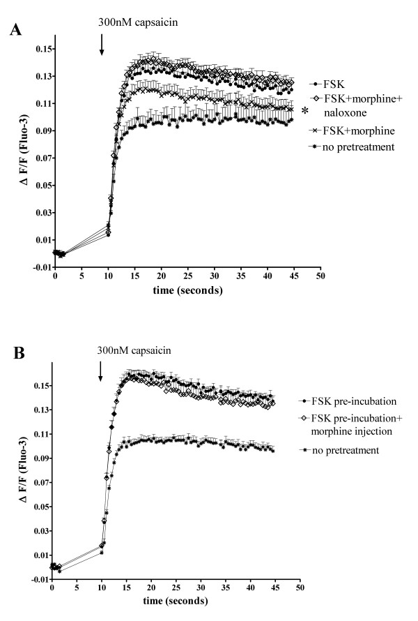
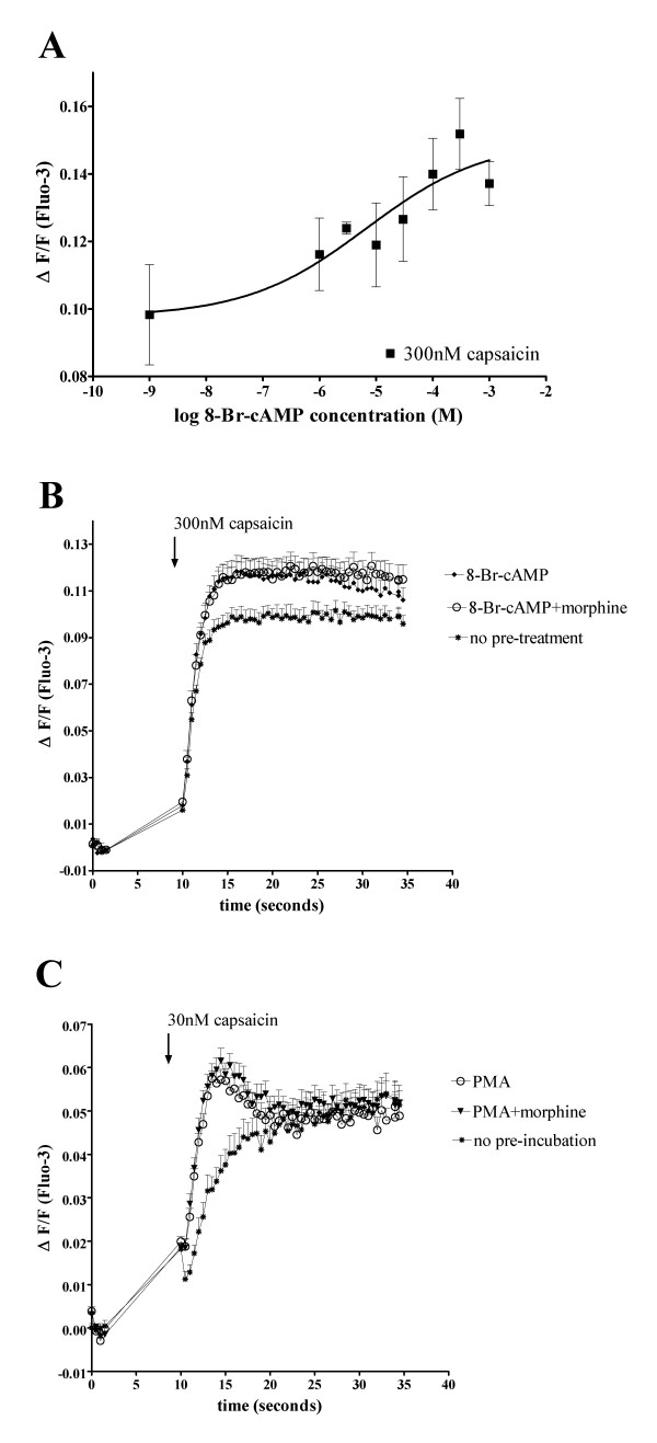
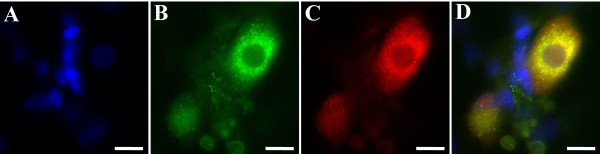
Results: In the present studies we investigated the hypothesis that the mu opioid receptor (MOP) agonist morphine can modulate forskolin-potentiated capsaicin responses through a cAMP-dependent PKA pathway. HEK293 cells were stably transfected with TRPV1 and MOP, and calcium (Ca2+) responses to injection of the TRPV1 agonist capsaicin were monitored in Fluo-3-loaded cells. Pre-treatment with morphine did not inhibit unpotentiated capsaicin-induced Ca2+ responses but significantly altered capsaicin responses potentiated by forskolin. TRPV1-mediated Ca2+ responses potentiated by the direct PKA activator 8-Br-cAMP and the PKC activator Phorbol-12-myristate-13-acetatewere not modulated by morphine. Immunohistochemical studies confirmed that the TRPV1 and MOP are co-expressed on cultured Dorsal Root Ganglion neurones, pointing towards the existence of a functional relationship between the G-protein coupled MOP and nociceptive TRPV1.

Conclusion: The results presented here indicate that the opioid receptor agonist morphine acts via inhibition of adenylate cyclase to inhibit PKA-potentiated TRPV1 responses. Targeting of peripheral opioid receptors may therefore have therapeutic potential as an intervention to prevent potentiation of TRPV1 responses through the PKA pathway in inflammation.


Figures

Figure 1
Figure 1
FLAG-MOP/TRPV1 double stable HEK293 cells express TRPV1 and MOP. (A) Western Blot Analysis of TRPV1 expression from HEK293 cells stably expressing TRPV1 and FLAG-tagged MOP. Precipitated cell proteins were subjected to SDS PAGE on a 7.5% gel, transferred to nitrocellulose membrane and probed for TRPV1 expression using a N-terminal directed TRPV1 antibody. β-actin was used as a loading control. Expected bands for the TRPV1 are at approximately 95 kDa for the unglycosylated and 113 kDa for the glycosylated receptor. Lane 1: Colony 13 FLAG-MOP/TRPV1 double expressant; Lane 2: Colony 21 FLAG-MOP/TRPV1 double expressant; Lane 3: untransfected HEK293 cells; Lane 4: FLAG-MOP only transfected HEK293 cells. (B) Western Blot showing FLAG-MOP protein in the same FLAG-MOP/TRPV1 and FLAG-MOP only cells, determined using an anti-FLAG M2 antibody. Expected protein size was a broad band at approximately 60 kDa representing multiple glyocsylated FLAG-MOP species. Lane 1: Colony 13 FLAG-MOP/TRPV1 double expressant; Lane 2: Colony 21 FLAG-MOP/TRPV1 double expressant; Lane 3: FLAG-MOP only transfected HEK293 cells; Lane 4: untransfected HEK293 cells. Representative blots from at least 3 independent experiments are presented. (C–E) FLAG-MOP/TRPV1 cells co-express TRPV1 and FLAG-MOP. Double stable FLAG-MOP/TRPV1 cells were stained for FLAG-MOP (D, green) and TRPV1 (E, red) expression with the overlay (C, yellow) clearly demonstrating co-expression of the two receptors. Specificity of the antibodies used was demonstrated by lack of non-specific binding (F, red and G, green). Scale bar, 40 μm.
Figure 2
Figure 2
Capsaicin dose-response curves from two independent FLAG-MOP/TRPV1 expressants. Ca2+ responses of Fluo-3-loaded cells to injection of capsaicin were measured using a fluorescent Microplate reader and maximum change in fluorescence, expressed as ΔF/F, was plotted as a function of capsaicin concentration. A 4-parameter Hill function was fitted to the data using GraphPad Prism (San Diego, California). (A) Capsaicin dose-response from FLAG-MOP/TRPV1 double expressant colony 13 (■). (B) Capsaicin dose-response from FLAG-MOP/TRPV1 double expressant colony 21 (△). Data are expressed as mean ± SEM with n = 8.
Figure 3
Figure 3
Inhibition of FSK-stimulated cAMP accumulation by morphine (1 μM). cAMP levels in FLAG-MOP/TRPV1 double stable colonies were measured using an enzyme-immunoassay kit either in unstimulated cells (no FSK), or in cells treated with FSK (50 μM) and IBMX (100 μM). Morphine (1 μM) significantly reduced cAMP levels in FSK-stimulated cells (morphine) compared to stimulated cells treated with water (vehicle). Morphine inhibited cAMP production by 67.0 ± 4.6% for FLAG-MOP/TRPV1 colony 13 (dotted bars) and 79.2 ± 7.4% for colony 21 (cross-hatched bars). Data are presented as mean ± SEM from n = 3 samples read in triplicate. ** p < 0.01 compared to vehicle.
Figure 4
Figure 4
FSK potentiates capsaicin-induced Ca2+ responses in FLAG-MOP/TRPV1 double stable HEK293 cells. Pre-incubation with varying concentrations of FSK (0.5–500 μM) and constant concentrations of IBMX (100 μM) caused a FSK-concentration-dependent increase in Ca2+ response to the injection of 300 nM capsaicin in colony 21 (△(A)) and colony 13 (■, (C)). (B and D) Individual Ca2+ traces after injection of 300 nM capsaicin from Fluo-3-loaded double colonies (Colony 21, (B) and Colony 13, (D)). Cells responded with a characteristic increase in intracellular Ca2+ after injection of capsaicin, which was increased after 15 minutes incubation with FSK and IBMX (100 μM). Data are presented as mean ± SEM with n = 4.
Figure 5
Figure 5
Morphine inhibits FSK-potentiated capsaicin responses in FLAG-MOP/TRPV1 double stable HEK293 cells but not capsaicin responses in absence of FSK. Double stable FLAG-MOP/TRPV1 colonies were loaded with Fluo-3 and pre-incubated with PSS for untreated or morphine (1 μM) for treated cells for 15 min followed by further incubation with buffer containing either no added drugs for unstimulated cells or FSK (50 μM) and IBMX (100 μM) stimulated cells. Ca2+ responses after injection of capsaicin were monitored for 45 s using a fluorescent microplate reader. (A and D) Capsaicin dose-response curves for colony 21 (A) and colony 13 (D) without pre-treatment (capsaicin, ▼), after morphine incubation (capsaicin+morphine, x), after pre-incubation with FSK+IBMX (capsaicin+FSK+IBMX, ●) and after treatment with FSK, IBMX and morphine (capsaicin+FSK+IBMX+morphine, ◊). A 4-parameter Hill equation was fitted using GraphPad Prism. Data was plotted as maximum increase in fluorescence (ΔF/F) after injection of capsaicin. FSK-potentiated capsaicin responses were inhibited by morphine (1 μM), while capsaicin responses in the absence of FSK were not affected by morphine. B and C, E and F: Representative Ca2+ responses evoked by capsaicin (300 nM) in FLAG-MOP/TRPV1 expressant 21 (B and C) and 13 (E and F). FLAG-MOP/TRPV1 colony 21 and 13 responded to injection with 300 nM capsaicin with a characteristic increase in Ca2+, represented as an increase in ΔF/F (B and E). Pre-incubation with morphine (1 μM) did not affect the magnitude of Ca2+ responses (B and E). Pre-incubation with FSK (50 μM) and IBMX (100 μM) increased capsaicin-evoked Ca2+ responses (C and F) and treatment with morphine (1 μM) inhibited FSK-potentiated capsaicin responses (C and F). Data are presented as mean ± SEM with n = 8 for each colony and capsaicin concentration and statistical significance were determined at peak Ca2+ responses after addition of capsaicin. * p < 0.05 compared to FSK alone.
Figure 6
Figure 6
(A) Naloxone reverses morphine-inhibition of FSK-potentiated TRPV1 Ca2+ responses. Fluo-3 loaded FLAG-MOP/TRPV1 double expressants (Colony 21) were pre-incubated with PSS for untreated cells (*) or morphine (1 μM) and naloxone (50 μM) as appropriate for treated cells for 15 min, followed by incubation with PSS for unstimulated cells or FSK (50 μM) and IBMX (100 μM) for stimulated cells. Pre-treatment with the opioid receptor antagonist naloxone (50 μM) (◊) prevented inhibition of FSK-potentiated capsaicin (300 nM) responses (●) by morphine (x). (B) Pre-incubation with morphine and activation of second messenger systems is required for morphine-inhibition of FSK-potentiated capsaicin responses. Fluo-3 loaded FLAG-MOP/TRPV1 double expressants (Colony 21) were pre-incubated with PSS for unstimulated cells (*) or FSK (50 μM) and IBMX (100 μM) for stimulated cells. Injection of morphine (◊) to a well concentration of 1 μM alongside capsaicin (300 nM) did not affect capsaicin (300 nM) responses in FSK-stimulated cells (●). Data are presented as mean ± SEM for n = 8 and statistical significance was determined at peak Ca2+ responses after addition of capsaicin. * p < 0.05 for FSK+morphine compared to FSK alone.
Figure 7
Figure 7
8-Br-cAMP stimulated capsaicin responses are not inhibited by morphine. (A) FLAG-MOP/TRPV1 double stable HEK293 cells (Colony 21) were loaded with the fluorescent Ca2+ dye Fluo-3 and Ca2+ responses to injection of 300 nM capsaicin were monitored using a fluorescent Microplate reader. Cells were pre-incubated for 15 min with IBMX (100 μM) as well as varying concentrations of the direct PKA activator 8-Br-cAMP (1 mM, 300 μM, 100 μM, 30 μM, 10 μM, 3 μM, 1 μM or PSS). Capsaicin (300 nM) was injected and Ca2+ responses plotted as the maximum increase in ΔF/F after capsaicin injection. Pre-incubation with 8-Br-cAMP and IBMX potentiated Ca2+ responses to 300 nM capsaicin in an 8-Br-cAMP-concentration-dependent manner. (B) Morphine does not inhibit capsaicin responses potentiated by the direct PKA activator 8-Br-cAMP. Fluo-3 loaded FLAG-MOP/TRPV1 cells were incubated with PSS for untreated cell (◆) or morphine (1 μM) for treated cells (○) for 15 min, followed by potentiated with IBMX (100 μM) and 8-Br-cAMP (300 μM) for 15 min. Untreated control responses are denoted by (*). Pre-incubation with morphine did not inhibit 8-Br-cAMP potentiated capsaicin responses (○). Data are presented as mean ± SEM for n = 4–8. (C) Morphine does not inhibit capsaicin responses potentiated by the PKC activator PMA. Fluo-3 loaded FLAG-MOP/TRPV1 cells were incubated with PSS for untreated cell (*). PMA-potentiation was achieved by incubation of cells for 10 minutes with PMA (100 nM), preceded by 15 min incubation with PSS for control responses (○) or morphine (1 μM) for treated cells (▼). Pre-incubation with morphine did not inhibit 8-Br-cAMP potentiated capsaicin responses.
Figure 8
Figure 8
Cultured Dorsal Root Ganglion Neurones co-express TRPV1 and MOP. (A) DAPI stain (blue) shows cell nuclei of neurones as well as accessory cells. (B) Labelling with anti-MOP antibody, visualised with a FITC-conjugated secondary antibody, shows expression of MOP protein in cells with neuronal morphology, but not accessory cells. (C) TRPV1 protein (red) was visualised using a TRPV1 antibody with Cy3-conjugated secondary antibody. (D) Overlay of panels A, B and C shows co-expression of TRPV1 and MOP (yellow) in cultured DRG neurones. Scale bar = 12 μm.

References

    1. Caterina MJ, Schumacher MA, Tominaga M, Rosen TA, Levine JD, Julius D. The capsaicin receptor: a heat-activated ion channel in the pain pathway. Nature. 1997;389:816–824. doi: 10.1038/39807. - DOI - PubMed
    1. Caterina MJ, Leffler A, Malmberg AB, Martin WJ, Trafton J, Petersen-Zeitz KR, Koltzenburg M, Basbaum AI, Julius D. Impaired nociception and pain sensation in mice lacking the capsaicin receptor. Science. 2000;288:306–313. doi: 10.1126/science.288.5464.306. - DOI - PubMed
    1. Amaya F, Oh-hashi K, Naruse Y, Iijima N, Ueda M, Shimosato G, Tominaga M, Tanaka Y, Tanaka M. Local inflammation increases vanilloid receptor 1 expression within distinct subgroups of DRG neurons. Brain Res. 2003;963:190–196. doi: 10.1016/S0006-8993(02)03972-0. - DOI - PubMed
    1. Tohda C, Sasaki M, Konemura T, Sasamura T, Itoh M, Kuraishi Y. Axonal transport of VR1 capsaicin receptor mRNA in primary afferents and its participation in inflammation-induced increase in capsaicin sensitivity. J Neurochem. 2001;76:1628–1635. doi: 10.1046/j.1471-4159.2001.00193.x. - DOI - PubMed
    1. Pomonis JD, Harrison JE, Mark L, Bristol DR, Valenzano KJ, Walker K. N-(4-Tertiarybutylphenyl)-4-(3-cholorphyridin-2-yl)tetrahydropyrazine -1(2H)-carbox-amide (BCTC), a novel, orally effective vanilloid receptor 1 antagonist with analgesic properties: II. in vivo characterization in rat models of inflammatory and neuropathic pain. J Pharmacol Exp Ther. 2003;306:387–393. doi: 10.1124/jpet.102.046268. - DOI - PubMed

Publication types

MeSH terms