

A volumetric MRI and magnetization transfer imaging follow-up study of patients with first-episode schizophrenia
- PMID: 16843641
- PMCID: PMC7617189
- DOI: 10.1016/j.schres.2006.06.019
A volumetric MRI and magnetization transfer imaging follow-up study of patients with first-episode schizophrenia
Abstract
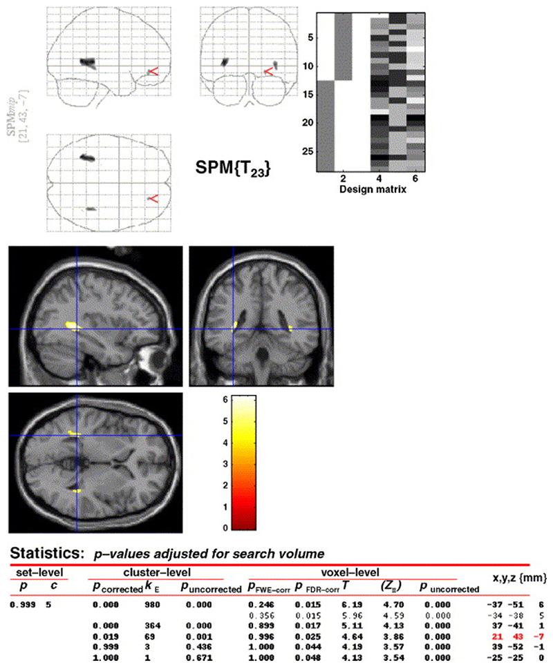
Conventional MRI studies have not provided definitive evidence of progressive loss of brain volume in the early stages of schizophrenia, although more subtle changes may have gone undetected. We have looked for such subtle changes using volumetric MRI and magnetization transfer imaging (MTI), an advanced MRI technique sensitive to subtle neuropathological abnormalities. Magnetization transfer images and high-resolution volumetric T1-weighted images were acquired from 16 patients with first-episode schizophrenia at the start of the study and 3.7 years later. A group of 12 healthy controls were also scanned on two occasions. Images were processed using a voxel-based approach that allows whole-brain analysis. There was a group difference with a significant volume loss in the patients' white matter adjacent to the lateral ventricles in the right and left temporal lobes, in medial temporal gyrus, and in the white matter in and around the right middle frontal gyrus. No cortical differences were detected between the groups using MTI or volumetric MRI. The absence of any time-by-group interaction suggests that these abnormalities do not progress in the early stages of the disease. The results of the study need to be interpreted in the light of the small sample size and of the limitations of current image analysis methods.
Figures
Similar articles
-
Brain pathology in first-episode psychosis: magnetization transfer imaging provides additional information to MRI measurements of volume loss.Neuroimage. 2010 Jan 1;49(1):185-92. doi: 10.1016/j.neuroimage.2009.07.037. Epub 2009 Jul 23. Neuroimage. 2010. PMID: 19632338 Free PMC article.
-
[Magnetization transfer imaging--the new method of brain tissue investigation in schizophrenia].Psychiatr Pol. 2007 May-Jun;41(3):309-18. Psychiatr Pol. 2007. PMID: 17900047 Review. Polish.
-
Progressive structural brain abnormalities and their relationship to clinical outcome: a longitudinal magnetic resonance imaging study early in schizophrenia.Arch Gen Psychiatry. 2003 Jun;60(6):585-94. doi: 10.1001/archpsyc.60.6.585. Arch Gen Psychiatry. 2003. PMID: 12796222
-
Gray and white matter brain abnormalities in first-episode schizophrenia inferred from magnetization transfer imaging.Arch Gen Psychiatry. 2003 Aug;60(8):779-88. doi: 10.1001/archpsyc.60.8.779. Arch Gen Psychiatry. 2003. PMID: 12912761
-
Regional deficits in brain volume in schizophrenia: a meta-analysis of voxel-based morphometry studies.Am J Psychiatry. 2005 Dec;162(12):2233-45. doi: 10.1176/appi.ajp.162.12.2233. Am J Psychiatry. 2005. PMID: 16330585 Review.
Cited by
-
Longitudinal brain changes in early-onset psychosis.Schizophr Bull. 2008 Mar;34(2):341-53. doi: 10.1093/schbul/sbm157. Epub 2008 Jan 29. Schizophr Bull. 2008. PMID: 18234701 Free PMC article. Review.
-
Brain pathology in first-episode psychosis: magnetization transfer imaging provides additional information to MRI measurements of volume loss.Neuroimage. 2010 Jan 1;49(1):185-92. doi: 10.1016/j.neuroimage.2009.07.037. Epub 2009 Jul 23. Neuroimage. 2010. PMID: 19632338 Free PMC article.
-
Brain Structural Changes in Schizophrenia Patients Compared to the Control: An MRI-based Cavalieri's Method.Basic Clin Neurosci. 2023 May-Jun;14(3):355-363. doi: 10.32598/bcn.2021.3481.1. Epub 2023 May 1. Basic Clin Neurosci. 2023. PMID: 38077177 Free PMC article.
-
The Longitudinal Course of Schizophrenia Across the Lifespan: Clinical, Cognitive, and Neurobiological Aspects.Harv Rev Psychiatry. 2016 Mar-Apr;24(2):118-28. doi: 10.1097/HRP.0000000000000092. Harv Rev Psychiatry. 2016. PMID: 26954596 Free PMC article. Review.
-
Magnetization transfer magnetic resonance imaging of the brain, spinal cord, and optic nerve.Neurotherapeutics. 2007 Jul;4(3):401-13. doi: 10.1016/j.nurt.2007.03.002. Neurotherapeutics. 2007. PMID: 17599705 Free PMC article. Review.
References
-
- Andreasen NC. Scale for the Assessment of Negative Symptoms (SANS) University of Iowa; Iowa City, IA: 1981.
-
- Andreasen NC. Scale for the Assessment of Positive Symptoms (SAPS) University of Iowa; Iowa City, IA: 1983.
-
- Antonova E, Kumari V, Morris R, Halari R, Anilkumar A, Mehrotra R, Sharma T. The relationship of structural alterations to cognitive deficits in schizophrenia: a voxel-based morphometry study. Biol Psychiatry. 2005;58(6):457–467. - PubMed
-
- Arnold SE, Trojanowski JQ, Gur RE, Blackwell P, Han LY, Choi C. Absence of neurodegenera-tion and neural injury in the cerebral cortex in a sample of elderly patients with schizophrenia. Arch Gen Psychiatry. 1998;55:225–232. - PubMed
-
- Ashburner J, Friston KJ. Voxel based morphometry—the methods. Neuroimage. 2000;11:805–821. - PubMed
Publication types
MeSH terms
Grants and funding
LinkOut - more resources
Full Text Sources
Medical
