. 2006 Aug;123(8):626-40.
doi: 10.1016/j.mod.2006.06.003. Epub 2006 Jul 14.

Notch signaling plays a key role in cardiac cell differentiation

Affiliations

Notch signaling plays a key role in cardiac cell differentiation

Mary D L Chau et al. Mech Dev. 2006 Aug.

Abstract

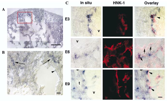
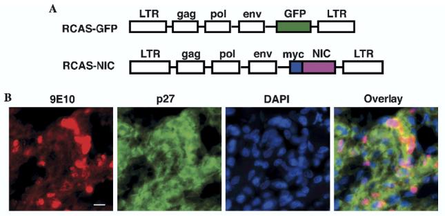
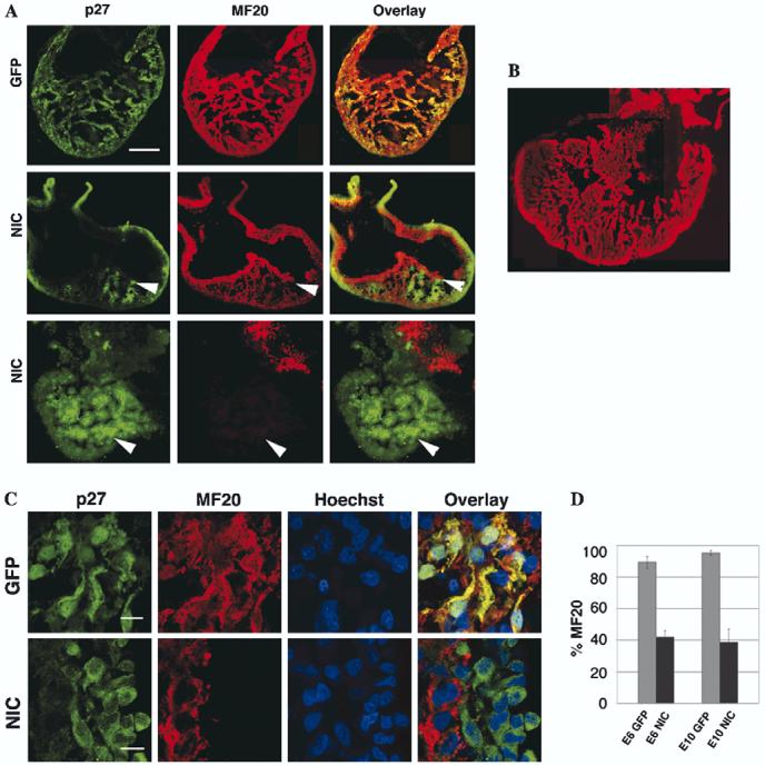
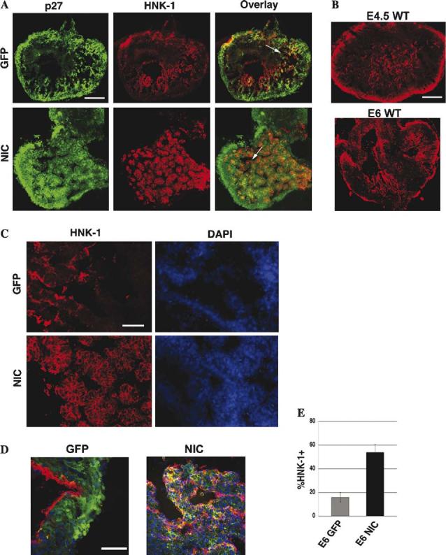
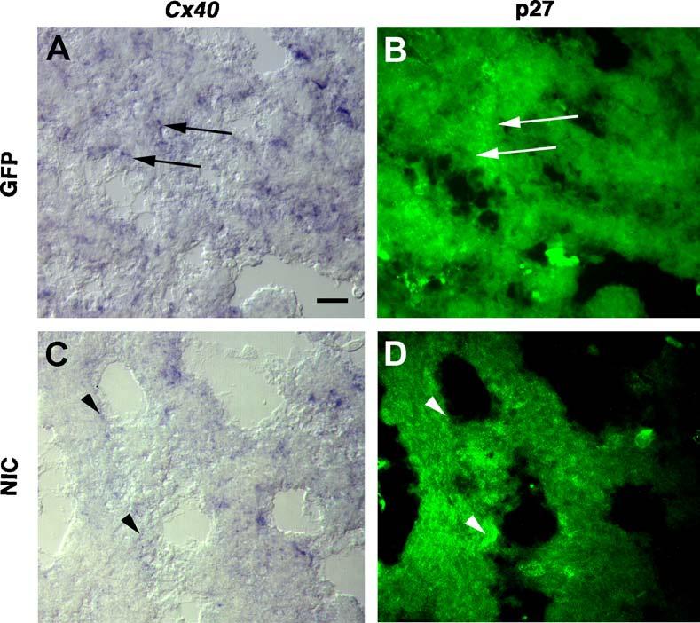
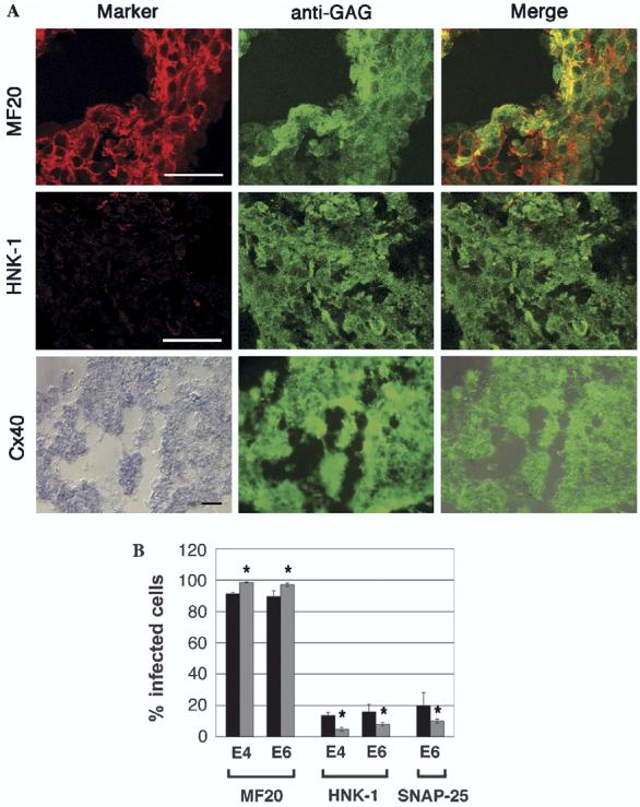
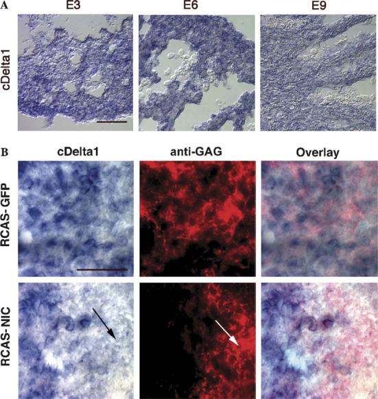
Results from lineage tracing studies indicate that precursor cells in the ventricles give rise to both cardiac muscle and conduction cells. Cardiac conduction cells are specialized cells responsible for orchestrating the rhythmic contractions of the heart. Here, we show that Notch signaling plays an important role in the differentiation of cardiac muscle and conduction cell lineages in the ventricles. Notch1 expression coincides with a conduction marker, HNK-1, at early stages. Misexpression of constitutively active Notch1 (NIC) in early heart tubes in chick exhibited multiple effects on cardiac cell differentiation. Cells expressing NIC had a significant decrease in expression of cardiac muscle markers, but an increase in expression of conduction cell markers, HNK-1, and SNAP-25. However, the expression of the conduction marker connexin 40 was inhibited. Loss-of-function study, using a dominant-negative form of Suppressor-of-Hairless, further supports that Notch1 signaling is important for the differentiation of these cardiac cell types. Functional studies show that the expression of constitutively active Notch1 resulted in abnormalities in ventricular conduction pathway patterns.


Figures

Fig. 1
Fig. 1
Expression patterns of notch1 mRNA in the embryonic chick heart, by in situ hybridizations on heart sections at embryonic days 3 (HH19), 6 (HH29), and 9 (HH35) (E3, E6, E9). (A) notch1 expression at E6 is shown at a low magnification in the ventricles. (B) The boxed area in A is shown at a higher magnification. Note the expression of notch1 in the myocardium (arrows), in addition to very weak signals in the endocardium (arrowhead). (C) To identify the cells expressing notch1, we immunostained the heart section with the antibody HNK-1 (red), after the completion of in situ hybridization. Note that notch1 in situ signals appear to be closely associated with cells positive for HNK-1 staining on the plasma membrane at E3 and E6 (arrows). The HNK-1 staining appears subendocardial. The sections shown are of oblique angles. At E9, many notch1-positive cells are no longer associated with HNK-1 staining. V, ventricles; t, trabecula. Scale bars in B, C = 20 μm; in A = 200 μm.
Fig. 2
Fig. 2
Expression of constitutively active Notch1 (NIC) via a retroviral construct in embryonic chick heart. (A) Retroviral constructs, RCAS-GFP and RCAS-NIC. LTR, long terminal repeat; gag, gene encoding the viral capsid proteins; env, gene encoding viral envelope protein; pol, gene encoding viral reverse transcriptase. (B) RCAS-NIC injected chick hearts were harvested at E6, sectioned and immunostained with p27 (anti-viral GAG protein, green) to identify the infected cells, and an antibody against the myc tag, 9E10 (red). DAPI (blue) was used to stain for the nuclei. Note that the myc-tag staining coincides with the nuclear stain. Scale bar, 10 μm.
Fig. 3
Fig. 3
Constitutively active Notch1 (NIC) inhibits cardiac muscle cell differentiation. RCAS-NIC or control RCAS-GFP injected hearts were harvested at E4.5 (A) and E6 (C), sectioned and immunostained with p27 (green) and the anti-sarcomeric myosin heavy chain marker, MF20 (red). Hoechst dye 34580 (blue) was used to stain for the nuclei. (A) E4.5 stained heart sections were analyzed at low magnification on an epifluorescence microscope. Note that, in the RCAS-GFP-infected sample (top panel), MF20 staining is widely distributed throughout the heart. RCAS-NIC-infected patches, however, show a substantial decrease in MF20 staining (arrowheads). In some heavily infected hearts, little MF-20 staining was seen in the ventricles (bottom panel). (B) Staining pattern of MF20 on a wild type E6 heart is shown. (C) To quantify the results, E6 heart samples were analyzed on confocal microscopy. Note that the control RCAS-GFP infected cells are mostly positive for MF20, showing characteristic banding patterns, whereas many RCAS-NIC infected cells are negative for MF20 staining. (D) Quantification of the results by scoring for the percent of infected cells stained with MF20 at E6 and E10, respectively. Scale bar in A common to B = 500 μm, and C = 10μm.
Fig. 4
Fig. 4
RCAS-NIC increases expression of the conduction system marker, HNK-1. RCAS-GFP or RCAS-NIC injected hearts were harvested at E4.5(A, C) or E6 (D), sectioned and immunostained with p27 (green), to identify infected cells, and the conduction lineage marker HNK-1 (red). Hoechst dye 34580 or DAPI (blue) were used to stain for the nuclei. (A) E4.5 stained heart sections were analyzed at low magnification on an epifluorescence microscope. Note that the RCAS-NIC infected sample shows a significant increase in HNK-1 staining as compared to the RCAS-GFP infected heart. (B) The staining patterns of HNK-1 in wild type E4.5 and E6 hearts are shown. Note that HNK-1 staining is concentrated around the trabeculae. (C) The areas of the hearts marked by arrows in (A) are shown in a higher magnification. The RCAS-NIC infected sample shows HNK-1 staining around almost every cell within the trabeculae, while HNK-1 staining mostly outlines the trabeculae in the RCAS-GFP infected heart. (D) The overlay images of the infected hearts at E6 are shown (red, HNK-1; green, P27; blue, Hoechst dye). Note that the trabeculae partially infected with RCAS-NIC has an increase in HNK-1 expression, especially within the trabeculae. (E) Quantification of the percentage of the cells infected with RCAS-GFP or RCAS-NIC expressing HNK-1 at E6. Scale bar in A common to B = 500 μm; C = 100 μm; D = 100 μm.
Fig. 5
Fig. 5
Increase in proportion of the RCAS-NIC infected cells expressing SNAP-25. RCAS-GFP or RCAS-NIC injected hearts were harvested at E6, sectioned and immunostained with anti-GAG antibody p27 (green), and the conduction marker SNAP-25 (red). Hoechst dye 34,580 (blue) was used to stain for the nuclei. (A) At low magnification, RCAS-NIC-infected cells are found to be largely localized to the trabeculae region where SNAP-25 expression is enriched. In contrast, the control RCAS-GFP-positive cells were largely distributed in the myocardium destined for the future compact zone. (B) The staining pattern of SNAP-25 antibody is shown on wild type E6 heart section. Note that the SNAP-25 antibody stains the tips of the trabeculae. (C) Stained heart sections were analyzed at higher magnification by confocal microscopy. Many RCAS-NIC-infected cells are SNAP-25 positive. High levels of expression of SNAP-25 were observed on the membrane and the cytoplasm of the RCAS-NIC expressing cells with some punctate staining among the myocardial fibers (top panels). (D) Quantification of the percentage of the cells infected with RCAS-GFP or RCAS-NIC expressing SNAP-25. Scale bar in A common to B = 500 μm and in C = 10 μm.
Fig. 6
Fig. 6
The expression of the conduction system marker connexin 40 (Cx40) is decreased by constitutively active Notch. RCAS-GFP (A, B) or RCAS-NIC (C, D) infected heart sections were analyzed by in situ hybridization with the Cx40 probe. Trabeculae areas were shown for both samples. Note the dark subendocardial Cx40 signals were not decreased by RCAS-GFP expression (arrows). The entire area shown was infected by RCAS-GFP. Because the dark precipitates of the in situ signals quench the fluorescence, small non-fluorescent areas overlapping exactly with the in situ signals are likely infected. In contrast, the RCAS-NIC-infected areas correlated with decreased Cx40 in situ signals, both in the subendocardial cells (arrowheads) and myocardial cells. Scale bar, 20 μm.
Fig. 7
Fig. 7
Expression of the dominant-negative form of Suppressor-of-Hairless alters the expression of cardiac cell type markers. Chick embryos were injected with the control virus, RCAS-GFP, or with the RCAS-Su(H)DN virus. (A) Hearts were harvested at E6 and E4 (not shown), sectioned and immunostained with anti-viral GAG antibody, and various cell type specific markers. Note that most of the RCAS-Su(H)DN infected are positive for MF20, but negative for HNK-1. Expression of Cx40 was also analyzed by in situ hybridization on the infected heart sections, followed by staining with anti-GAG antibody. In the RCAS-Su(H)DN infected areas, the Cx40 expression appears to be at a low level uniformly, unlike the control hearts which show relatively high levels of Cx40 in some subendocardial cells (Fig. 6A, B). Scale bars, 20 μm. (B) Quantification of the results by scoring the percentage of the RCAS-Su(H)DN infected cells that express various markers. Note that, compared to the GFP control (black bar), expression of Su(H)DN (gray bar) increased the percentage of cells positive for MF20, and decreased the percentage of cells positive for HNK-1 or SNAP-25. Asterisks indicate statistical significance.
Fig. 8
Fig. 8
Constitutively active Notch1 alters the conduction propagation pattern in embryonic chick hearts. (A) Optical mapping was performed on uninjected control hearts at E4.5-5. Images were collected at 2 ms/frame, and processed using a custom software. The first derivative was computed and the maximum upstroke velocity was defined as dF/dt max and depicted as red in the color scale. Note, in the top panel, the impulse propagates along the myocardial wall from the atrium towards the outflow tract in the immature propagation pattern (red arrows) in about 8–10 ms. Also note, in the bottom panel, the impulse travels from the apex to the base (red arrows) within 8 ms in the mature activation sequence. (B) Optical mapping was similarly performed on RCAS-NIC-injected hearts at E4.5-5. After imaging, the hearts were fixed, sectioned and stained with anti-P27 to visualize the extent of infection (shown in green fluorescence). Note, the impulse fails to advance towards the base and dissipates within 4ms in the top two panels, and a diffuse activation pattern in the bottom panel. The heart in the bottom panel was more extensively infected with the RCAS-NIC virus.
Fig. 9
Fig. 9
Expression of Delta1 is decreased in the cells infected with RCAS-NIC. (A) Expression of the chick Delta1 transcripts in the developing heart. In situ hybridizations were performed on cardiac sections of E3, E6, and E9 chick embryos. Note that Delta1 is widely expressed in the myocardium. (B) Expression of Delta1 was reduced in the areas infected with the RCAS-NIC virus. Heart tubes were injected with control RCAS-GFP or RCAS-NIC virus at HH9-10 and the infected hearts were harvested at E6. In situ hybridizations were performed on the infected tissues with the Delta1 probe, followed by anti-viral GAG staining. Note that Delta1 transcript expression was inhibited by the expression of NIC (arrows), but not GFP. Scale bars, 50 μm.

Similar articles

Cited by

References

    1. Aoyama N, Kikawada R, Yamashina S. Immunohistochemical study on the development of the rat heart conduction system using anti-Leu-7 antibody. Arch. Histol. Cytol. 1993;56:303–315. - PubMed
    1. Aoyama N, Tamaki H, Kikawada R, Yamashina S. Development of the conduction system in the rat heart as determined by Leu-7 (HNK-1) immunohistochemistry and computer graphics reconstruction. Lab. Invest. 1995;72:355–366. - PubMed
    1. Artavanis-Tsakonas S, Rand MD, Lake RJ. Notch signaling: cell fate control and signal integration in development. Science. 1999;284:770–776. - PubMed
    1. Bao ZZ, Bruneau BG, Seidman JG, Seidman CE, Cepko CL. Regulation of chamber-specific gene expression in the developing heart by Irx4. Science. 1999;283:1161–1164. - PubMed
    1. Bastide B, Neyses L, Ganten D, Paul M, Willecke K, Traub O. Gap junction protein connexin40 is preferentially expressed in vascular endothelium and conductive bundles of rat myocardium and is increased under hypertensive conditions. Circ. Res. 1993;73:1138–1149. - PubMed

Publication types

LinkOut - more resources