. 2006 Jul 25;103(30):11323-8.
doi: 10.1073/pnas.0604772103. Epub 2006 Jul 14.

Benefit of farnesoid X receptor inhibition in obstructive cholestasis

Affiliations

Benefit of farnesoid X receptor inhibition in obstructive cholestasis

Catherine Stedman et al. Proc Natl Acad Sci U S A. .

Abstract

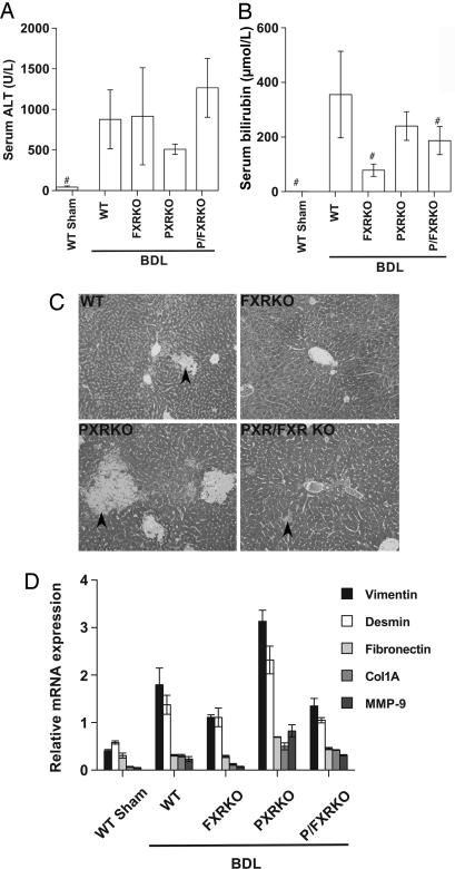
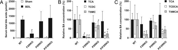
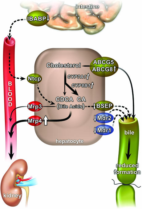
The nuclear hormone receptors farnesoid X receptor (FXR) and pregnane X receptor have been implicated in regulating bile acid, lipid, carbohydrate, and xenobiotic metabolism. Bile duct ligation was used to increase endogenous bile acids and evaluate the roles of these receptors in modulating cholestatic liver injury. FXR knockout (KO) mice were found to be protected from obstructive cholestasis. Concurrent deletion of FXR also could ameliorate an increase in liver injury that is seen usually in pregnane X receptor KO mice with cholestasis. Mechanisms proposed for this protection include the lowering of bile acid concentrations and altered expression of the hepatic transporters Mdr1, Mdr2, BSEP, and Mrp4. FXR KO mice also exhibit a biphasic lipid profile after bile duct ligation, with an increase in high-density lipoprotein cholesterol and triglycerides by day 6. The expression of apolipoprotein AV was reduced in these mice, implicating FXR in triglyceride regulation. We show that FXR modulates cholestasis by controlling bile acids within the hepatocyte and is involved in bile acid synthesis, bile excretion via BSEP, and serum export via Mrp4. This study strongly suggests a potential clinical role for FXR antagonists in the treatment of obstructive cholestatic liver disorders.


Conflict of interest statement

Conflict of interest statement: No conflicts declared.

Figures

Fig. 1.
Fig. 1.
Deletion of PXR or FXR results in differential patterns of liver injury after BDL. Serum and liver samples were collected 6 days after BDL or sham operation in WT, FXRKO, PXRKO, and P/FXRKO mice. (A) Serum ALT was significantly elevated in all BDL animals compared with shams. (B) Total serum bilirubin was significantly elevated in WT, PXRKO, and P/FXRKO BDL mice relative to sham and was significantly lower in FXR-null mice than WT after BDL. #, P < 0.01 relative to WT BDL. (C) Livers were fixed for histological examination and stained with Gomori’s Trichrome to evaluate liver injury and necrosis. WT livers show modest areas of bile infarct/necrosis (arrowheads); PXRKO mice have increased areas of bile infarct/necrosis compared to WT; FXRKO and P/FXRKO mice have a reduction in bile infarcts, but disseminated liver cell necrosis and mild to moderate hepatocyte steatosis is still observed. (D) Effects of BDL on hepatic expression of genes involved in fibrosis and tissue remodeling. Relative mRNA expression of vimentin, desmin, fibronectin, collagen 1A (Col1A), and matrix metalloproteinase 9 (MMP-9) were examined by real-time RT-PCR and normalized for U36B4 expression.
Fig. 2.
Fig. 2.
Relative bile acid concentrations of CDCA and its metabolites decrease in FXR-null BDL mice. Bile acid concentrations were measured 6 days after BDL or sham operation. (A) Total serum bile acids increased after BDL in all genotypes but were reduced in FXRKO relative to WT BDL mice. (B) Individual serum bile acids were quantitated by liquid chromatography/MS and expressed as a percentage of WT BDL concentration. Serum tauro-cholic acid (TCA), Tβ-MCA, and tauro-chenodeoxycholic acid (TCDCA) were significantly reduced in FXRKO mice; TCDCA and Tβ-MCA were reduced in P/FXRKO mice, but TCA concentrations were preserved in this genotype. (C) Individual hepatic bile acids were quantitated by liquid chromatography/MS and expressed as a percentage of WT BDL concentration. All FXR-null genotypes had a significant relative reduction in TCDCA and Tβ-MCA relative to WT BDL, whereas concentrations of TCA were preserved. #, P < 0.01 relative to WT BDL.
Fig. 3.
Fig. 3.
FXRKO mice have a biphasic lipid profile after BDL with marked hypertriglyceridemia. Blood samples were collected from mice before BDL or sham operation on day 0 and day 6. (A) Total cholesterol concentrations were similar in all sham mice. (B) After BDL, total cholesterol increased significantly in WT mice (P < 0.01). In FXRKO mice, a transient increase at day 3 was followed by a decline to less than one-half of WT levels by day 6. PXRKO mice also had an attenuated increase in total cholesterol. (C) Serum HDL cholesterol was significantly higher at baseline in FXRKO mice. (D) After BDL, FXRKO mice had a biphasic HDL cholesterol profile, with a transient decrease in HDL on day 3 and subsequent increase by day 6. (E) Fasting serum triglycerides in sham animals showed no genotype-specific differences by using the Cholestech LDX Analyzer. (F) After BDL, a marked increase in serum triglycerides was observed in FXR-null mice. (G) FXR regulates expression of apoAV. Relative mRNA expression of apolipoprotein genes in liver were examined by real-time RT-PCR. apoAV is induced in WT BDL mice, and its expression depends on FXR.
Fig. 4.
Fig. 4.
Model showing the benefit of FXR deletion or antagonism on FXR target genes in obstructive cholestasis. Although overall bile acid synthesis is increased, there is reduced synthesis of CDCA and reduced absorption of bile acids from the intestine. Canalicular transporters involved in bile formation are down-regulated, reducing pressure in the obstructed bile ducts, and the basolateral transporter Mrp4 is up-regulated, facilitating export of bile acids into blood where they are available for renal excretion. Dashed arrows represent down-regulation; continuous or block arrows depict up-regulation.

References

    1. Russell D. W. Annu. Rev. Biochem. 2003;72:137–174. - PubMed
    1. Sirvent A., Claudel T., Martin G., Brozek J., Kosykh V., Darteil R., Hum D. W., Fruchart J. C., Staels B. FEBS Lett. 2004;566:173–177. - PubMed
    1. Sinal C. J., Tohkin M., Miyata M., Ward J. M., Lambert G., Gonzalez F. J. Cell. 2000;102:731–744. - PubMed
    1. Staudinger J. L., Madan A., Carol K. M., Parkinson A. Drug Metab. Dispos. 2003;31:523–527. - PubMed
    1. Sonoda J., Xie W., Rosenfeld J. M., Barwick J. L., Guzelian P. S., Evans R. M. Proc. Natl. Acad. Sci. USA. 2002;99:13801–13806. - PMC - PubMed

Publication types