. 2006 Jul 25;103(30):11282-7.
doi: 10.1073/pnas.0602131103. Epub 2006 Jul 14.

Drusen, choroidal neovascularization, and retinal pigment epithelium dysfunction in SOD1-deficient mice: a model of age-related macular degeneration

Affiliations

Drusen, choroidal neovascularization, and retinal pigment epithelium dysfunction in SOD1-deficient mice: a model of age-related macular degeneration

Yutaka Imamura et al. Proc Natl Acad Sci U S A. .

Abstract

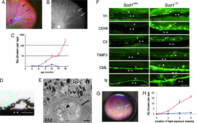
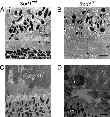
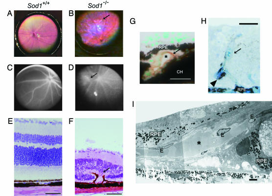
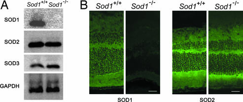
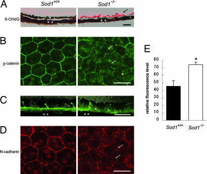
Oxidative stress has long been linked to the pathogenesis of neurodegenerative diseases; however, whether it is a cause or merely a consequence of the degenerative process is still unknown. We show that mice deficient in Cu, Zn-superoxide dismutase (SOD1) have features typical of age-related macular degeneration in humans. Investigations of senescent Sod1(-/-) mice of different ages showed that the older animals had drusen, thickened Bruch's membrane, and choroidal neovascularization. The number of drusen increased with age, and exposure of young Sod1(-/-) mice to excess light induced drusen. The retinal pigment epithelial cells of Sod1(-/-) mice showed oxidative damage, and their beta-catenin-mediated cellular integrity was disrupted, suggesting that oxidative stress may affect the junctional proteins necessary for the barrier integrity of the retinal pigment epithelium. These observations strongly suggest that oxidative stress may play a causative role in age-related retinal degeneration, and our findings provide evidence for the free radical theory of aging. In addition, these results demonstrate that the Sod1(-/-) mouse is a valuable animal model to study human age-related macular degeneration.


Conflict of interest statement

Conflict of interest statement: No conflicts declared.

Figures

Fig. 1.
Fig. 1.
Senescent Sod1−/− mice showing drusen. (A) Fundus photograph from a 15-month-old Sod1−/− mouse. White and slightly yellowish deposits are evident (arrows). (B) Fluorescein angiogram showing hyperfluorescent areas corresponding to the sites of the drusen (arrows). (C) Plot of the number of drusen in Sod1−/− mice at 3–18 months of age, showing an increase in the number of drusen with age. Values for Sod1−/− are plotted in red, and those for Sod1+/+ are plotted in blue. (D) Dome-shaped deposit (arrow) between the RPE (∗) and choroid (∗∗) in a 12-month-old Sod1−/− mouse. (E) Electron micrograph of drusen (arrow) from a 12-month-old Sod1−/− mouse. Heterogeneous materials separated by a septum (arrowhead) are observed. The drusen appears to contain debris-like material of outer segments. BM, Bruch’s membrane. (F) Immunohistochemistry with markers for vitronectin (Vn), CD46, C5, TIMP3, CML, and Ig. Drusen and/or their surrounding tissues from 12-month-old Sod1−/− mice (Right) are positive for vitronectin, CD46, C5, TIMP3, CML, and Ig in contrast to the results for age-matched wild-type mice (Left). ∗, RPE; ∗∗, choroid; arrow, positive signal; arrowhead, positive signal of CML in RPE cell layer. (G) Fundus changes in a 5-month-old Sod1−/− mouse after continuous light exposure for 8 weeks (white fluorescent light of 10,000 lux). Drusen are present (circles and arrows). (H) The number of drusen increases with the length of light exposure in Sod1−/− mice, as compared with the minimal appearance of drusen in wild-type mice. Values for Sod1−/− are plotted in red, and those for Sod1+/+ are plotted in blue. (Scale bars: D and F, 50 μm; E, 2 μm.)
Fig. 2.
Fig. 2.
Degenerated RPE and thickened Bruch’s membrane in Sod1−/− mice. (A) Ultrastructure of Bruch’s membrane (BM) in a 12-month-old wild-type mouse. (B) Thickened Bruch’s membrane of an age-matched Sod1−/− mouse. RPE cells show marked vacuolization (∗). (C) Ultrastructure of the outer segments (OS) of the photoreceptor cells and RPE of a 12-month-old wild-type mouse. (D) Destruction of inner segments (IS), outer segments, and RPE in an age-matched Sod1−/− mouse. (Scale bars: B and D, 2 μm.)
Fig. 3.
Fig. 3.
CNVs in Sod1−/− mice. (A) Fundus of a 15-month-old wild-type mouse. (B) Fundus of a 15-month-old Sod1−/− mouse showing exudative retinal changes (arrow) adjacent to the optic disk. (C) Fluorescein angiogram of the wild-type mouse reveals no abnormal findings. (D) Fluorescein angiogram reveals dye leakage (arrow) from the CNV in a 15-month-old Sod1−/− mouse. (E) Hematoxylin and eosin staining of a retinal section of a wild-type mouse. (F) Fibrovascular tissue (arrow) involving RPE in the thinned retina of the Sod1−/− mouse. A thinning of the outer nuclear layer can be seen. Hematoxylin and eosin staining. (G) CNV in a 12-month-old Sod1−/− mouse. CNV is present beneath the RPE, and the vascular cavity (∗) is surrounded by CD31-positive (diaminobenzidine) endothelial cells (arrow). Methyl green counterstaining was used. CH, choroid. (H) CNV with vessel cavity filled with erythrocytes (arrow) proliferates spirally into the sensory retina in a 12-month-old Sod1−/− mouse. RPE is attached within the intraretinal vascular structure (arrowhead). Toludine blue was used for staining. (I) Electron micrograph of CNV. CNV (∗) surrounded by endothelial cells (E) pass through a defect in Bruch’s membrane. RPE is severely disrupted. (Scale bars: EH, 50 μm; I, 2 μm.)
Fig. 4.
Fig. 4.
Expression of SOD1, SOD2, and SOD3 in the eyes of Sod1−/− mice. (A) Western blot analysis reveals the absence of SOD1 protein expression in the eyes of Sod1−/− mice. The protein levels of SOD2 (Mn-SOD) and SOD3 (extracellular SOD) appear to be the same in wild-type and Sod1−/− mice. (B) SOD1 and SOD2 expression in the retinas of wild-type and Sod1−/− mice. (Scale bars: 50 μm.)
Fig. 5.
Fig. 5.
Oxidatively damaged RPE and its disrupted β-catenin-mediated integrity in Sod1−/− mice. (A) 8-hydroxy-2′-deoxyguanosine (8-OHdG) was minimally present in the RPE of a 10-month-old wild-type mouse; however, it is markedly up-regulated in the RPE of a 10-month-old Sod1−/− mouse (arrow). ∗, RPE; ∗∗, choroid. (B) Irregular distribution of junctional proteins in RPE. β-Catenin is located at the cell walls of RPE cells in a flat-mount preparation of a 12-month-old wild-type mouse. The cytoplasmic level of β-catenin increased (∗), with reduced expression at the junctional sites (arrows). (C) A cross-section of the RPE of a 10-month-old wild-type mouse showing staining of the cell walls by β-catenin. An absence of β-catenin in the periphery and the increasing cytoplasmic level (∗) are observed in a 10-month-old Sod1−/− mouse. (D) Distribution of N-cadherin in a 12-month-old Sod1−/− mouse is severely disrupted, which is in contrast to the hexagonal expressions of N-cadherin in an age-matched wild-type mouse. Incomplete expression at the junctional sites can be seen (arrows). (E) Quantification of cytoplasmic fluorescence intensity of β-catenin in RPE cells by using the digitalized images of the flat mounts of the wild-type and the Sod1−/− mice. Randomized selected cells (n = 49) of each group are compared. Results are shown as mean ± SEM. ∗, Statistical difference between the two groups (P < 0.001, Student’s t test). (Scale bars: A, B, and D, 50 μm; C, 10 μm.)

Similar articles

Cited by

References

    1. Sommer A., Tielsch J. M., Katz J., Quigley H. A., Gottsch J. D., Javitt J. C., Martone J. F., Royall R. M., Witt K. A., Ezrine S. N. Engl. J. Med. 1991;325:1412–1417. - PubMed
    1. Smith W., Assink J., Klein R., Mitchell P., Klaver C. C., Klein B. E., Hofman A., Jensen S., Wang J. J., de Jong P. T. Ophthalmology. 2001;108:697–704. - PubMed
    1. Attbo K., Mitchell P., Smith W. Ophthalmology. 1996;103:357–364. - PubMed
    1. VanNewkirk M. R., Weih L., McCarty C. A., Taylor H. R. Ophthalmology. 2001;108:960–967. - PubMed
    1. Oshima Y., Ishibashi T., Murata T., Tahara Y., Kiyohara Y., Kubota T. Br. J. Ophthalmol. 2001;39:367–372. - PMC - PubMed

Publication types