. 2006 Jul 25;103(30):11124-9.
doi: 10.1073/pnas.0601924103. Epub 2006 Jul 14.

The 2.7-Angstrom crystal structure of a 194-kDa homodimeric fragment of the 6-deoxyerythronolide B synthase

Affiliations

The 2.7-Angstrom crystal structure of a 194-kDa homodimeric fragment of the 6-deoxyerythronolide B synthase

Yinyan Tang et al. Proc Natl Acad Sci U S A. .

Abstract

The x-ray crystal structure of a 194-kDa fragment from module 5 of the 6-deoxyerythronolide B synthase has been solved at 2.7 Angstrom resolution. Each subunit of the homodimeric protein contains a full-length ketosynthase (KS) and acyl transferase (AT) domain as well as three flanking "linkers." The linkers are structurally well defined and contribute extensively to intersubunit or interdomain interactions, frequently by means of multiple highly conserved residues. The crystal structure also reveals that the active site residue Cys-199 of the KS domain is separated from the active site residue Ser-642 of the AT domain by approximately 80 Angstrom. This distance is too large to be covered simply by alternative positioning of a statically anchored, fully extended phosphopantetheine arm of the acyl carrier protein domain from module 5. Thus, substantial domain reorganization appears necessary for the acyl carrier protein to interact successively with both the AT and the KS domains of this prototypical polyketide synthase module. The 2.7-Angstrom KS-AT structure is fully consistent with a recently reported lower resolution, 4.5-Angstrom model of fatty acid synthase structure, and emphasizes the close biochemical and structural similarity between polyketide synthase and fatty acid synthase enzymology.


Conflict of interest statement

Conflict of interest statement: No conflicts declared.

Figures

Fig. 1.
Fig. 1.
Chain elongation cycle catalyzed by a minimal PKS module. In this example, the crystallized KS–AT fragment is shown in color, whereas the ACP domains that interact with the KS and AT are in black and white or grayscale. (1) A methylmalonyl unit that is added to a pentaketide substrate to yield the hexaketide product. KS is primed with a growing polyketide chain by the upstream ACP. AT is acylated with a methylmalonyl extender unit from its CoA derivative (2), which is transferred to the downstream ACP (3) and condensation takes place in the active site of KS with the release of carbon dioxide (4). The extended polyketide chain is anchored on the downstream ACP. Domains are drawn in the order they appear in amino acid sequence of the module. Orange, N-terminal docking domain; blue, ketosynthase (KS); green, acyl transferase (AT); gray, acyl carrier protein (ACP); yellow, the KS–to–AT linker; red, post-AT linker peptide. The phosphopantetheine prosthetic group of the ACP is drawn as a curly line. The same coloring scheme is used in subsequent figures.
Fig. 2.
Fig. 2.
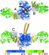
X-ray crystal structure of the KS–AT didomain from DEBS module5. The KS–AT protein forms a homodimer. Orange, N-terminal coiled coil linker domain; blue, ketosynthase (KS) domain; green, acyl transferase (AT) domain; yellow, KS–to–AT linker; red, post-AT linker peptide. Residues 458–465, which lack electron density, are manually modeled and shown in gray. Interdomain interactions detailed in Fig. 4 (vide infra) are highlighted in gray boxes. (A and B) Top view. (C and D) Side view.
Fig. 3.
Fig. 3.
Electron density map of the post-AT linker peptide. The 2FoFc map is contoured at 1.5 σ.
Fig. 4.
Fig. 4.
Protein–protein interactions in the KS–AT didomain (for clarity, only selected residues are labeled). Locations of the interacting regions are shown in Fig. 2. (A) Highly conserved hydrophobic core at the interface between the KS domain, the KS–to–AT linker, and the post-AT linker peptide. (B) Highly conserved hydrophobic core at the interface between the AT domain, the KS–to–AT linker, and the post-AT linker peptide. (C) Two key hydrogen bonds between the AT domain and the KS–to–AT linker. (D) Hydrogen bonds between the KS domain, the KS–to–AT linker, and the post-AT linker peptide.
Fig. 5.
Fig. 5.
Proposed substrate binding sites of the KS and AT domains. (A) The surfaces of the individual KS domains are shown in blue and purple. The KS active site is separated from the AT active site by ≈80 Å. (B) Model of the pentaketide-phosphopantetheine group in the KS active site near the KS:KS dimer interface. (C) Model of the (2S)-methylmalonyl-phosphopantetheine group in the active site of the AT domain.

References

    1. Walsh C. T. Science. 2004;303:1805–1810. - PubMed
    1. Cane D. E., Walsh C. T., Khosla C. Science. 1998;282:63–68. - PubMed
    1. Asturias F. J., Chadick J. Z., Cheung I. K., Stark H., Witkowski A., Joshi A. K., Smith S. Nat. Struct. Mol. Biol. 2005;12:225–232. - PubMed
    1. Cortes J., Haydock S. F., Roberts G. A., Bevitt D. J., Leadlay P. F. Nature. 1990;348:176–178. - PubMed
    1. Donadio S., Staver M. J., McAlpine J. B., Swanson S. J., Katz L. Science. 1991;252:675–679. - PubMed

Publication types

Substances

Associated data