. 2006 Sep;142(1):265-79.
doi: 10.1104/pp.106.084657. Epub 2006 Jul 14.

The Medicago truncatula lysin [corrected] motif-receptor-like kinase gene family includes NFP and new nodule-expressed genes

Affiliations

The Medicago truncatula lysin [corrected] motif-receptor-like kinase gene family includes NFP and new nodule-expressed genes

Jean-François Arrighi et al. Plant Physiol. 2006 Sep.

Erratum in

  • Plant Physiol. 2007 Feb;143(2):1078

Abstract

Rhizobial Nod factors are key symbiotic signals responsible for starting the nodulation process in host legume plants. Of the six Medicago truncatula genes controlling a Nod factor signaling pathway, Nod Factor Perception (NFP) was reported as a candidate Nod factor receptor gene. Here, we provide further evidence for this by showing that NFP is a lysin [corrected] motif (LysM)-receptor-like kinase (RLK). NFP was shown both to be expressed in association with infection thread development and to be involved in the infection process. Consistent with deviations from conserved kinase domain sequences, NFP did not show autophosphorylation activity, suggesting that NFP needs to associate with an active kinase or has unusual functional characteristics different from classical kinases. Identification of nine new M. truncatula LysM-RLK genes revealed a larger family than in the nonlegumes Arabidopsis (Arabidopsis thaliana) or rice (Oryza sativa) of at least 17 members that can be divided into three subfamilies. Three LysM domains could be structurally predicted for all M. truncatula LysM-RLK proteins, whereas one subfamily, which includes NFP, was characterized by deviations from conserved kinase sequences. Most of the newly identified genes were found to be expressed in roots and nodules, suggesting this class of receptors may be more extensively involved in nodulation than was previously known.


Figures

Figure 1.
Figure 1.
Amino acid alignment of NFP (M. truncatula), SYM10 (pea), and NFR5 (L. japonicus), showing conserved domain structure and conserved Cys residues in the predicted extracellular parts (boxed). Sites where the P-loops, activation loops, and the DFG motifs are missing are indicated. In NFP, positions of mutations found in nfp-1 (residue no. 1) and nfp-2 (residue no. 67) are boxed and indicated by arrowheads. SP, Signal peptide; LysM1, LysM2, and LysM3, three LysM domains; TM, transmembrane segment.
Figure 2.
Figure 2.
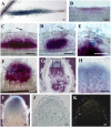
Localization of NFP gene expression throughout nodulation in M. truncatula. A to H, Expression of a ProNFP-GUS fusion in transgenic roots, inoculated or not with S. meliloti. A, Whole, noninoculated lateral root showing constitutive GUS activity (blue) in root hairs. B to F, Root segments showing GUS activity (magenta) in the epidermis 1 d postinoculation (dpi; B), in the inner and middle cortex where cell divisions are occurring under a curled root hair (arrow) at 2 dpi (C), in the inner and middle cortex 2 dpi, and in outer cortical cells (D), in outer cortical cells through which an infection thread (arrow) is passing (E), and in an emerging nodule 5 dpi (F). G and H, Longitudinal section of an 18-d-old nodule showing GUS activity (blue) in the apical region and bacteria (magenta) in infection threads and the nitrogen-fixing zones. H, Close-up of the box in G. I, GUS activity (blue) in an 18-d-old whole nodule, with bacteria not stained. J and K, In situ hybridization of NFP gene expression on a sectioned 14-d-old nodule using a 35S-UTP labeled antisense NFP probe. Bright-field image of nodule section with silver grains visible in black (J); epipolarization image of I (K). Arrows indicate points of infection in root hair curls (C and D) and an infection thread (E). Bars = 100 μm.
Figure 3.
Figure 3.
Analysis of the role of NFP in controlling infection thread formation. Roots of wild-type M. truncatula plants were transformed with either the vector alone (A) or an RNAi construct for NFP (B and C). Confocal images of root hairs in which GFP-expressing S. meliloti are seen in infection threads, which are tubular for the control and sac-like in NFPi root hairs. Root hairs in A and C were counterstained by propidium iodide (0.2 μg/mL). Bars = 10 μm.
Figure 4.
Figure 4.
Autophosphorylation tests on the intracellular kinase domains of NFP and LYK3. Purified GST-tagged intracellular domains of NFP, LYK3, and BAK1 (GST-NFP, GST-LYK3, and GST-BAK1) were assayed for autophosphorylation activity using [γ-32P]ATP. Reactions were subjected to SDS-PAGE and proteins visualized with Coomassie Blue (A). Phosphorylated proteins were visualized by phosphor imaging (B). Molecular mass markers of 55 and 72 kD are shown.
Figure 5.
Figure 5.
Schematic representation of the domain structure of 16 M. truncatula LysM-RLKs, based on the amino acid alignment presented in Supplemental Figure 1 and protein domain predictions explained in the text. SP, Signal peptide; LysM1, LysM2, and LysM3, three LysM domains; CXC, Cys-X-Cys motif; TM, transmembrane segment; PL, P-loop; AL, activation loop; Ser-Thr kinase, predicted intracellular kinase domain.
Figure 6.
Figure 6.
Phylogenetic tree of kinase domains of LysM-RLK proteins of M. truncatula (16), rice (six), Arabidopsis (five), and NFR1 and NFR5 of L. japonicus, showing bootstrap values. NFP and LYK3 are underlined and the three groups are labeled I, II, and III.
Figure 7.
Figure 7.
Structural predictions for LysM domains of NFP. 1 and 3, HCA plots showing homologous hydrophobic residues (circled orange) and secondary structural features (boxed; 1) and electropositively (circled blue) and electronegatively (circled red) charged residues (3). 2, Ribbon diagrams showing the N- and C-terminal ends of the LysM domain polypeptides. 4, 3-D models with electrostatic potentials displayed on the molecular surface of LysM domains at −5 kT and +5 kT levels; red (electronegative), blue (electropositive), and white (neutral). A, LysM domain of E. coli murein hydrolase. B to D, Structural predictions for NFP LysM domains built from the atomic coordinates of this E. coli LysM domain: LysM1 (B); LysM2 (C); LysM3 (D). Note the strongly electropositive cavity in LysM2 of NFP (4C).
Figure 8.
Figure 8.
Genetic map positions of M. truncatula LysM-RLK genes. LYK and LYR genes are positioned between microsatellite markers on six of the eight M. truncatula linkage groups (LG). The three cases of tandemly arranged genes (NFP/LYR3, LYK17, and LYR5/LYR6) are each indicated by a single map position. LYR4 and LYK17 also have a single map position, but are not physically linked (see text). The drawing was made using MapChart (Voorips, 2002). Distances are in cm Kosambi.

References

    1. Amon P, Haas E, Sumper M (1998) The sex-inducing pheromone and wounding trigger the same set of genes in the multicellular green alga Volvox. Plant Cell 10: 781–789 - PMC - PubMed
    1. Ané JM, Kiss GB, Riely BK, Penmetsa RV, Oldroyd GE, Ayax C, Lévy J, Debellé F, Baek JM, Kalo P, et al (2004) Medicago truncatula DMI1 required for bacterial and fungal symbioses in legumes. Science 30: 1364–1367 - PubMed
    1. Ardourel M, Demont N, Debelle F, Maillet F, de Billy F, Promé JC, Dénarié J, Truchet G (1994) Rhizobium meliloti lipooligosaccharide nodulation factors: different structural requirements for bacterial entry into target root hair cells and induction of plant symbiotic developmental responses. Plant Cell 6: 1357–1374 - PMC - PubMed
    1. Bateman A, Bycroft M (2000) The structure of a LysM domain from E. coli membrane-bound lytic murein transglycosylase D (MltD). J Mol Biol 299: 1113–1119 - PubMed
    1. Ben Amor B, Shaw SL, Oldroyd GE, Maillet F, Penmetsa RV, Cook D, Long SR, Dénarié J, Gough C (2003) The NFP locus of Medicago truncatula controls an early step of Nod factor signal transduction upstream of a rapid calcium flux and root hair deformation. Plant J 34: 495–506 - PubMed

Publication types

MeSH terms

Associated data