. 2006 Sep;142(1):254-64.
doi: 10.1104/pp.106.080119. Epub 2006 Jul 14.

ZmPIN1a and ZmPIN1b encode two novel putative candidates for polar auxin transport and plant architecture determination of maize

Affiliations

ZmPIN1a and ZmPIN1b encode two novel putative candidates for polar auxin transport and plant architecture determination of maize

Nicola Carraro et al. Plant Physiol. 2006 Sep.

Abstract

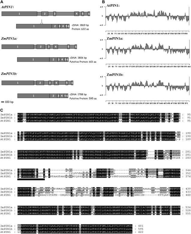
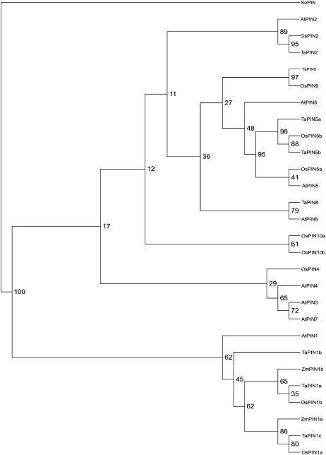
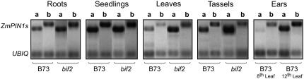
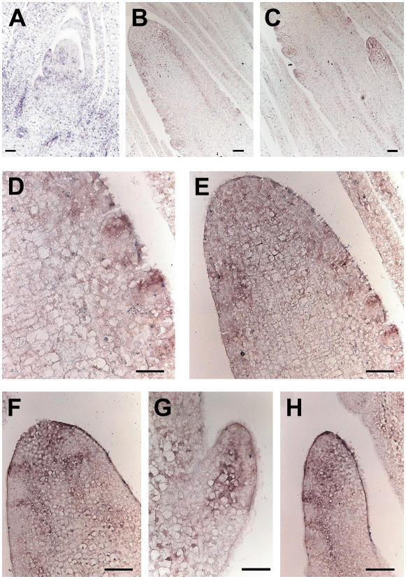
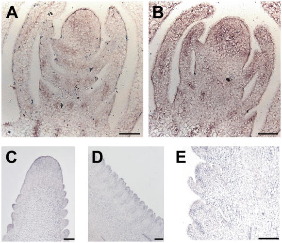
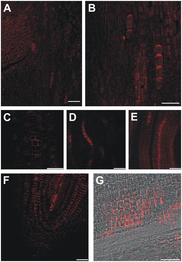
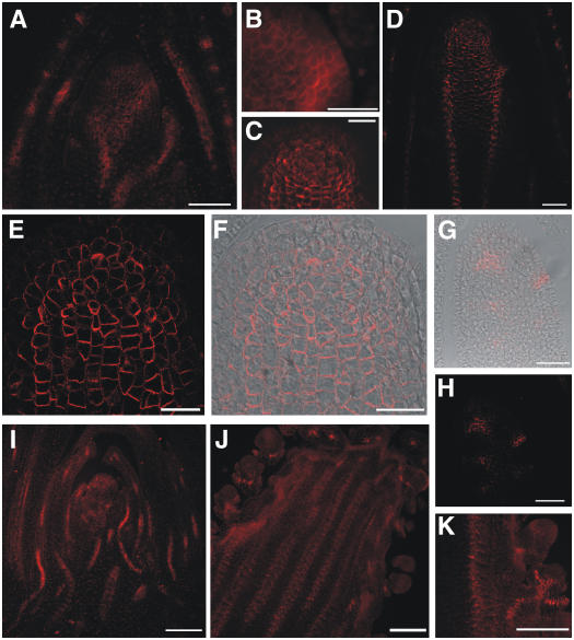
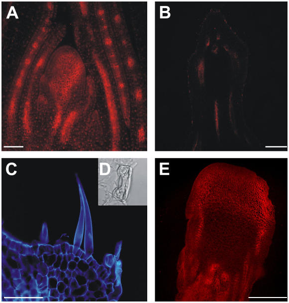
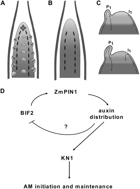
Shoot apical meristems produce organs in a highly stereotypic pattern that involves auxin. Auxin is supposed to be actively transported from cell to cell by influx (AUXIN/LIKE AUXIN proteins) and efflux (PIN-FORMED proteins) membrane carriers. Current hypotheses propose that, at the meristem surface, PIN proteins create patterns of auxin gradients that, in turn, create patterns of gene expression and morphogenesis. These hypotheses are entirely based on work in Arabidopsis (Arabidopsis thaliana). To verify whether these models also apply to other species, we studied the behavior of PIN proteins during maize (Zea mays) development. We identified two novel putative orthologs of AtPIN1 in maize and analyzed their expression pattern during development. The expression studies were complemented by immunolocalization studies using an anti-AtPIN1 antibody. Interestingly, the maize proteins visualized by this antibody are almost exclusively localized in subepidermal meristematic layers. Both tassel and ear were characterized by a compact group of cells, just below the surface, carrying PIN. In contrast to or to complement what was shown in Arabidopsis, these results point to the importance of internally localized cells in the patterning process. We chose the barren inflorescence2 (bif2) maize mutant to study the role of auxin polar fluxes in inflorescence development. In severe alleles of bif2, the tassel and the ear present altered ZmPIN1a and ZmPIN1b protein expression and localization patterns. In particular, the compact groups of cells in the tassel and ear of the mutant were missing. We conclude that BIF2 is important for PIN organization and could play a role in the establishment of polar auxin fluxes in maize inflorescence, indirectly modulating the process of axillary meristem formation and development.


Figures

Figure 1.
Figure 1.
Structural comparison between AtPIN1 and ZmPIN1 genes and proteins. A, Structures of the AtPIN1, ZmPIN1a, and ZmPIN1b genes (drawn to scale), with larger boxes with numbers representing exons and the smaller ones representing introns. ORFs are also depicted. The region coding for the oligopeptide recognized by the anti-AtPIN1 antibody is also reported. Scale bar = 100 bp. B, Hydropathy analysis of AtPIN1, ZmPIN1a, and ZmPIN1b proteins. The hydropathy plot was generated using Lasergene software (DNASTAR) and the method of Kyte and Doolittle, with a window size of nine amino acids. C, Amino acid sequence alignment of Arabidopsis and maize putative PIN1 proteins. AtPIN1 (At1g73590) and putative ZmPIN1a (accession no. DQ836239) and ZmPIN1b (accession no. DQ836240) were aligned using ClustalX 1.81 and then edited with Genedoc (http://www.psc.edu/biomed/genedoc). The number of amino acids is indicated on the right. Below the multiple sequence alignment, a black box indicates the oligopeptide targeted by the anti-AtPIN1 antibody.
Figure 2.
Figure 2.
Neighbor-joining phylogenetic tree showing the predicted relationship between ZmPIN1a, ZmPIN1b, and the Arabidopsis, rice, and wheat PIN proteins, according to Paponov et al. (2005). The tree is based on an alignment prepared using ClustalX 1.81. The phylograms were drawn using Tree-View 1.6.6 (http://darwin.zoology.gla.ac.uk/wrpage/treeviewx). Amino acid sequences for Arabidopsis were taken from http://www.Arabidopsis.org: AtPIN1, At1g73590; AtPIN2, At5g57090; AtPIN3, At1g70940; AtPIN4, At2g01420; AtPIN5, At5g16530; AtPIN6, At1g77110; AtPIN7, At1g23080; and AtPIN8, At5g15100. Data used were predicted amino acid sequences based on EST clones. The full cDNA sequences for wheat are unknown, so ESTs from the 5′ end were used. The sequences were taken from TIGR (http://www.tigr.org). Genes are named according to the cluster of the Arabidopsis PIN family to which they belong. The gene nomenclature corresponds to the following TIGR gene index: OsPIN1a, NP895789; OsPIN1b, TC250501; OsPIN2, Np897806; OsPIN4, TC259719; OsPIN5a, TC255589; OsPIN5b, TC272668; OsPIN9, TC256882; OsPIN10a, Tc260564; OsPIN10b, NP1102328; TaPIN1a, TC224207; TaPIN1b, Ck208849; TaPIN1c, Tc224208; TaPIN2, TC200857; TaPIN5a, BQ484087; TaPIN5b, CA722918; TaPIN8, CB307721; and TaPIN9, Cd895017. SEPIN (gi:57636762) from Staphylococcus epidermidis was selected as the outgroup.
Figure 3.
Figure 3.
Semiquantitative RT-PCR analysis of two putative PIN1 orthologous genes from different maize tissues. B73 and bif2 show similar levels of expression in roots and seedlings, whereas in leaves ZmPIN1b displays a weaker expression than ZmPIN1a. No alteration of the expression levels is observed in bif2 roots and seedlings compared to wild type. In wild-type male inflorescences, the two genes share the same levels of expression, whereas in young ears, no ZmPIN1a expression was detected. Later, in developed female inflorescences, ZmPIN1a is expressed too. In bif2 tassels, ZmPIN1a shows high levels of expression compared to the wild type. a and b, Expression levels of ZmPIN1a and ZmPIN1b, respectively. Ubiquitin (UBIQ) was used as a control.
Figure 4.
Figure 4.
In situ hybridization of ZmPIN1 mRNA in maize apex and tassel. ZmPIN1 mRNA is expressed during maize apex development. The mRNA probe is supposed to hybridize to both ZmPIN1a and ZmPIN1b. All images represent longitudinal sections of B73 inbred line apexes. A, In the apex, the hybridization signal is mainly present in the vascular tissues; in the SAM, the signal is visible in the dome of the meristem, in the lateral outgrowing primordia, and in the tips of young leaves. B and C, In the differentiating tassel, the PIN1 probe hybridized to the corresponding mRNA in the vascular bundles and at the site of forming BM. D and E, The signal stains the whole top part of the tassel and also the upper cells of the growing SMs. G, In the lateral branches, which arise from the base of the tassel, the signal is first localized in the central part of the branch. F and H, It later follows the distichous pattern of SMs. Bars: A to E, G, and H = 100 μm; F = 50 μm.
Figure 5.
Figure 5.
ZmPIN1 expression in the developing ear. In situ hybridization reveals that mRNA of ZmPIN1a and ZmPIN1b is localized in the young and adult female inflorescence. All images represent longitudinal B73 ear sections. A and B, In young ears, the signal is present in the vascular cells of the elongating axis and in the developing bracts. Also, the whole upper part, which includes the inflorescence meristem, presents diffuse staining. C, At a later stage of development, mRNA is localized in the position of growing SMs and again in the cells forming the upper part of the ear. D and E, During spikelet development, mRNA is found in the cells between the outer glumes and the differentiating SM. Bars = 100 μm.
Figure 6.
Figure 6.
ZmPIN1 protein localization in maize vegetative tissues. The putative auxin efflux carriers are polarly localized in the vegetative organs of maize. All images portray maize B73 sections labeled with the anti-PIN1 antibody plus a secondary antibody carrying the Alexa568 fluorochrome. A to C, F, and G, Laser confocal images. D and E, Epifluorescence images acquired with a Leica DC300F camera. A and B, In the mature embryo, the anti-PIN1 antibody recognizes a polar signal in a few cells located along the embryo axis. The labeling suggests auxin fluxes directed toward the upper embryo tissues. C to E, In the leaves, putative efflux carriers are inserted into the basal membrane of vascular cell files. The labeling suggests a PAT toward the leaf base. C, In cross sections of maize vascular bundles, the signal is clearly polarized in the lateral membranes of the outer cells and indicates that auxin is loaded into the vascular bundles from the outer tissues. F and G, In the primary root, proteins are inserted into the basal membrane of the cells, suggesting an acropetal auxin flux directed toward the root tip. The labeling is present in the central cylinder and also in the inner cortex cells. Bars: A = 80 μm; B and C = 40 μm; D to G = 30 μm.
Figure 7.
Figure 7.
ZmPIN1 protein localization in maize wild-type apex and reproductive structures. All images portray maize B73 longitudinal sections labeled with the anti-PIN1 antibody plus a secondary antibody carrying the Alexa568 fluorochrome. C to H, Laser confocal images. A, B, I, J, and K, Epifluorescence images acquired with a Leica DC300F camera. A, At the vegetative stage, the maize SAM shows polar labeling in cells that form the middle part of its dome, but no labeling is present in the outer layers. B, When a primordium is initiated, the auxin efflux carriers are expressed in its outer cell layers. In all cells, the signal is present in the basal or lateral membranes, suggesting an overall auxin flux directed toward the base of the plant. C to F, When the SAM switches from the vegetative to the reproductive phase, ZmPIN1 proteins are present in the inner tissues of the developing tassel and along the tassel axis in the vasculature. G and H, When the tassel forms SMs, the protein localizes in the incipient meristems, reaching the surface of the tassel. I, In the young ear, the pattern of the putative auxin efflux carriers is repeated, with labeling on the basal membranes and in the L1 at the place of outgrowing primordia. J, Again, the vascular bundles are stained in the ear. K, In the developing spikelets, the polar proteins are located in continuity with the main axis vasculature and in the place of the forming glumes. An interaction between the internal tissues of the meristems and the surface of outgrowing primordia is suggested by the immunolabeling results. The vascular tissue shows the same polarization in the tassel, ear, and leaves, where the auxin flux seems directed downward. Bars: A to C, G, and H = 50 μm; D = 75 μm; E and F = 30 μm; I to K = 100 μm.
Figure 8.
Figure 8.
ZmPIN1 localization in bif2 vegetative and reproductive tissues. The bif2 mutant presents wild-type vegetative development, but abnormal reproductive structures. During the vegetative phase, bif2 plants develop a normal phyllotaxis and the putative auxin efflux carrier localization appears the same as in wild-type maize. After the switch to the reproductive phase, the tassel and ear show several developmental defects and PAT seems to be completely impaired. A, Longitudinal section of a bif2 SAM, where ZmPIN1 presents a wild-type localization. The 7-d after germination bif2 SAM section was labeled with the anti-AtPIN1 antibody coupled to an Alexa568 fluorochrome (red). B, In the fully developed tassels of severe bif2 mutants, the ZmPIN1 is completely absent. This image shows a longitudinal tassel section, which presents no polar signal; tissue autofluorescence appears red. C, Epifluorescence image of a longitudinal section detail of a mutant tassel at flowering. Structures appear blue as autofluorescence was observed with a 4′,6-diamino-phenylindole filter. In the tassel of severe bif2 mutants, the antibody does not recognize any membrane protein. The male inflorescence presents no AMs and a completely disrupted vasculature. The developmental defects include the production of leaf-like epidermis, which carries stomata (D) and trichomes. E, Epifluorescence image of a bif2 adult ear. Red color codes for the Alexa568 fluorochrome. In female reproductive structures, AMs are absent and the vascular bundles are disrupted, although the defects seem less severe than in the tassel. The anti-PIN1 antibody labels polarly localized proteins in the vasculature. Bars: A = 100 μm; B and E = 75 μm; C = 30 μm.
Figure 9.
Figure 9.
Model for the role of PAT in maize and Arabidopsis meristem differentiation. Arrows indicates polar auxin fluxes. A, Schematic representation of a maize wild-type tassel. The image was adapted from McSteen et al. (2000). ZmPIN1 is polarized in a way that suggests auxin fluxes spreading at 360° around a small group of cells at the top of the tassel and along the tassel axis in two parallel cell files. The polarization pattern suggests an auxin flux directed downward. The existence of an interaction between the internal tissues of the tassel and the surface of the outgrowing primordia is hypothesized. B, Schematic representation of a bif2 mutant tassel with alteration in AM initiation. The image was modified from McSteen et al. (2000). Whereas bif2 mutant plants still show vascular tissues with basally oriented PIN1, the subapical cells showing fluxes spreading at 360° were completely absent from the mutant tassel, which fails to initiate and maintain AMs. C, Schematic representation of two Arabidopsis SAMs in early (top) and later (bottom) stage of development during primordium formation (adapted from Reinhardt et al., 2003). The primordium P1 accumulates acropetally transported auxin, preventing accumulation on the left flank of the meristem, whereas auxin can reach the right flank (I1). Here, auxin promotes the formation of an organ initium and the establishment of a new auxin sink. D, A causal link between bif2 and ZmPIN1 is hypothesized. BIF2 could regulate the expression of ZmPIN1 and other PIN family members in the inflorescence, thus indirectly modulating auxin distribution at the site of AM formation.

Similar articles

Cited by

References

    1. Benkova E, Michniewicz M, Sauer M, Teichmann T, Seifertova D, Jurgens G, Friml J (2003) Local, efflux-dependent auxin gradients as a common module for plant organ formation. Cell 115: 591–602 - PubMed
    1. Bennett MJ, Marchant A, Green HG, May ST, Ward SP, Millner PA, Walker AR, Schulz B, Feldmann KA (1996) Arabidopsis AUX1 gene: a permease-like regulator of root gravitropism. Science 273: 948–950 - PubMed
    1. Blilou I, Xu J, Wildwater M, Willemsen V, Paponov I, Friml J, Heidstra R, Aida M, Palme K, Scheres B (2005) The PIN auxin efflux facilitator network controls growth and patterning in Arabidopsis roots. Nature 433: 39–44 - PubMed
    1. Bortiri E, Chuck G, Vollbrecht E, Rocheford T, Martienssen R, Hake S (2006) ramosa2 encodes a LATERAL ORGAN BOUNDARY domain protein that determines the fate of stem cells in branch meristems of maize. Plant Cell 18: 574–585 - PMC - PubMed
    1. Boutté Y, Crosnier MT, Carraro N, Traas J, Satiat-Jeunemaitre B (2006) The plasma membrane recycling pathway and cell polarity in plants: studies on PIN proteins. J Cell Sci 119: 1255–1265 - PubMed

Publication types

Associated data

LinkOut - more resources