. 2006 Aug;18(8):1991-2004.
doi: 10.1105/tpc.106.043224. Epub 2006 Jul 14.

Arabidopsis CULLIN4 Forms an E3 Ubiquitin Ligase with RBX1 and the CDD Complex in Mediating Light Control of Development

Affiliations

Arabidopsis CULLIN4 Forms an E3 Ubiquitin Ligase with RBX1 and the CDD Complex in Mediating Light Control of Development

Haodong Chen et al. Plant Cell. 2006 Aug.

Abstract

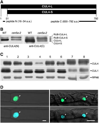
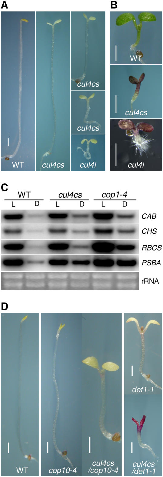
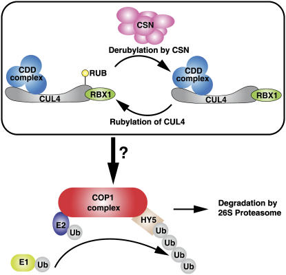
Repression of photomorphogenesis in Arabidopsis thaliana requires activity of the COP9 signalosome (CSN), CDD, and COP1 complexes, but how these three complexes work in concert to accomplish this important developmental switch has remained unknown. Here, we demonstrate that Arabidopsis CULLIN4 (CUL4) associates with the CDD complex and a common catalytic subunit to form an active E3 ubiquitin ligase both in vivo and in vitro. The partial loss of function of CUL4 resulted in a constitutive photomorphogenic phenotype with respect to morphogenesis and light-regulated gene expression. Furthermore, CUL4 exhibits a synergistic genetic interaction with COP10 and DET1. Therefore, this CUL4-based E3 ligase is essential for the repression of photomorphogenesis. This CUL4-based E3 ligase appears to associate physically with COP1 E3 ligase and positively regulates the COP1-dependent degradation of photomorphogenesis-promoting transcription factors, whereas the CSN controls the biochemical modification of CUL4 essential for E3 activity. Thus, this study suggests a biochemical activity connection between CSN and CDD complexes in their cooperation with COP1 in orchestrating the repression of photomorphogenesis.


Figures

Figure 1.
Figure 1.
Arabidopsis CUL4 Protein Forms, Expression Pattern, and Subcellular Localization. (A) Schemes depicting the two putative CUL4 forms in Arabidopsis and the locations of the two antigens for antibody preparation. CUL4-L and CUL4-S refer to the putative large and small CUL4 proteins, respectively, with translation beginning from different start codons in the same ORF. Peptides N and C show the positions of the two antigens used for antibody preparation. a.a., amino acids. (B) Unrubylated and rubylated CUL4 in Arabidopsis. Total soluble protein extracts from wild-type Arabidopsis and the csn5a-2 mutant were examined by protein gel blot analysis using anti-CUL4(N) and anti-CUL4(C) antibodies. (C) CUL4 protein is expressed extensively in Arabidopsis tissues. Total soluble protein extracts from inflorescences (lane 1), stems (lane 2), roots (lane 3), siliques (lane 4), light-grown seedlings (lane 5), dark-grown seedlings (lane 6), cauline leaves (lane 7), and rosette leaves (lane 8) were examined by protein gel blot analysis using anti-CUL4 and anti-CUL1 antibodies. An anti-RPN6 antibody was used as a sample loading control. (D) CUL4 is likely a nuclear protein. The left two panels show the localization of CUL4 in onion cells in a transient assay. The top left panel shows sGFP-CUL4 localization in epidermal cells, and the bottom left panel shows 4′,6-diamidino-2-phenylindole (DAPI) staining of the same cell to visualize the position of the nucleus (arrowheads). The right two panels show the localization of CUL4 in stable transgenic plants. The top right panel shows EGFP-CUL4 localization in light-grown seedling roots, and the bottom right panel shows DAPI staining of the same field to visualize the position of the nucleus (arrowheads). All of the images were taken using the GFP or DAPI channel of a confocal microscope. Bars = 20 μm.
Figure 2.
Figure 2.
Evidence for a CUL4-RBX1-CDD E3 Ligase in Arabidopsis. (A) CUL4 interacts with RBX1, DDB1a, and COP10, as shown in a yeast two-hybrid assay. The previously known interaction of DDB1a and COP10 was used as a positive control. The β-galactosidase activity resulting from the interaction is shown. Error bars represent sd (n = 4). (B) CUL4 associates with TAP-RBX1 but not TAP-ASK1 in vivo. Total flower protein extracts prepared from wild-type Arabidopsis, 35S:TAP-ASK1, and 35S:TAP-RBX1 transgenic Arabidopsis plants were incubated with IgG-coupled Sepharose. The precipitates and total extracts were subjected to immunoblot analysis with antibodies against CUL4, CUL1, Myc, and RPN6. Arrowheads indicate protein positions, and T indicates total protein extract. Anti-RPN6 antibody was used as a pull-down control. (C) CUL4 protein level is different among independent 35S:flag-CUL4 transgenic plants. Total protein was extracted from light-grown seedlings of the wild type and six independent transgenic lines. Protein gel blot analysis was subsequently performed using anti-flag, anti-CUL4, and anti-CUL1 antibodies. An anti-RPN6 antibody was used as a sample loading control. (D) Flag-CUL4 associates with COP10 in vivo. Total flower protein extracts prepared from wild-type and 35S:flag-CUL4 transgenic Arabidopsis (line 10) were incubated with anti-flag antibody–conjugated agarose (α-flag). The precipitates and total extracts were subjected to immunoblot analysis with antibodies against flag and COP10. An unspecific band was used as a pull-down control. COP10(F) and COP10(P) indicate the full-length and partially degraded COP10 protein forms, respectively. (E) CUL4 associates with flag-COP10 in vivo. Total flower protein extracts prepared from wild-type and 35S:flag-COP10 transgenic Arabidopsis were incubated with anti-flag antibody–conjugated agarose (α-flag). The precipitates and total extracts were subjected to immunoblot analysis with antibodies against flag and CUL4. An unspecific band was used as a pull-down control.
Figure 3.
Figure 3.
CUL4-RBX1-CDD E3 Ligase Can Be Reconstituted in Vitro. (A) Glutathione affinity purification of the recombinant CDD complex overexpressed in the baculovirus–insect cell system. The purified complex was analyzed by SDS-PAGE. (B) and (C) Further purification of the recombinant CDD complex by size exclusion chromatography. The TEV-cleaved GST tag and the excess COP10 protein were completely separated from the CDD complex, which migrated as a single peak species on the Superdex 200 gel filtration column (B). The fractions from the CDD complex peak were examined by SDS-PAGE (C) and appear to be approximately equimolar for the three components. (D) and (E) CUL4-RBX1 can interact with the CDD complex to form a holo E3 ligase complex in vitro. The five-protein complex CUL4-RBX1-COP10-DDB1a-DET1 migrated as a large single-peak species on a Superdex 200 gel filtration column (D). The star indicates where COP10-DDB1a-DET1 migrates (as see in [B]). The fractions from the holo E3 ligase complex peak appear to have equimolar ratios of the five proteins, as revealed by SDS-PAGE analysis (E). (F) and (G) In vitro ubiquitination assays with recombinant GST-RBX1-CUL4 and CDD complexes. GST-RBX1-CUL4 mediates ubiquitin chain formation, and the CDD complex can significantly enhance this process. The identical blots were probed with antibodies against flag epitope (F) or GST (G). (H) A proposed Arabidopsis CUL4-RBX1-CDD E3 ligase supported by our data.
Figure 4.
Figure 4.
CUL4-Containing E3 Ligases Are Regulated by CSN and CAND1 through Physical Association. (A) Evidence for direct CUL4 interaction with CSN and CAND1 by yeast two-hybrid assay. The previously known interaction of DDB1a and COP10 was used as a positive control. The β-galactosidase activity resulting from the interaction is shown. Error bars represent sd (n = 4). (B) The flag-CUL4 associates with three representative CSN subunits (CSN3, CSN4, and CSN5) in vivo. Total flower protein extracts prepared from wild-type and 35S:flag-CUL4 transgenic Arabidopsis were incubated with anti-flag antibody–conjugated agarose (α-flag). The precipitates and total extracts were subjected to immunoblot analysis with antibodies against flag, CSN3, CSN4, and CSN5. An unspecific band was used as a pull-down control. (C) The flag-CAND1 associates with CUL4 but not CSN in vivo. Total flower protein extracts prepared from wild-type and 35S:flag-CAND1 transgenic Arabidopsis were incubated with anti-flag antibody–conjugated agarose (α-flag). The precipitates and total extracts were subjected to immunoblot analysis with antibodies against flag, CUL4, CSN3, and CSN5. An unspecific band was used as a pull-down control. (D) The CUL4 protein is subjected to modification by RUB in vivo. Total protein was extracted from light-grown seedlings of the wild type, csn5a-2, csn1-1 (fus6-1), csn3-1 (fus11-1), cand1-1, cop10-1, cop1-6, and det1-1, and protein gel blot analysis was subsequently performed using anti-CUL4 and anti-CUL1 antibodies. Anti-RPN6 was used as a sample loading control.
Figure 5.
Figure 5.
CUL4 Is Involved in Repressing Photomorphogenesis. (A) CUL4 mediates a cop-like phenotype in the dark. The three CUL4 cosuppression (cul4cs) seedlings are 7-d-old dark-grown 35S:flag-CUL4 transgenic seedlings exhibiting different length hypocotyls and different degrees of opening and expansion of cotyledons (lines 1, 2, 7, and 8 in Figure 2C). The CUL4 RNAi (cul4i) yields dark-grown 35S:CUL4i transgenic seedlings exhibiting short hypocotyls and open and fully expanded cotyledons. A wild-type (Columbia-0) seedling is shown at left. Bar = 1 mm. (B) CUL4 mediates a fusca phenotype in the light. The middle and bottom panels show 5-d-old light-grown cul4cs and cul4i seedlings, respectively, with hyperphotomorphogenic morphology and a high level of anthocyanin accumulation. The top panel shows a wild-type (Columbia-0) seedling. Bars = 1 mm. (C) RNA gel blot analysis of steady state RNA levels of nucleus- and plastid-encoded genes. RNA levels from wide-type, cul4cs, and cop1-4 plants were analyzed. Total RNA was isolated from seedlings grown for 7 d in the light (L) or dark (D). Equal amounts of total RNA from the different plant samples were used, and four identical blots were hybridized and labeled with gene-specific probes for four different genes: CAB, CHS, RBCS, and PSBA. The rRNA band pattern was used to show equal loading. (D) Reduction of CUL4 enhances the phenotypes of weak cop10 and det1 alleles in the dark. Different Arabidopsis lines (labeled at bottom) were grown in complete darkness for 7 d. Bars = 1 mm.
Figure 6.
Figure 6.
The CUL4-RBX1-CDD E3 Ligase Can Enhance COP1-Mediated HY5 Degradation. (A) The flag-CUL4 associates with COP1 in vivo. Total flower protein extracts prepared from wild-type and 35S:flag-CUL4 transgenic Arabidopsis were incubated with anti-flag antibody–conjugated agarose (α-flag). The precipitates and total extracts were subjected to immunoblot analysis with antibodies against flag and COP1. A nonspecific band was used as a pull-down control. (B) A possible direct COP10 and COP1 interaction is supported by a yeast two-hybrid assay. The previously known interaction of DDB1a and COP10 was used as a positive control. The β-galactosidase activity resulting from the interaction is shown. Error bars represent sd (n = 4). (C) HY5 is degraded less efficiently in the cul4cs mutants than in wild-type Arabidopsis. Four-day-old light-grown seedlings of wild-type Arabidopsis and cul4cs plants were transferred to complete darkness. Samples were collected at different time points starting from the transfer (indicated at top) and blotted with anti-HY5 and anti-RPN6 antibodies.
Figure 7.
Figure 7.
Multifaceted Developmental Defects of cul4cs Mutant Plants. (A) Seven-day-old light- and dark-grown wild-type and cul4cs seedlings. L and D indicate light and dark, respectively. (B) Three-week-old wild-type and cul4cs plants under the 16L/8D (16 h of light/8 h of dark each day) condition. (C) Eight-week-old wild-type plants under the 16L/8D condition and cul4cs plants under both the 16L/8D and 12L/12D conditions. For (A) to (C), photographs in the same panel were taken at the same magnification. (D) Rosette leaves from 5-week-old cul4cs plants. The bottom right image was taken with a twofold magnification compared with the others. (E) Comparison of wild-type and cul4cs flowers. (F) Comparison of wild-type and cul4cs siliques.
Figure 8.
Figure 8.
A Working Model of CUL4 and the Three-Protein Complex in Mediating the Repression of Photomorphogenesis. In the dark, the CUL4-RBX1-CDD complex positively regulates COP1-mediated degradation of light-regulated transcriptional factors such as HY5. CUL4-RBX1-CDD E3 ligase activity is regulated through the rubylation and derubylation cycle, with CSN mediating derubylation. It remains unknown how this CUL4-based E3 ligase positively regulates COP1 E3 ligase activity.

Similar articles

Cited by

References

    1. Ang, L.H., Chattopadhyay, S., Wei, N., Oyama, T., Okada, K., Batschauer, A., and Deng, X.W. (1998). Molecular interaction between COP1 and HY5 defines a regulatory switch for light control of Arabidopsis development. Mol. Cell 1 213–222. - PubMed
    1. Castle, L.A., and Meinke, D.W. (1994). A FUSCA gene of Arabidopsis encodes a novel protein essential for plant development. Plant Cell 6 25–41. - PMC - PubMed
    1. Chamovitz, D.A., Wei, N., Osterlund, M.T., von Arnim, A.G., Staub, J.M., Matsui, M., and Deng, X.W. (1996). The COP9 complex, a novel multisubunit nuclear regulator involved in light control of a plant developmental switch. Cell 86 115–121. - PubMed
    1. Chiu, W., Niwa, Y., Zeng, W., Hirano, T., Kobayashi, H., and Sheen, J. (1996). Engineered GFP as a vital reporter in plants. Curr. Biol. 6 325–330. - PubMed
    1. Chory, J., Peto, C., Feinbaum, R., Pratt, L., and Ausubel, F. (1989). Arabidopsis thaliana mutant that develops as a light-grown plant in the absence of light. Cell 58 991–999. - PubMed

Publication types

MeSH terms

Associated data