. 2006 Aug;18(8):1833-45.
doi: 10.1105/tpc.106.042978. Epub 2006 Jul 14.

Functional analyses of two tomato APETALA3 genes demonstrate diversification in their roles in regulating floral development

Affiliations

Functional analyses of two tomato APETALA3 genes demonstrate diversification in their roles in regulating floral development

Gemma de Martino et al. Plant Cell. 2006 Aug.

Abstract

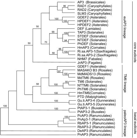
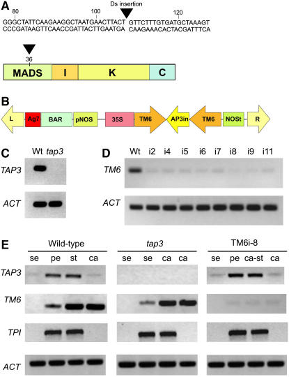
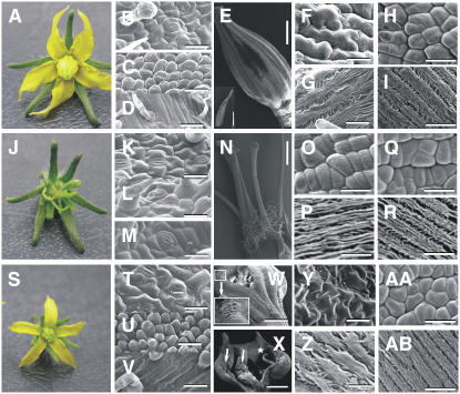
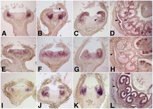
The floral homeotic APETALA3 (AP3) gene in Arabidopsis thaliana encodes a MADS box transcription factor required for specifying petal and stamen identities. AP3 is a member of the euAP3 lineage, which arose by gene duplication coincident with radiation of the core eudicots. Although Arabidopsis lacks genes in the paralogous Tomato MADS box gene 6 (TM6) lineage, tomato (Solanum lycopersicum) possesses both euAP3 and TM6 genes, which have functionally diversified. A loss-of-function mutation in Tomato AP3 (TAP3) resulted in homeotic transformations of both petals and stamens, whereas RNA interference-induced reduction in TM6 function resulted in flowers with homeotic defects primarily in stamens. The functional differences between these genes can be ascribed partly to different expression domains. When overexpressed in an equivalent domain, both genes can partially rescue the tap3 mutant, indicating that relative levels as well as spatial patterns of expression contribute to functional differences. Our results also indicate that the two proteins have differing biochemical capabilities. Together, these results suggest that TM6 and TAP3 play qualitatively different roles in floral development; they also support the ideas that the ancestral role of AP3 lineage genes was in specifying stamen development and that duplication and divergence in the AP3 lineage allowed for the acquisition of a role in petal specification in the core eudicots.


Figures

Figure 1.
Figure 1.
Neighbor-Joining Analysis of euAP3 and TM6 Lineage Genes. Representative AP3 lineage genes from core eudicots and basal eudicots were included in the analysis; the basal eudicot Pn AP3 gene was used as the outgroup. Bootstrap values of ≥50% are shown.
Figure 2.
Figure 2.
Characterization of Loss-of-Function Lines for TAP3 and TM6. (A) Location of the insertion of a Ds element in the first exon of TAP3 at position +108 within the MADS domain. (B) pTCSH1-TM6 RNAi construct used for RNAi-induced silencing of the TM6 gene. (C) RT-PCR using TAP3-specific primers on wild-type and tap3 inflorescence tissue (stages 9 to 18); no amplification could be detected in the tap3 tissue. Amplification of the tomato ACTIN gene (ACT) was used as a control. (D) RT-PCR using TM6-specific primers on wild-type and TM6i lines. Flowers from stages 15 to 20 showing a strong phenotype were analyzed for all transgenic lines. As expected, the level of the TM6 transcript was highly reduced in the transgenic lines. (E) RT-PCR analyses to determine the organ-specific expression of TAP3, TM6, and TPI genes in wild-type, tap3 mutant, and TM6i8 transgenic lines. Floral organs were dissected from stage 9 to 18 flowers and pooled, and the resulting RNA was used for RT-PCR. se, sepals; pe, petals; st, stamens; ca, carpels; ca-st, carpelloid stamens.
Figure 3.
Figure 3.
Phenotypic Analyses of the tap3 Mutant and the TM6i-8 Line. Light and scanning electron microscopy of wild-type (Micro-Tom) flowers ([A] to [I]), tap3 flowers ([J] to [R]), and TM6i-8 flowers ([S] to [AB]). (B), (K), and (T) Adaxial epidermal cells of the first whorl organs. (C), (L), and (U) Adaxial epidermal cells of the second whorl organs. (D), (M), and (V) Abaxial epidermal cells of the second whorl organs. (E) Wild-type staminal cone. The inset shows the adaxial surface of the anther. (N) tap3 third whorl organs showing carpel-like morphology. (W) Abaxial view of a TM6i-8 staminal cone showing partial lateral fusion (inset). (X) Adaxial view of a TM6i-8 carpelloid stamen showing naked ovules (arrows) and stigmatic tissue (star). (F), (O), and (Y) Abaxial epidermal cells of the proximal region of the third whorl organs. (G), (P), and (Z) Abaxial epidermal cells of the distal region of the third whorl organs. (H), (Q), and (AA) Epidermal cells of the proximal region of the fourth whorl organs. (I), (R), and (AB) Epidermal cells of the distal region of the fourth whorl organs. Bars = 40 μm ([B], [C], [G] to [I], [K] to [M], [O] to [R], [T], [U], [Z], [AA], and [AB]), 90 μm ([D], [F], and [V]), 20 μm (Y), 2 mm (inset in [E] and [N]), 1 mm ([W] and [X]), and 1.5 mm (E).
Figure 4.
Figure 4.
In Situ Expression Analyses of TAP3, TM6, and TPI. Expression in wild type (Micro-Tom) flower buds of TAP3 ([A] to [D]), TM6 ([E] to [H]), and TPI ([I] to [L]). (A) TAP3 expression is apparent by stage 2, with expression seen predominantly in the petal primordia. (B) By stage 3, expression can be seen throughout the petal primordia as well as in the subepidermal cell layers of the incipient stamen primordia (arrowhead). (C) TAP3 expression in the petal and stamen primordia (arrowhead) at stage 5. (D) Cross section of a flower at stage 9. TAP3 expression becomes restricted to the vascular bundle and tapetal cells of the stamens, whereas expression in the petals is seen in the lateral edges (arrowhead). (E) Relatively low levels of TM6 expression can be seen at stage 2 throughout the presumptive petal, stamen, and carpel primordia. (F) and (G) This pattern of expression persists through stage 3 (F) and stage 5 (G). (H) By stage 9, TM6 expression is most prominent in the developing ovules, localized predominantly to the inner integuments (inset). Low levels of expression can also be detected in the lateral margins of the petals (arrowhead). (I) TPI expression can be detected by early stage 2 in the presumptive petal primordia. (J) and (K) TPI expression is observed in developing petal and stamen primordia at stage 4 (J) and stage 5 (K). (L) By stage 9, TPI expression is seen mainly in the stamens, with high levels of expression in the tapetal cells. TPI expression can also be detected in the lateral margins of the petals (arrowhead).
Figure 5.
Figure 5.
Rescue of the Phenotype Conferred by tap3 by Overexpression of TM6 or TAP3. (A) 35S:TM6;tap3 flower. (B) and (C) Adaxial (B) and abaxial (C) epidermal cells of the 35S:TM6;tap3 second whorl organs. (D) and (E) Epidermal cells of proximal (D) and distal (E) regions of third whorl organs of the 35S:TM6;tap3 flower. (F) 35S:TAP3;tap3 flower. (G) and (H) Adaxial (G) and abaxial (H) epidermal cells of the 35S:TAP3;tap3 second whorl organs. (I) and (J) Epidermal cells of proximal (I) and distal (J) regions of third whorl organs of the 35S:TAP3;tap3 flower. Bars in scanning electron micrographs = 20 μm.

Similar articles

Cited by

References

    1. Ambrose, B.A., Lerner, D.R., Ciceri, P., Padilla, C.M., Yanofsky, M.F., and Schmidt, R.J. (2000). Molecular and genetic analyses of the Silky1 gene reveal conservation in floral organ specification between eudicots and monocots. Mol. Cell 5 569–579. - PubMed
    1. Angenent, G.C., Busscher, M., Franken, J., Dons, H.J., and van Tunen, A.J. (1995). Functional interaction between the homeotic genes fbp1 and pMADS1 during petunia floral organogenesis. Plant Cell 7 507–516. - PMC - PubMed
    1. Benfey, P.N., and Chua, N.-H. (1990). The cauliflower mosaic virus 35S promoter: Combinatorial regulation of transcription in plants. Science 250 959–966. - PubMed
    1. Bowman, J.L., Smyth, D.R., and Meyerowitz, E.M. (1989). Genes directing flower development in Arabidopsis. Plant Cell 1 37–52. - PMC - PubMed
    1. Brukhin, V., Hernould, M., Gonzalez, N., Chevalier, C., and Mouras, A. (2003). Flower development schedule in tomato Lycopersicon esculentum cv. Sweet Cherry. Sex. Plant Reprod. 15 311–320.

Publication types

Associated data

LinkOut - more resources