. 2006 Aug;7(8):831-7.
doi: 10.1038/sj.embor.7400752. Epub 2006 Jul 14.

Growth differentiation factor 11 signals through the transforming growth factor-beta receptor ALK5 to regionalize the anterior-posterior axis

Affiliations

Growth differentiation factor 11 signals through the transforming growth factor-beta receptor ALK5 to regionalize the anterior-posterior axis

Olov Andersson et al. EMBO Rep. 2006 Aug.

Abstract

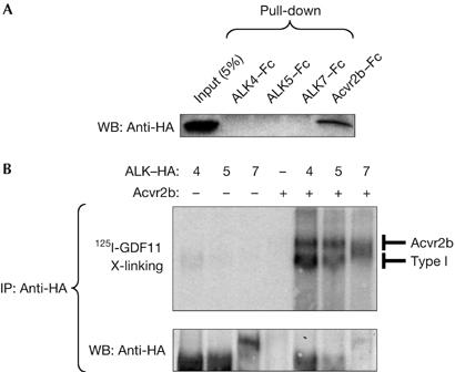
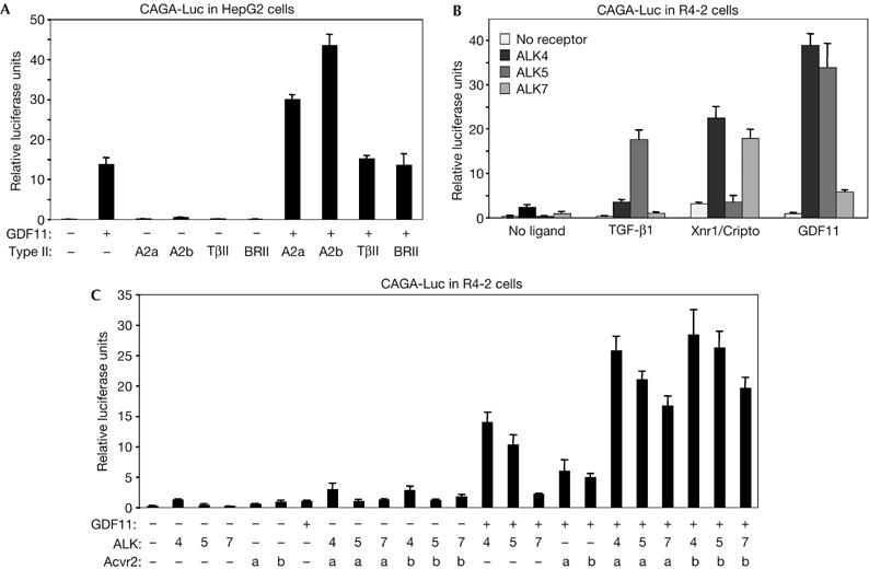
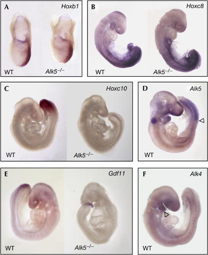
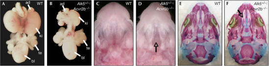
Growth differentiation factor 11 (GDF11) contributes to regionalize the mouse embryo along its anterior-posterior axis by regulating the expression of Hox genes. The identity of the receptors that mediate GDF11 signalling during embryogenesis remains unclear. Here, we show that GDF11 can interact with type I receptors ALK4, ALK5 and ALK7, but predominantly uses ALK4 and ALK5 to activate a Smad3-dependent reporter gene. Alk5 mutant embryos showed malformations in anterior-posterior patterning, including the lack of expression of the posterior determinant Hoxc10, that resemble defects found in Gdf11-null mutants. A heterozygous mutation in Alk5, but not in Alk4 or Alk7, potentiated Gdf11(-/-)-like phenotypes in vertebral, kidney and palate development in an Acvr2b(-/-) background, indicating a genetic interaction between the two receptor genes. Thus, the transforming growth factor-beta (TGF-beta) receptor ALK5, which until now has only been associated with the biological functions of TGF-beta1 to TGF-beta3 proteins, mediates GDF11 signalling during embryogenesis.


Figures

Figure 1
Figure 1
GDF11 binding to Acvr2b and type I receptors ALK4, ALK5 and ALK7. (A) Pull-down assay of haemagglutinin (HA)-tagged GDF11 with soluble Fc-fusion proteins of Acvr2b, ALK4, ALK5 and ALK7. The first lane in the western blot (WB) corresponds to 5% input of HA-tagged GDF11 (50 μl conditioned media). (B) Crosslinking binding assay in COS cells transfected with the indicated constructs and incubated with 125I-GDF11. After crosslinking, receptor complexes were immunoprecipitated (IP) with anti-HA antibodies. The lower panel shows reprobing with anti-HA antibodies. GDF11, growth differentiation factor 11.
Figure 2
Figure 2
Characterization of GDF11 signalling through type I and type II receptors. Gene reporter assays in (A) HepG2 and (B,C) R4-2 cells. The results are relative luciferase activity of triplicate determinations ±s.d. A2a, Acvr2; A2b, Acvr2b; BRII, bone morphogenetic protein type II receptor; GDF11, growth differentiation factor 11; TβII, transforming growth factor-β type II receptor.
Figure 3
Figure 3
Genetic interaction between Acvr2b and Alk5 during vertebral patterning. Representative skeleton preparations of (A,C) Acvr2b−/− and (B,D) Alk5+/−;Acvr2b−/− littermate mutants in frontal (A,B) and lateral (C,D) views. The numbers of thoracic vertebrae (A,B) and vertebrosternal ribs (C,D) are indicated. T1/T2 denotes fused first and second ribs.
Figure 4
Figure 4
Expression of Hox genes, ALK5, GDF11 and ALK4 in Alk5−/− mutants. (A) Hoxb1 messenger RNA expression in the posterior streak of wild-type (WT) and Alk5−/− mutant embryos at embryonic day (E)7.5. (B) Expression of Hoxc8 mRNA in E8.5 WT and Alk5−/− mutant embryos. (C) Hoxc10 mRNA expression in the paraxial mesoderm of an E9 WT embryo (left). Note the lack of expression in the Alk5−/− mutant embryo (right). (D) Alk5 mRNA expression in the paraxial mesoderm in a WT E9.5 embryo. The arrowhead marks the anterior limit of Alk5 mRNA expression. The signal in the otic vesicle is background staining caused by trapping of antibody. (E) Expression of Gdf11 mRNA in WT and Alk5−/− mutant embryos at E9.5. (F) Lack of Alk4 mRNA expression in the paraxial mesoderm in a WT E9.5 embryo. Note the expression of Alk4 mRNA in the first branchial arch (arrowhead). GDF11, growth differentiation factor 11.
Figure 5
Figure 5
Genetic interaction between Acvr2b and Alk5 during kidney development and palate closure. (A,B) Whole-mount urogenital system from wild type (WT) (A) and Alk5+/−;Acvr2b−/− mutant (B). Note the unilateral kidney agenesis in the Alk5+/−;Acvr2b−/− mutant. (C,D) Macroscopic view of WT (C) and Alk5+/−;Acvr2b−/− mutant (D) heads. Arrow in (D) points at the cleft palate. (E,F) Skeleton staining of skulls from embryos shown in (C,D). Arrow in (F) points to the cleft palate, which leads to absence of secondary palate. ad, adrenal gland; bl, bladder; ki, kidney; te, testis.

References

    1. Andersson O, Reissmann E, Jörnvall H, Ibáñez CF (2006) Synergistic interaction between Gdf1 and Nodal during anterior axis development. Dev Biol 293: 370–381 - PubMed
    1. Chang H, Brown CW, Matzuk MM (2002) Genetic analysis of the mammalian transforming growth factor-β superfamily. Endocr Rev 23: 787–823 - PubMed
    1. Chen C, Ware SM, Sato A, Houston-Hawkins DE, Habas R, Matzuk MM, Shen MM, Brown CW (2006) The Vg1-related protein Gdf3 acts in a Nodal signaling pathway in the pre-gastrulation mouse embryo. Development 133: 319–329 - PubMed
    1. Deschamps J, van Nes J (2005) Developmental regulation of the Hox genes during axial morphogenesis in the mouse. Development 132: 2931–2942 - PubMed
    1. Dubrulle J, Pourquie O (2004) Coupling segmentation to axis formation. Development 131: 5783–5793 - PubMed

Publication types

MeSH terms

Substances