. 2006 Aug;7(8):883-9.
doi: 10.1038/ni1366. Epub 2006 Jul 16.

Remodeling of the lectin-EGF-like domain interface in P- and L-selectin increases adhesiveness and shear resistance under hydrodynamic force

Affiliations

Remodeling of the lectin-EGF-like domain interface in P- and L-selectin increases adhesiveness and shear resistance under hydrodynamic force

Uyen T Phan et al. Nat Immunol. 2006 Aug.

Abstract

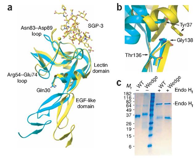
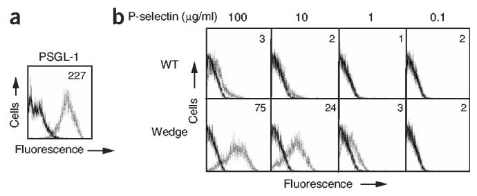
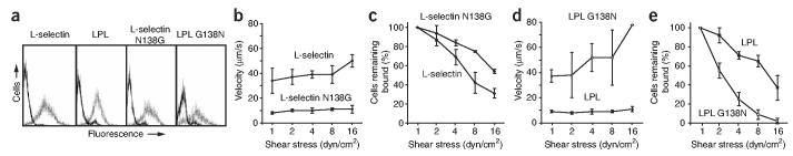
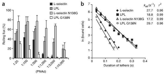
Crystal structures of the lectin and epidermal growth factor (EGF)-like domains of P-selectin show 'bent' and 'extended' conformations. An extended conformation would be 'favored' by forces exerted on a selectin bound at one end to a ligand and at the other end to a cell experiencing hydrodynamic drag forces. To determine whether the extended conformation has higher affinity for ligand, we introduced an N-glycosylation site to 'wedge open' the interface between the lectin and EGF-like domains of P-selectin. This alteration increased the affinity of P-selectin for its ligand P-selectin glycoprotein 1 (PSGL-1) and thereby the strength of P-selectin-mediated rolling adhesion. Similarly, an asparagine-to-glycine substitution in the lectin-EGF-like domain interface of L-selectin enhanced rolling adhesion under shear flow. Our results demonstrate that force, by 'favoring' an extended selectin conformation, can strengthen selectin-ligand bonds.


Figures

Figure 1
Figure 1
Selectin variants. (a) Superposition on the lectin domain of lectin and EGF-like domain fragments of P-selectin. Non-ligand-bound, cyan; bound to sulfoglycopeptide SGP-3, yellow. Structure includes side chains of Gln30 in both conformations; the calcium ion of the lectin domain is a yellow sphere. (b) Structures of lectin and EGF-like domain fragments of P-selectin, presented as in a but superimposed on the EGF domain. Structure includes side chains of Thr136, Gly138 and Tyr37 for both P-selectin conformations. (c) Reducing SDS-PAGE of soluble wild-type (WT) and mutant (Wedge) P-selectin containing the lectin, EGF-like and the first SCR domains, produced in 293S GnT1 cells. Wild-type and mutant P-selectin migrate with an apparent Mr of 36 and 39 kDa, respectively, and deglycosylation with endoglycosidase Hf (Endo Hf) produces material with an apparent Mr of 33 kDa.
Figure 2
Figure 2
Wedge mutant P-selectin has increased adhesiveness to HL-60 cells. (a) Flow cytometry of CHO-K1 transfectants expressing wild-type or wedge mutant P-selectin, stained with control mAb X63 (solid lines) or various mAbs to lectin and SCR domains (dotted lines). Numbers in plots indicate mean channel fluorescence values. (b) Adhesion of CHO-K1 transfectants to HL-60 cells expressing PSGL-1. Adhesion, quantified as a percentage of input fluorescence. Each experiment included six replicates. Data represent the average of three experiments (error bars, mean ± s.e.m.). P = 0.025, wedge compared with wild-type. Mock, transfected with empty vector pcDNA3.1(+).
Figure 3
Figure 3
Rolling and transient tethering of wedge mutant P-selectin. (a–f) Mean velocity (a–c) and cells remaining bound (d–f), calculated at each shear stress, for CHO-K1 cells expressing WT or mutant P-selectin, perfused in a flow chamber over 5 μg/ml of biotinylated sLeX (a,d), a 1:25 dilution of PNAd (b,e) or 10 ng/ml of biotinylated PSGL-1.19ek (c,f). Cells were perfused at 0.3 dyn/cm2 for 30 s and shear stress was increased every 10 s. (g,h) Frequency of transient tether formation (g) and duration of transient tethers (h) for wild-type and mutant P-selectin, determined at a shear stress of 0.5 dyn/cm2 in conditions in which rolling was not observed (PSGL-1.19ek, 5 ng/ml for g and 3 ng/ml for h). Data (a–g) represent the average of three experiments (error bars, mean ± s.d.).
Figure 4
Figure 4
Increased affinity of wedge mutant P-selectin. (a) Reducing SDS-PAGE of WT and wedge mutant P-selectin containing the lectin, EGF-like and first SCR domains, purified by size-exclusion chromatography. (b,c) SPR of WT monomeric material (0.026–3.3 μM; b) and wedge mutant monomeric material (0.012–1.5 μM, c), injected at twofold dilutions over immobilized PSGL-1–Fc and reference cell. Sensograms show the specific responses, as response units (RU; PSGL-1 Fc flow cell – ethanolamine-blocked reference cell). (c,d) Req for WT (c) and wedge mutant (d) plotted against their concentrations; curves are nonlinear fits to a 1:1 binding isotherm. Steady-state Kd values are the average ± s.d. of three experiments.
Figure 5
Figure 5
Increased binding of wedge mutant soluble P-selectin to PSGL-1 transfectants. (a) Flow cytometry of CHO-K1–PSGL-1–C2GnT1–FucT VII cells, stained with control mAb X63 (solid line) and PSGL-1-specific mAb PL2 (dotted line). (b) Flow cytometry of PSGL-1 transfectants incubated with 0.1, 1, 10 and 100 μg/ml of soluble WT or wedge P-selectin (dotted lines) or buffer alone (solid lines), then washed and stained with fluorescein isothiocyanate–conjugated P-selectin SCR–specific mAb AC1.2. Numbers in plots indicate mean channel fluorescence values.
Figure 6
Figure 6
Enhancement of rolling adhesiveness by the N138G substitution in L-selectin. (a) Flow cytometry showing similar cell surface expression of L-selectin, LPL, L-selectin N138G and LPL G138N. K562 transfectants were stained with control mAb X63 (solid lines) and lectin domain–specific mAb LAM1-3 (dotted lines). (b–e) Velocity (b,d) and cells remaining bound (c,e) of transfectants perfused into a flow chamber for 30 s at a shear stress of 0.3 dyn/cm2 over a 1:25 dilution of PNAd; shear stress was increased every 10 s. Mean velocity and percentage of cells remaining bound were calculated at each shear stress. Data represent the average of three experiments (error bars, mean ± s.d.).
Figure 7
Figure 7
The N138G substitution in L-selectin increases accumulation in shear flow and decreases transient tether koff. Transfectants expressing native L-selectin, LPL, L-selectin N138G or LPL G138N were perfused into a flow chamber at a shear stress of 0.6 dyn/cm2 over varying concentrations of PNAd. (a) After 1 min of perfusion, the percentage of cells in the field of view that were rolling was calculated. Data represent the average of three experiments (error bars, mean ± s.d.). (b) Durations of transient tethers determined at a shear stress of 0.6 dyn/cm2 in conditions in which rolling was not observed. Transient tethers were measured at the following PNAd dilutions: 1:300 for L-selectin; 1:10,000 for LPL and L-selectin N138G; and 1:150 for LPL G138N.

References

    1. Rosen SD, Bertozzi CR. The selectins and their ligands. Curr. Opin. Cell Biol. 1994;6:663–673. - PubMed
    1. Springer TA. Traffic signals for lymphocyte recirculation and leukocyte emigration: the multi-step paradigm. Cell. 1994;76:301–314. - PubMed
    1. Kansas GS. Selectins and their ligands: Current concepts and controversies. Blood. 1996;88:3259–3287. - PubMed
    1. Vestweber D, Blanks JE. Mechanisms that regulate the function of the selectins and their ligands. Physiol. Rev. 1999;79:181–213. - PubMed
    1. Butcher EC, Picker LJ. Lymphocyte homing and homeostasis. Science. 1996;272:60–66. - PubMed

Publication types