Comparative Study
. 2006 Jul 25;45(29):8894-902.
doi: 10.1021/bi060534y.

Spectroscopic analysis of benzylidene anabaseine complexes with acetylcholine binding proteins as models for ligand-nicotinic receptor interactions

Affiliations
Comparative Study

Spectroscopic analysis of benzylidene anabaseine complexes with acetylcholine binding proteins as models for ligand-nicotinic receptor interactions

Todd T Talley et al. Biochemistry. .

Abstract

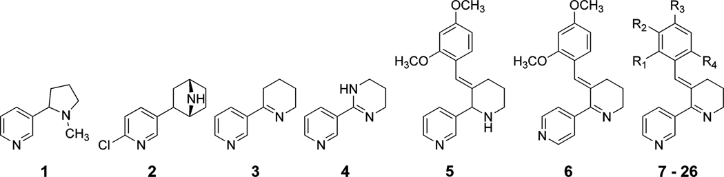
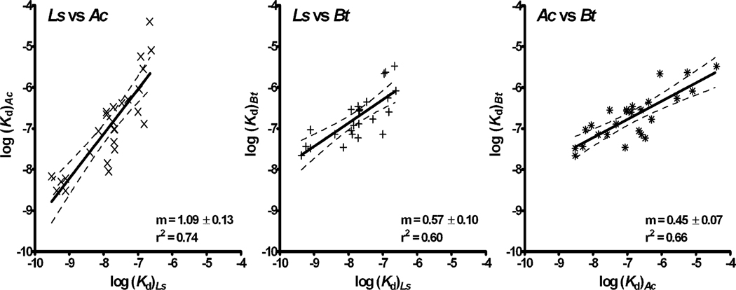
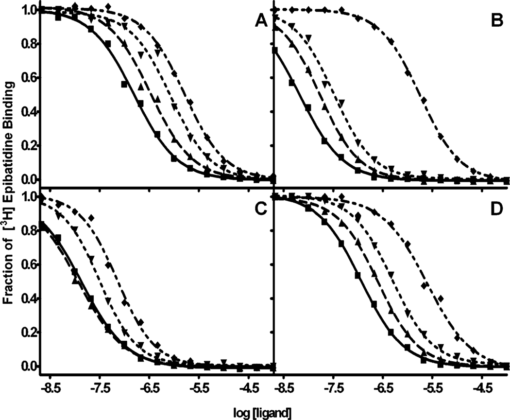
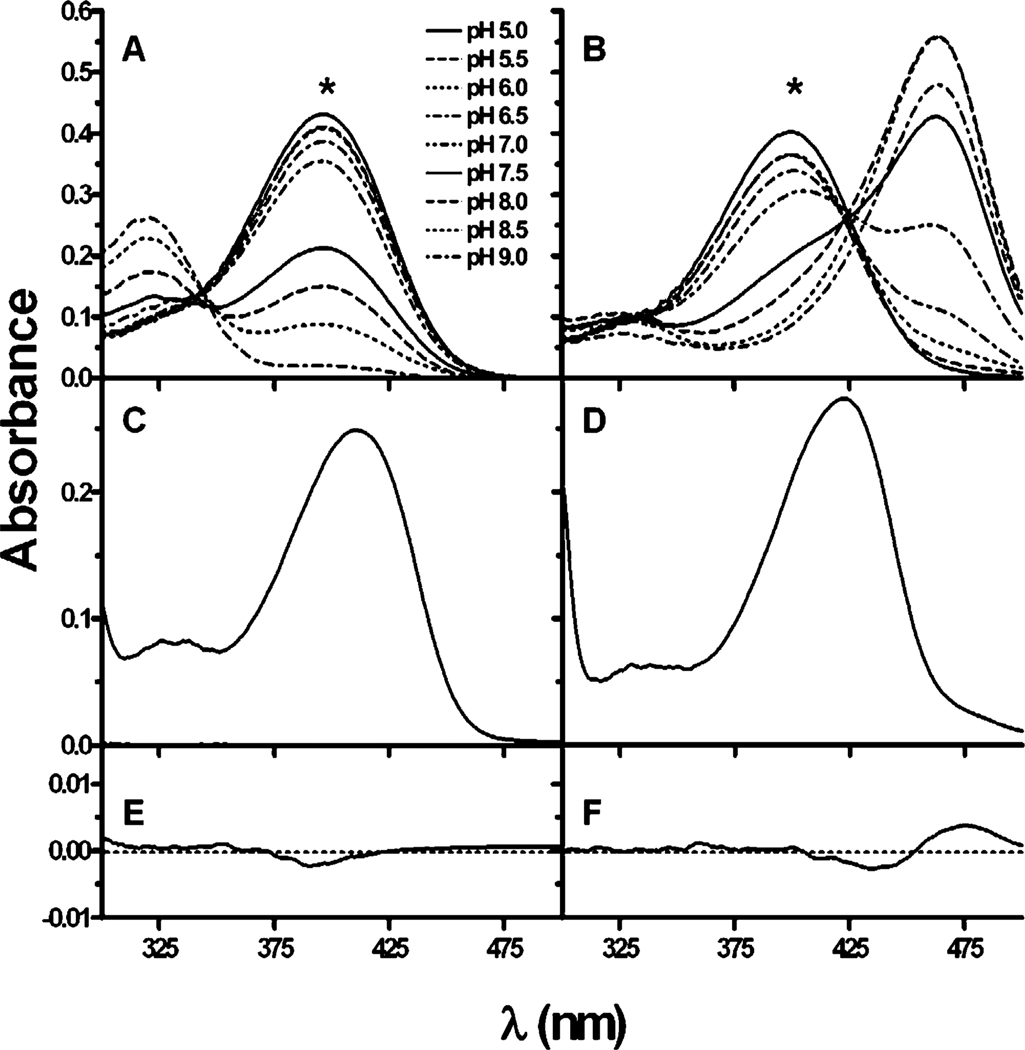
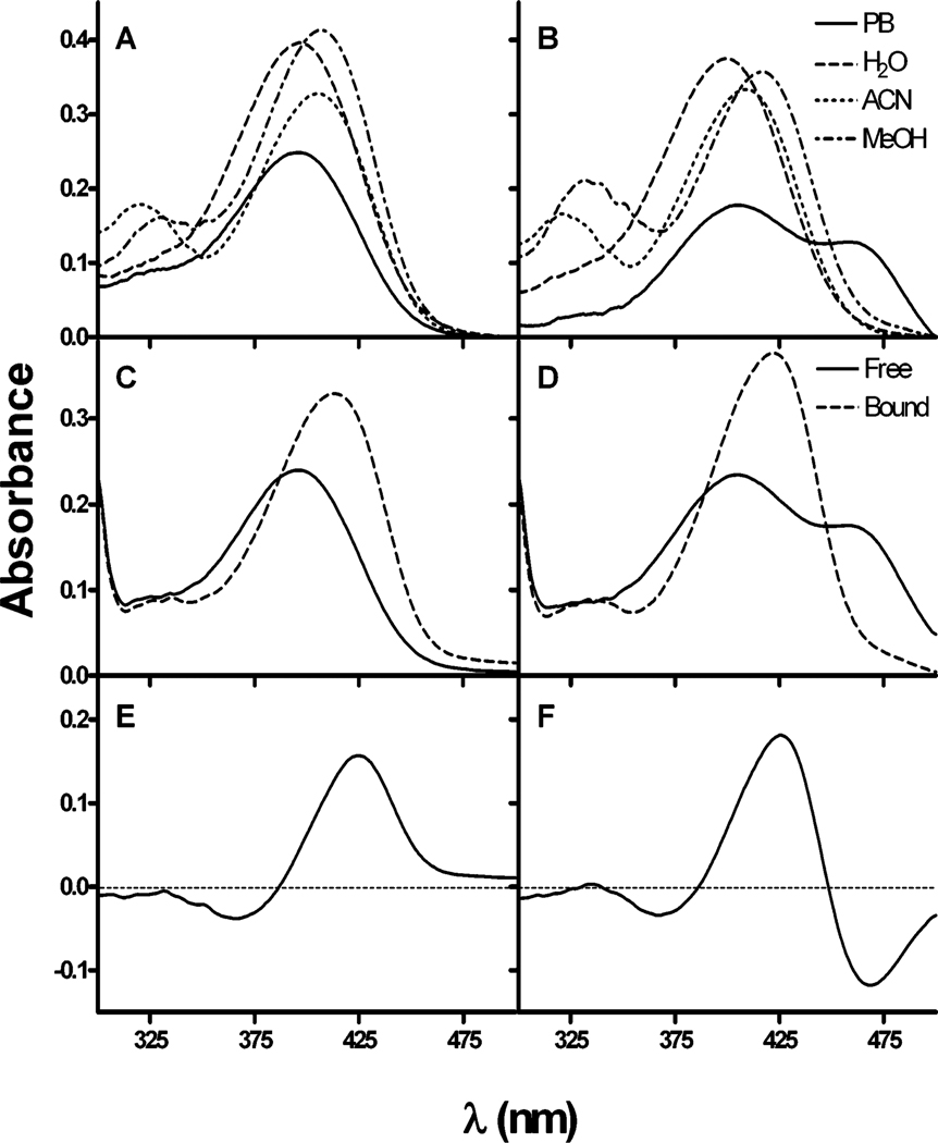
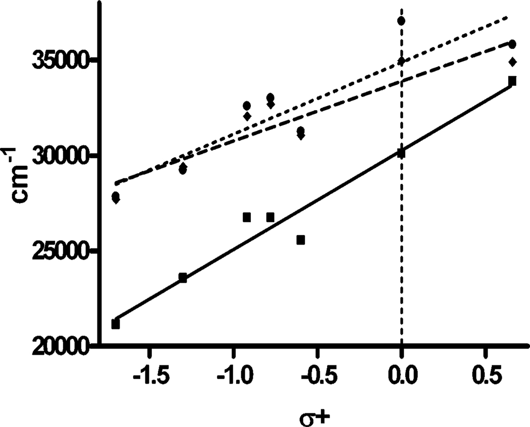
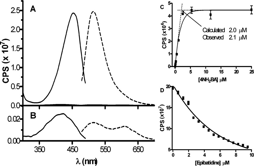
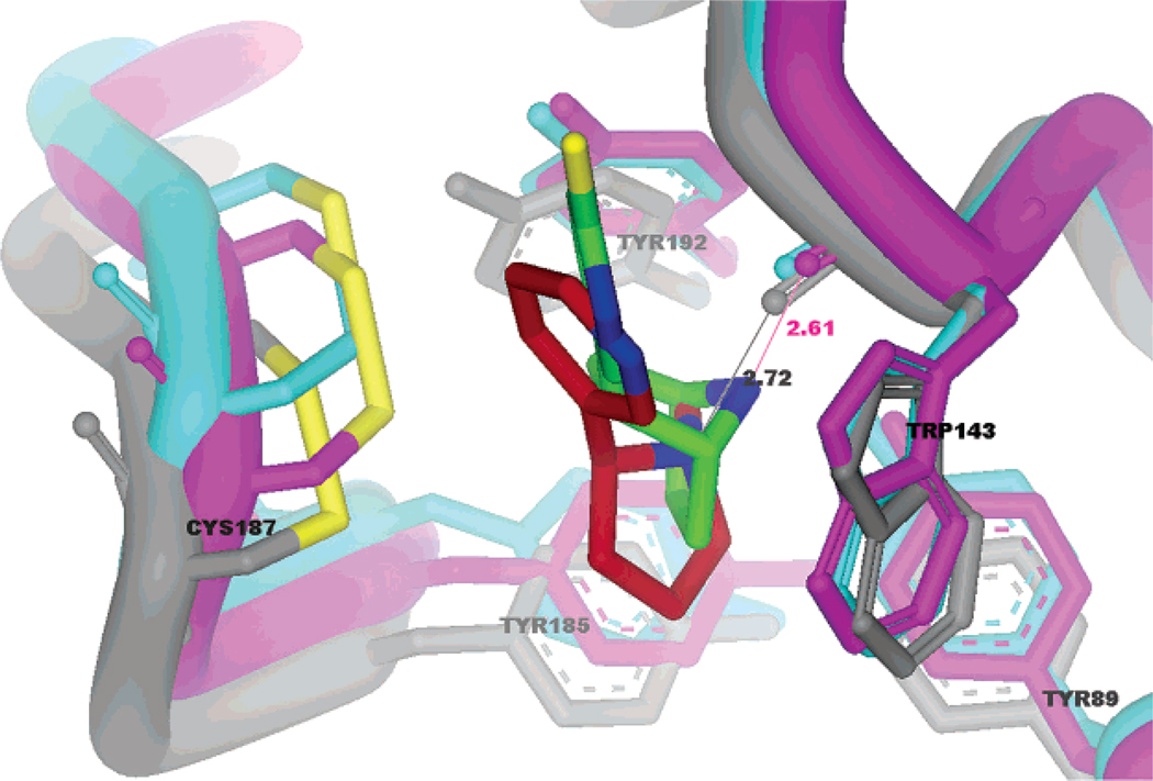
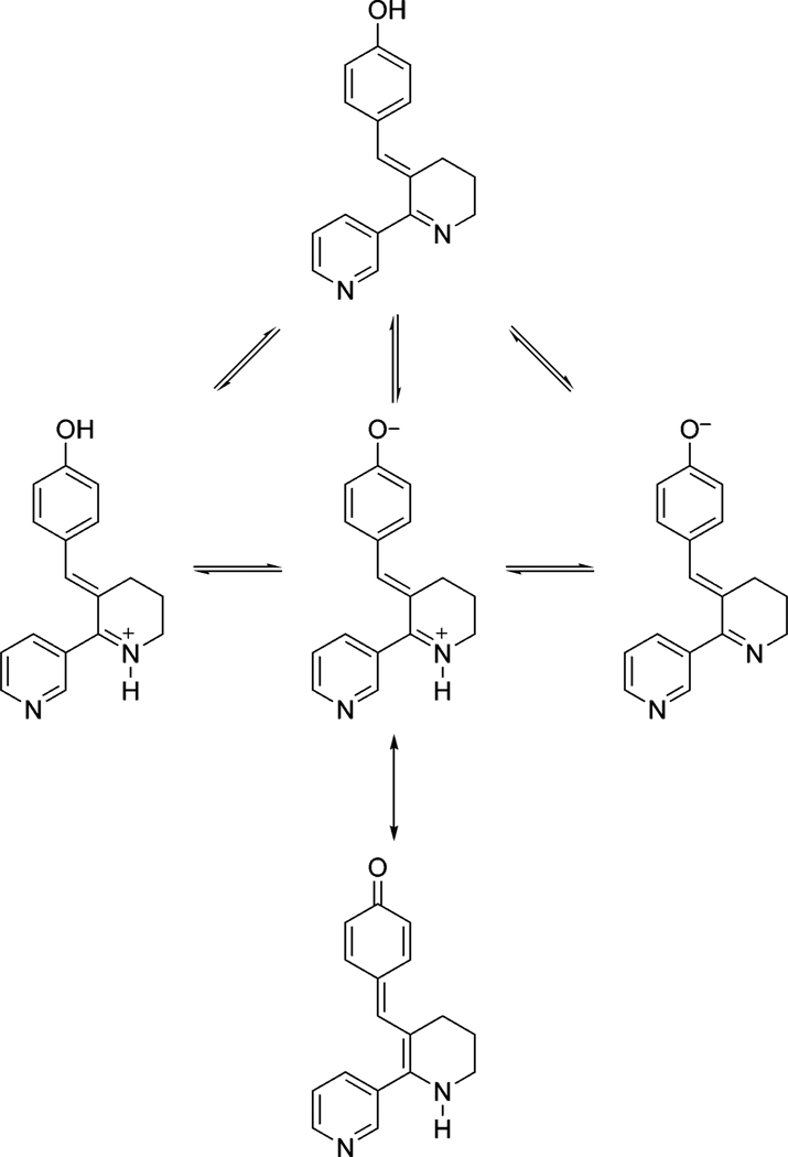
The discovery of the acetylcholine binding proteins (AChBPs) has provided critical soluble surrogates for examining structure and ligand interactions with nicotinic receptors and related pentameric ligand-gated ion channels. The multiple marine and freshwater sources of AChBP constitute a protein family with substantial sequence divergence and selectivity in ligand recognition for analyzing structure-activity relationships. The purification of AChBP in substantial quantities in the absence of a detergent enables one to conduct spectroscopic studies of the ligand-AChBP complexes. To this end, we have examined the interaction of a congeneric series of benzylidene-ring substituted anabaseines with AChBPs from Lymnaea, Aplysia, and Bulinus species and correlated their binding energetics with spectroscopic changes associated with ligand binding. The anabaseines display agonist activity on the alpha7 nicotinic receptor, a homomeric receptor with sequences similar to those of the AChBPs. Substituted anabaseines show absorbance and fluorescence properties sensitive to the protonation state, relative permittivity (dielectric constant), and the polarizability of the surrounding solvent or the proximal residues in the binding site. Absorbance difference spectra reveal that a single protonation state of the ligand binds to AChBP and that the bound ligand experiences a solvent environment with a high degree of polarizability. Changes in the fluorescence quantum yield of the bound ligand reflect the rigidification of the ring system of the bound ligand. Hence, the spectral properties of the bound ligand allow a description of the electronic character of the bound state of the ligand within its aromatic binding pocket and provide information complementary to that of crystal structures in defining the determinants of interaction.


Figures

Figure 1
Figure 1
Structures of nicotine 1, epibatidine 2, anabaseine 3, 2-(3-pyridyl)-3,4,5,6-tetrahydropyrimidine (PTHP) (4), 3-(2,4-dimethoxybenzyl)-(±) anabaseine (DMXBzAi) (5), 2-(4-pyridyl)-3-(2,4-dimethoxybenzylidene)-4,5,6,-tetrahydropyridine (DMXB IsoA) (6), and benzylidene anabaseines 7–26.
Figure 2
Figure 2
Relationship between the dissociation constants, Kd, for ligand binding to the three acetylcholine binding proteins from Lymnaea stagnalis (Ls), Aplysia californica (Ac), and Bulinus truncatus (Bt). Data from Table 1 are plotted logarithmically to reflect the proportionate relationship of the free energies of binding, log(K) = −ΔG/(2.3RT), for the various ligands.
Figure 3
Figure 3
Competition between the binding of the benzylidene anabaseine derivatives and [3H]-epibatidine (pKa = 10.1) to Lymnaea AChBP at various pH values. (A) DMXBA 18; (B) 2-MeO, 4-OHBA 21; (C) 4-NH2BA 15; (D) BA 7. The pH of the 0.1 M phosphate/pyrophosphate buffer used: (■) pH 6, (▲) pH 7, (▼) pH 8, (♦) pH 9. The structures are shown in Figure 1.
Figure 4
Figure 4
Absorption spectra of 3-(2,4-dimethoxybenzylidene)-anabaseine (DMXBA, 18) (A, C, E) and 3-(2-methoxy-4-hydroxybenzylidene)-anabaseine (2-MeO, 4-OHBA, 21) (B, D, F) as a function of pH. (A) and (B) Representative absorption spectra as a function of pH (0.5 increments from pH 5.0 to 9.0) for DMXBA and 2-MeO, 4-OHBA. Note the position of the wavelength peaks and that the long wavelength peak decreases in intensity with increasing pH for DMXBA, whereas it increases for 2-MeO, 4-OHBA. (C) and (D) DMXBA and 2-MeO, 4-OHBA, respectively, bound to Ls-AChBP in pH 8.1 and a 7.6 M NaPO4 buffer. (E) and (F) Difference spectra recorded for the complexes of the ligands with stoichiometric amounts of AChBP (30 µM of binding sites and 20 µM ligand). The spectra taken at pH values of 0.5 pH units above and below the apparent pKa value of each compound overlay closely, indicating the presence of a single ionization state.
Figure 5
Figure 5
Absorption spectra of DMXBA 18 and 2-MeO and 4-OHBA 21 in the presence of different solvents and bound to the AChBP. (A) and (B) DMXBA and 2-MeO and 4-OHBA in the presence of methanol (εr = 33.0), acetonitrile (εr = 37.5), H2O (εr = 80.0), and a 0.1 M phosphate buffer (PB) at pH 7.0 (εr ~ 80.0). (C) and (D) DMXBA and 2-MeO and 4-OHBA absorbance of ligands in a 0.1 M phosphate buffer at pH 7.0 (−), bound to AChBP. (E) and (F) Difference spectra reflecting DMXBA (E) and 2-MeO, 4-OHBA (F) in mixed (sample cuvette) and unmixed (reference cuvette) tandem compartments. Substituted BAs (20 µM) were used in the presence of 1.2 M excess of AChBP sites in 0.42 path length cuvettes. The difference spectra were measured comparing mixed and unmixed 20 µM BA and 24 µM AChBP in the same tandem cuvettes. The absorption values in the difference spectra are corrected for a 1 cm path length.
Figure 6
Figure 6
Relationship between the absorption maximum of the substituted anabaseines and the Hammett substituent parameter (σ+) (22) in the presence of various solvents (r2 > 0.8). Values of σ+ used: 0.0 for BA 7, −1.7 for 4-DMABA 16, −1.3 for 4-NH2BA 15, −0.92 for 4-OHBA 12, −0.78 for 4-MeOBA 13, −0.6 for 4-MeSBA 14, and 0.66 for 4-CNBA 17. Solvents: 0.1 M pH 7.0 phosphate buffer (■, solid line), DMSO (♦, dashed line), DMF (●, dotted line). The dashed vertical line shows the position of the unsubstituted BA.
Figure 7
Figure 7
Fluorescence properties of the free and Ls bound anabaseine derivative 4-NH2BA 15 in a 0.1 M phosphate buffer at pH 7.0. (A) Excitation (—) and emission (---) spectra of 10 µM 4-NH2BA bound to Ls-AChBP shows prominent peaks; (B) expansion of the fluorescence of the free species, also at 10 µM concentration. (C) Titration of 2 µM (binding sites) Ls-AChBP with varying concentrations of 4-NH2BA (λex = 420 nm, λem = 520 nm). Because the AChBP concentration greatly exceeds the Kd value (0.77 nM), the titration gives an estimate of the stoichiometry of binding. (D) Dissociation of 4-NH2BA from Ls-AChBP with increasing concentrations of epibatidine demonstrating the complete loss of fluorescence by the competing ligand (λex = 420 nm, λem = 520 nm).
Figure 8
Figure 8
Structural alignment of the crystallographic coordinates for Ls-AChBP (gray) with nicotine (red) bound (pdb ID 1UW6 (5)), Ac-AChBP (magenta) with epibatidine (green) bound (pdb ID 2BYQ (6)), and Bt-AChBP (cyan) with CHAPS (not shown) in the binding site (pdb ID 2BJ0 (3)). The bound ligand is enveloped by the side chains of Trp 143 and Tyr 192 using Ls numbering with other proximal aromatic side chains. Bond distances between the carbonyl oxygen of Trp 143 and the protonatable nitrogen of nicotine and epibatidine are shown.
Scheme 1
Scheme 1. Ionization Scheme for Hydroxyl Benzylidene Anabaseinesa
a The equilibrium arrows denote proton addition and removal.

References

    1. Brejc K, van Dijk WJ, Klaassen RV, Schuurmans M, van Der Oost J, Smit AB, Sixma TK. Crystal structure of an ACh-binding protein reveals the ligand-binding domain of nicotinic receptors. Nature. 2001;411:269–276. - PubMed
    1. Smit AB, Syed NI, Schaap D, van Minnen J, Klumperman J, Kits KS, Lodder H, van der Schors RC, van Elk R, Sorgedrager B, Brejc K, Sixma TK, Geraerts WP. A glia-derived acetylcholine-binding protein that modulates synaptic transmission. Nature. 2001;411:261–268. - PubMed
    1. Celie PH, Klaassen RV, van Rossum-Fikkert SE, van Elk R, van Nierop P, Smit AB, Sixma TK. Crystal structure of acetylcholine-binding protein from Bulinus truncatus reveals the conserved structural scaffold and sites of variation in nicotinic acetylcholine receptors. J. Biol. Chem. 2005;280:26457–26466. - PubMed
    1. Hansen SB, Talley TT, Radic Z, Taylor P. Structural and ligand recognition characteristics of an acetylcholine-binding protein from Aplysia californica. J. Biol. Chem. 2004;279:24197–24202. - PMC - PubMed
    1. Celie PH, van Rossum-Fikkert SE, van Dijk WJ, Brejc K, Smit AB, Sixma TK. Nicotine and carbamylcholine binding to nicotinic acetylcholine receptors as studied in AChBP crystal structures. Neuron. 2004;41:907–914. - PubMed

Publication types