. 2006 Jul 25;45(29):8978-87.
doi: 10.1021/bi060440o.

Selective inactivation of glutaredoxin by sporidesmin and other epidithiopiperazinediones

Affiliations

Selective inactivation of glutaredoxin by sporidesmin and other epidithiopiperazinediones

Usha Srinivasan et al. Biochemistry. .

Abstract

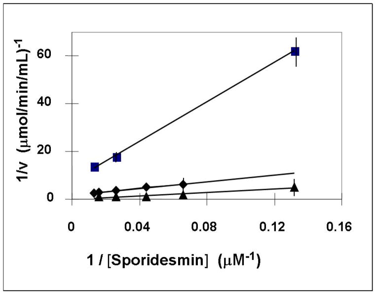
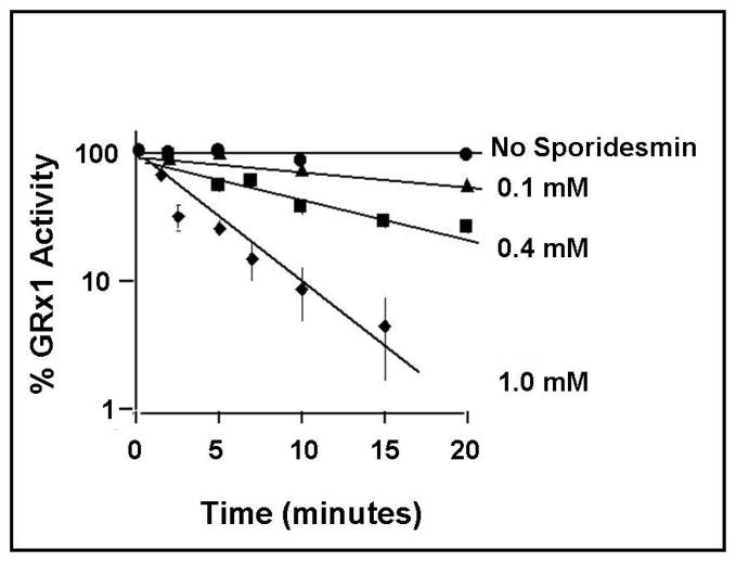
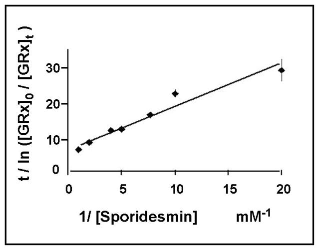
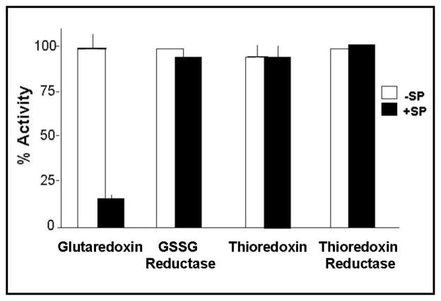
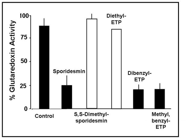
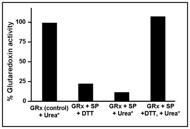
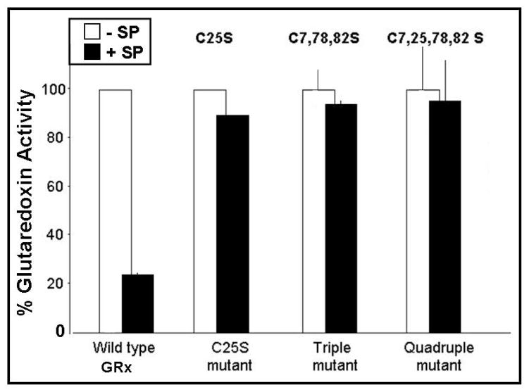
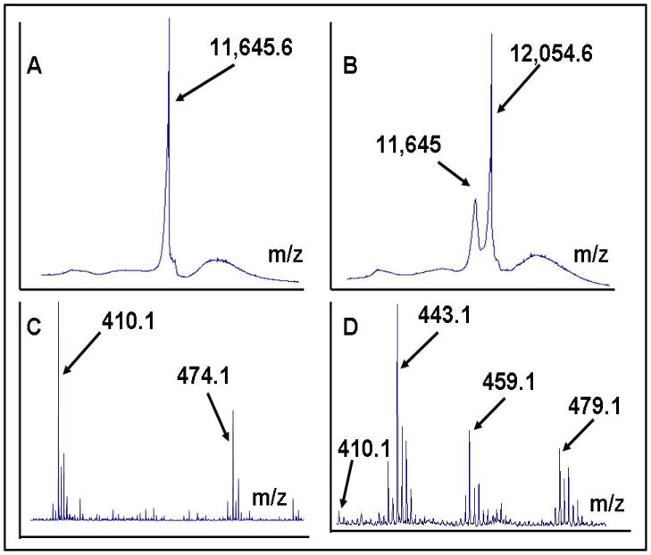
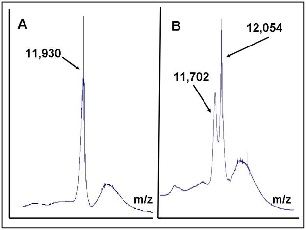
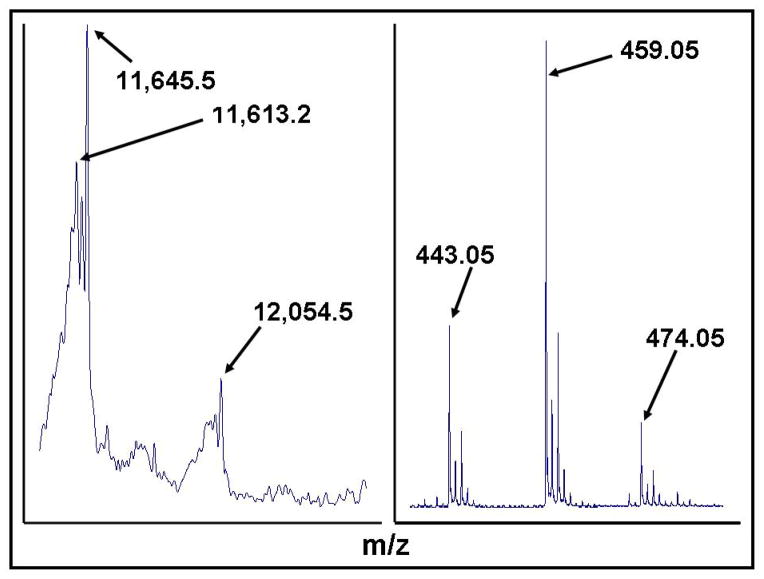
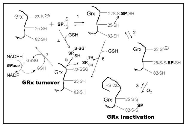
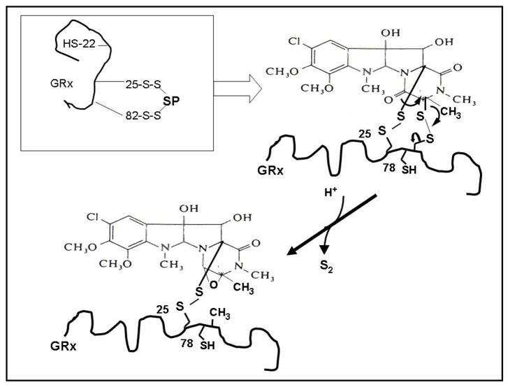
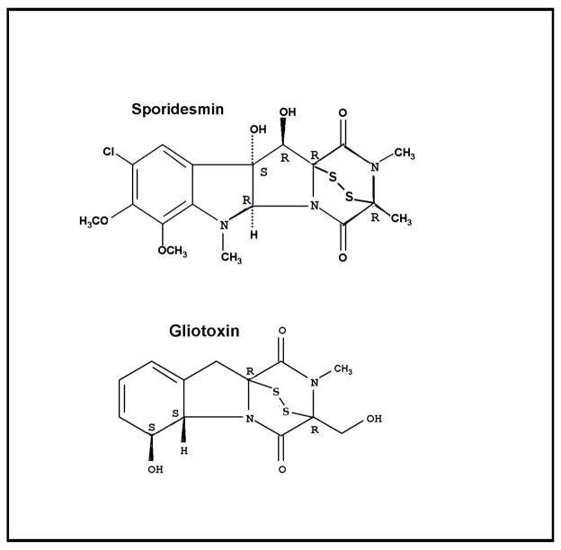
Glutaredoxin (thioltransferase) is a thiol-disulfide oxidoreductase that displays efficient and specific catalysis of protein-SSG deglutathionylation and is thereby implicated in homeostatic regulation of the thiol-disulfide status of cellular proteins. Sporidesmin is an epidithiopiperazine-2,5-dione (ETP) fungal toxin that disrupts cellular functions likely via oxidative alteration of cysteine residues on key proteins. In the current study sporidesmin inactivated human glutaredoxin in a time- and concentration-dependent manner. Under comparable conditions other thiol-disulfide oxidoreductase enzymes, glutathione reductase, thioredoxin, and thioredoxin reductase, were unaffected by sporidesmin. Inactivation of glutaredoxin required the reduced (dithiol) form of the enzyme, the oxidized (intramolecular disulfide) form of sporidesmin, and molecular oxygen. The inactivated glutaredoxin could be reactivated by dithiothreitol only in the presence of urea, followed by removal of the denaturant, indicating that inactivation of the enzyme involves a conformationally inaccessible disulfide bond(s). Various cysteine-to-serine mutants of glutaredoxin were resistant to inactivation by sporidesmin, suggesting that the inactivation reaction specifically involves at least two of the five cysteine residues in human glutaredoxin. The relative ability of various epidithiopiperazine-2,5-diones to inactivate glutaredoxin indicated that at least one phenyl substituent was required in addition to the epidithiodioxopiperazine moiety for inhibitory activity. Mass spectrometry of the modified protein is consistent with formation of intermolecular disulfides, containing one adducted toxin per glutaredoxin but with elimination of two sulfur atoms from the detected product. We suggest that the initial reaction is between the toxin sulfurs and cysteine 22 in the glutaredoxin active site. This study implicates selective modification of sulfhydryls of target proteins in some of the cytotoxic effects of the ETP fungal toxins and their synthetic analogues.


Figures

Figure 1
Figure 1. Two substrate kinetics for GSH-dependent GRx1-catalyzed reduction of sporidesmin
Two substrate kinetics for sporidesmin and glutathione with GRx1. Concentrations of GSH and sporidesmin were varied in the presence and absence of GRx1. The two substrate kinetic analysis of sporidesmin (7.6–76 μM) and GSH (0.4 mM, solid square), (2 mM, solid diamond), (5 mM, solid triangle) with GRx1 (0.035 μM) shows an intersecting pattern of lines consistent with a sequential mechanism. Each point represents the mean value, plus and minus the standard error, for at least three determinations. Where error bars are not evident they are within the size of the symbol.
Figure 2
Figure 2. Inactivation of GRx1. Time and concentration dependent loss of GRx1 activity
GRx1 (2-10 μM) was treated with different concentrations (0.1–1 mM) of sporidesmin in 0.1 M K phosphate, pH 7.5, 10 % ethanol. At different times, aliquots were tested for GRx1 activity according to the spectrophotometric assay (GSSG-reductase mediated NADPH oxidation coupled to GSSG formation) using cys-SSG as the prototype substrate. Rates of GSSG formation were also determined with sporidesmin as the disulfide substrate and are designated as non-cys-SSG rates. Since residual sporidesmin is transferred to the assay with the deactivated GRx1, non-cys-SSG rates were subtracted from cys-SSG rates for each time point. Each point represents the mean value, plus and minus the standard error, for at least three determinations. Where error bars are not evident they are within the size of the symbol.
Figure 3
Figure 3. Modified Kitz-Wilson plot for GRx1 inactivation by sporidesmin
GRx1 (0.9 μM) was incubated with different concentrations of sporidesmin (0.05–1 mM) for 5 min at 30 °C, 0.1 M K phosphate, pH 7.5, and residual activities were determined relative to ethanol treated control as described in Figure 2. KI and kinact were determined according to the relationship: ln (Eo/Et)/ t = kinact [I] / (KI + [I]), where Eo and Et refer to GRx1 concentrations at times 0 and t, [I] refers to sporidesmin concentration, KI is the concentration of inactivator that gives half the maximal rate of inactivation, and kinact is the net rate constant for inactivation. The KI of sporidesmin for GRx1 is 0.15 mM and the kinact is 0.72 min−1. Each point represents the mean value, plus and minus the standard error, for at least three determinations. Where error bars are not evident they are within the size of the symbol.
Figure 4
Figure 4. Selective inactivation of GRx1 by sporidesmin
Thiol-disulfide oxidoreductase enzymes were incubated with 1 mM sporidesmin at 30 °C, 0.1 M K phosphate (5% ethanol), pH 7.5 for 20 min. The remaining enzyme activity was expressed as percent of the control activity. Human GRx1 (1.8 μM) activity was determined by GSSG-reductase mediated NADPH oxidation coupled to GSSG formation. Yeast GSSG reductase (0.125 μM) activity was measured by the loss of NADPH absorbance at 340 nm. E.coli thioredoxin (0.2 mg.ml−1) and thioredoxin reductase (0.1 mg.ml−1) were assayed by NADPH oxidation coupled to thioredoxin oxidation using GSSG as the substrate. Mammalian thioredoxin was assayed by a turbidometric assay using insulin as the substrate. Each bar represents the mean value, plus and minus the standard error, for at least three determinations. Where errors are not shown the value represents a single experiment carried out after optimization of conditions.
Figure 5
Figure 5. Reaction of ETP analogs with GRx1: Structural requirements for inactivation
GRx1 (2 μM) was treated with different ETP analogues at 1 mM final concentration. The synthetic ETPs were dissolved in dimethyl sulfoxide to a final concentration of 100 mM and diluted 1/100 into the GRx1 –containing solution at 0.1 M K phosphate, pH 7.5, 30 °C. GRx1 was treated with 1 % ethanol or dimethyl sulfoxide as controls for these experiments. After incubation for 20 min, the samples were assayed for GRx1 activity using cys-SSG as the substrate. Each bar represents the mean value, plus and minus the standard error, for at least three determinations. In the case of Diethyl-ETP, no error is shown because this represents a single experiment after optimization of conditions, due to limited supply of this ETP derivative.
Figure 6
Figure 6. Requirements for reactivation of sporidesmin-inactivated GRx1
GRx1 (100 μM) was treated either with 1 mM sporidesmin or 5 % ethanol at 30 °C, 0.1 M K phosphate, pH 7.5. Enzyme activity was measured after incubation for 15 min. The sporidesmin-treated sample was approximately 90% inactivated. The control was treated with 8 M urea plus 50 mM DTT. The inactivated GRx1 was divided into two aliquots and treated either with either 8 M urea or 50 mM DTT and 8 M urea. The samples (including urea plus DTT treated control) were each loaded on a gel filtration column and separated from excess small molecules. The GRx1 protein was pooled and analyzed for activity and protein content. The results shown in the figure are for a single experiment, after conditions were optimized. The recovery of protein after size exclusion chromatography was 55–60% for all three samples. GRx1 inactivated with sporidesmin and exposed to urea alone or DTT alone showed no reactivation, while treatment with sporidesmin then urea plus DTT gave 60% recovery of activity, i.e. full recovery of GRx activity when normalized to 55–60% protein recovery.
Figure 7
Figure 7. Study of sporidesmin reaction with cysteine-to-serine mutants of GRx1
Site-directed mutagenesis was utilized to produce the cys to ser mutant GRx1 enzymes, as described in Methods. Each enzyme (3–5 μM) was incubated with 1 mM sporidesmin for 20 min in 0.1 M K phosphate, pH 7.5, 10% ethanol at 30 °C. GRx1 activity was then measured using cys-SSG substrate. Each point represents the mean value, plus and minus the standard error, for at least three determinations. Where error bars are not evident they are within the size of the symbol.
Figure 8
Figure 8. Mass spectrometry of GRx1, sporidesmin and reaction products
MALDI spectra of GRx1 and the reaction product with sporidesmin show ions at m/z 11,645 corresponding to native GRx1 (A), and m/z 12,054 consistent with formation of a mixed disulfide between GRx1 and sporidesmin followed by elimination of two sulfur atoms (B). ESI spectra of sporidesmin (C) and the corresponding low mass regions in the product of reaction with GRx1 (D) contain ions consistent with the presence of sporidesmin (calculated m/z 474.0555), sporidesmin minus two sulfur atoms (calculated mass 410.1113) and in the reaction product ions corresponding to sporidesmin in which a sulfur has been replaced by hydrogen (calculated m/z 443.0912) and the product of monooxygenation of the remaining sulfur atom (calculated m/z 459.0862). The identity of the m/z 479 species is unknown. Low mass regions of the spectra were internally calibrated using the sporidesmin ion.
Figure 9
Figure 9. Alkylation of GRx1 cysteines with iodoacetamide (IAM)
MALDI spectra of GRx1 reacted with iodoacetamide (A), and GRx1 first reacted with sporidesmin, then with iodoacetamide (B). IAM-treated GRx1 contained a major ion m/z 11,930 +/− 3 consistent with alkylation of all five cysteines. MALDI analysis of the products of reaction of GRx1 with sporidesmin, then treated with IAM, only showed major ions corresponding to the SP-GRx1 adduct (m/z 12054) and GRx1 modified by carboxamidomethylation of a single cysteine (calculated m/z 11,702).
Figure 10
Figure 10. Reaction of GRx1 and adduct with DTT
GRx1 was pretreated with sporidesmin, followed by incubation in the presence of 35 mM DTT for 1 h at room temperature, then subjected to mass spectral analysis. MALDI ions at m/z 11,613 +/− 2 and 443 were consistent with release of GRx1 and sporidesmin, respectively, each less a sulfur atom, as would be expected if two sulfurs had been eliminated from the adduct. Ions consistent with the presence of unreacted GRx1 (11,645) and SP-GRx1 adduct minus 64 Da (12,054), reflecting possible loss of two sulfur atoms, are also evident.
Figure 11
Figure 11. Mechanistic models of sporidesmin as a substrate and inactivator of GRx1
This scheme shows two alternative explanations for sporidesmin as a substrate of GRx1: (a) steps 4, 5, 7 depict an initial non-enzymatic formation of glutathionyl mixed disulfide of sporidesmin, sporidesmin-S-SG, which is then reduced by GSH in a GRx1 catalyzed reaction to form reduced sporidesmin. This postulated mechanism is consistent with the two-substrate kinetics (Figure 1) and previous reports of the substrate specificity of GRx1 and the mode of substrate activity of non-GSH containing disulfides (1,2,4,5). (b) Steps 1, 6, 7 depict the direct reaction of sporidesmin with GRx1 to form GRx1 sporidesmin mixed disulfide (GRx1-S-S-sporidesmin). GSH then displaces sporidesmin from this adduct to form GRx1-S-SG which can enter the normal catalytic cycle of GRx1. (c) When GSH is scarce or absent the GRx1-S-S-sporidesmin complex goes on to an irreversibly inactivated form according to steps 1, 2, 3 involving molecular oxygen.
Figure 12
Figure 12. Hypothetical scheme for spontaneous de-sulfuration of the GRx1-sporidesmin adduct
This scheme depicts one plausible mechanism by which the adduct of GRx1 and sporidesmin might spontaneously lose 2 sulfur atom equivalents yet remain intact as a disulfide-bonded bimolecular complex. (As indicated in the text, there are other conceivable ways to lose 64 Da of mass, e.g., loss of four oxygens, or 2 oxygens and 1 sulfur).
Chart 1
Chart 1. Structures of sporidesmin and gliotoxin
“R” and “S” notations on the molecular structures refer to the configurations at the respective chiral centers.

Similar articles

Cited by

References

    1. Gravina SA, Mieyal JJ. Thioltransferase is a Specific Glutathionyl Mixed Disulfide Oxidoreductase. Biochemistry. 1993;32:3368–3376. - PubMed
    1. Mieyal JJ, Gravina SA, Mieyal PA, Srinivasan U, Starke DW. Glutathionyl Specificity of Thioltransferases: Mechanistic and Physiological Implications. In: Packer LEC, editor. Biothiols in Health and Disease. Marcel Dekker, Inc; New York: 1995. pp. 305–372.
    1. Jung CH, Thomas JA. S-Glutathiolated Hepatocyte Proteins and Insulin Disulfides as Substrates for Reduction by Glutaredoxin, Thioredoxin, Protein Disulfide Isomerase, and Glutathione. Arch Biochem Biophys. 1996;335:61–72. - PubMed
    1. Srinivasan U, Mieyal PA, Mieyal JJ. pH Profiles indicative of Rate Limiting Nucleophilic Displacement in Thioltransferase (Glutaredoxin) Catalysis. Biochemistry. 1997;36:3199–3206. - PubMed
    1. Yang Y, Jao S, Nanduri S, Starke DW, Mieyal JJ, Qin J. Reactivity of the Human Thioltransferase (C7S, C25S, C78S, C82S) Mutant and NMR Solution Structure of its Glutathionyl Mixed Disulfide Intermediate Reflect Catalytic Specificity. Biochemistry. 1998;37:17145–17156. - PubMed

Publication types

MeSH terms