Review
. 2006 Jun;2(6):e62.
doi: 10.1371/journal.ppat.0020062.

The thymus is a common target organ in infectious diseases

Affiliations
Review

The thymus is a common target organ in infectious diseases

Wilson Savino. PLoS Pathog. 2006 Jun.

Abstract

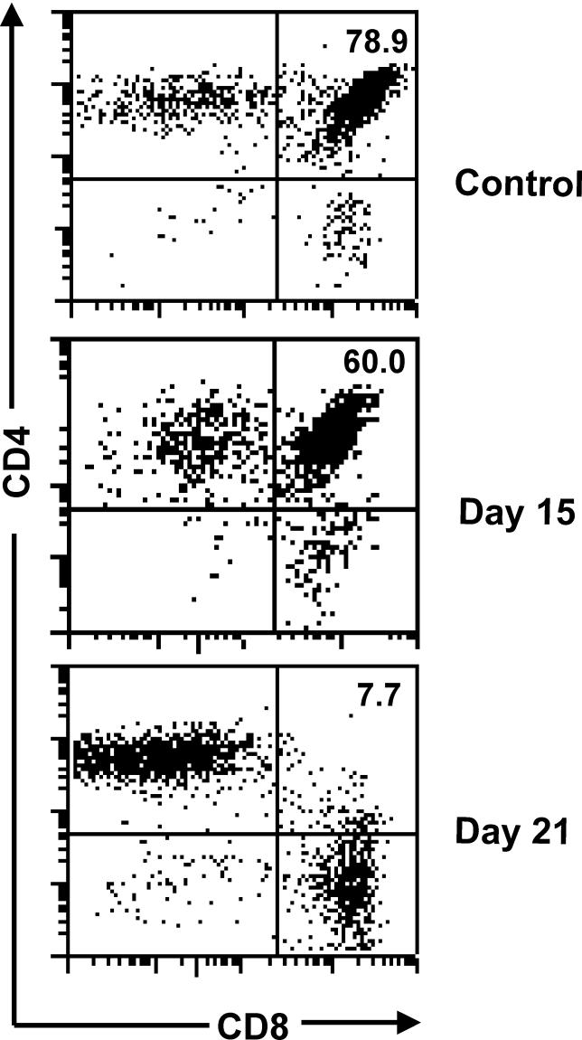
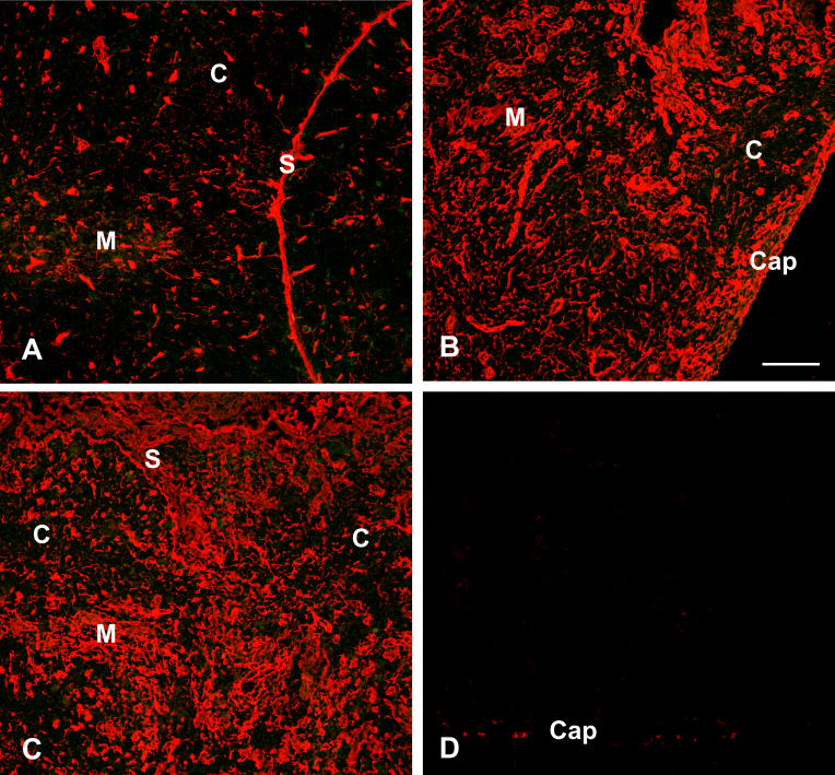
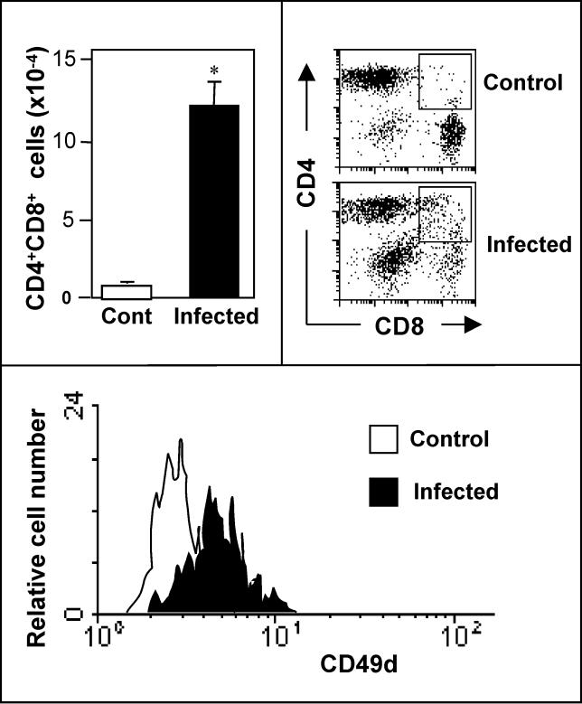
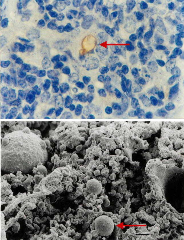
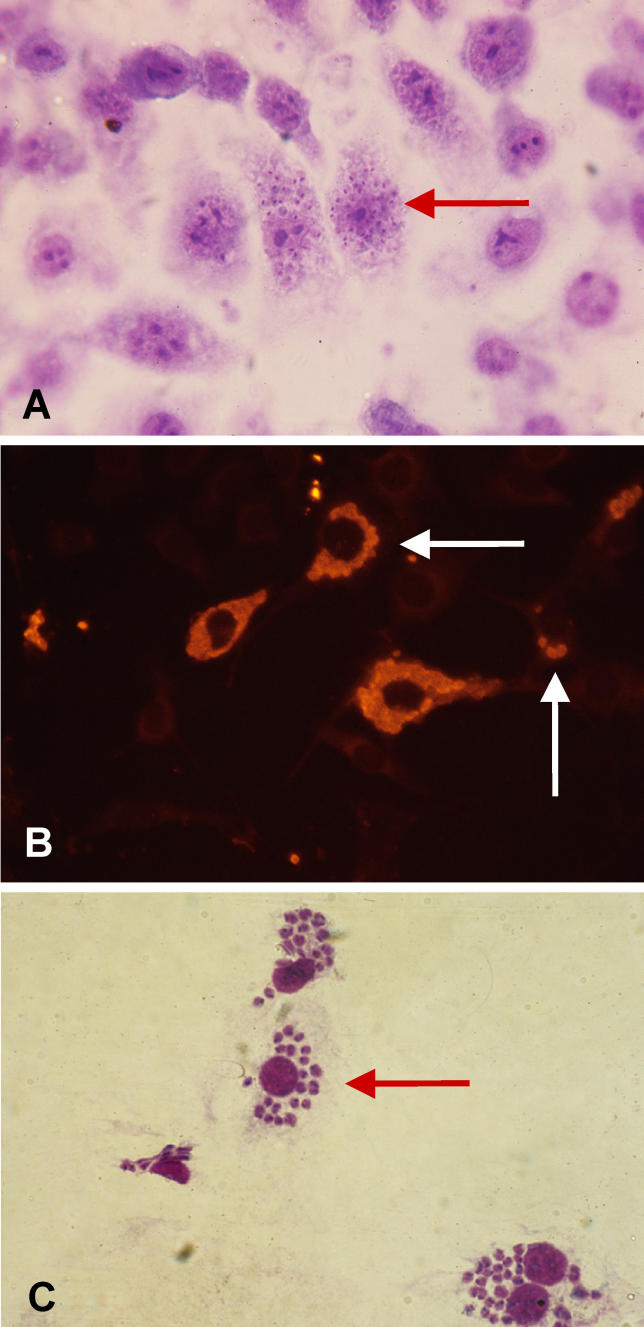
Infectious disease immunology has largely focused on the effector immune response, changes in the blood and peripheral lymphoid organs of infected individuals, and vaccine development. Studies of the thymus in infected individuals have been neglected, although this is progressively changing. The thymus is a primary lymphoid organ, able to generate mature T cells that eventually colonize secondary lymphoid organs, and is therefore essential for peripheral T cell renewal. Recent data show that normal thymocyte development and export can be altered as a result of an infectious disease. One common feature is the severe atrophy of the infected organ, mainly due to the apoptosis-related depletion of immature CD4+CD8+ thymocytes. Additionally, thymocyte proliferation is frequently diminished. The microenvironmental compartment of the thymus is also affected, particularly in acute infectious diseases, with a densification of the epithelial network and an increase in the deposition of extracellular matrix. In the murine model of Chagas disease, intrathymic chemokine production is also enhanced, and thymocytes from Trypanosoma cruzi-infected mice exhibit greater numbers of cell migration-related receptors for chemokines and extracellular matrix, as well as increased migratory responses to the corresponding ligands. This profile is correlated with the appearance of potentially autoreactive thymus-derived immature CD4+CD8+ T cells in peripheral organs of infected animals. A variety of infectious agents--including viruses, protozoa, and fungi--invade the thymus, raising the hypothesis of the generation of central immunological tolerance for at least some of the infectious agent-derived antigens. It seems clear that the thymus is targeted in a variety of infections, and that such targeting may have consequences on the behavior of peripheral T lymphocytes. In this context, thymus-centered immunotherapeutic approaches potentially represent a new tool for the treatment of severe infectious diseases.


Conflict of interest statement

Competing interests. The author has declared that no competing interests exist.

Figures

Figure 1
Figure 1. The Normal Process of Intrathymic T Cell Differentiation
This diagram shows that most immature thymocytes localized in the subcapsular cortical region of the thymic lobules do not express CD4 or CD8 accessory molecules, nor the CD3/TCR complex, and are known as double-negative (DN, for CD4 and CD8) cells. As they progress in differentiation, they begin to express on their cell membranes the TCR/CD3 complex as well as CD4 and CD8, becoming double-positive (DP) thymocytes, which occupy most of the cortical region. These cells are then submitted to the processes of positive and negative selection, as a consequence of the interaction with the thymic microenvironment (gray network) through MHC-TCR interactions. Those cells undergoing negative selective die by apoptosis, whereas the small percentages of positively selected thymocytes progress in their differentiation, moving toward the medulla and becoming single-positive cells (SP) for either CD4 or CD8, both expressing high densities of CD3/TCR complex. These mature thymocytes can be exported from the thymus into the peripheral lymphoid organs. Finally, this overall process of thymocyte differentiation occurs in the context of the three-dimensional thymic microenvironment (gray network) through membrane interactions as well as soluble products (represented by red stars) released by microenvironmental cells. Modified from [9].
Figure 2
Figure 2. The Thymic Microenvironment and Its Role in Thymopoiesis
(A) A simplified model of thymocyte migration includes two compartments. On the left is depicted the entrance site of precursor cells into the thymus through blood vessels. Having entered the thymus, thymocytes migrate during differentiation to ultimately leave the organ, bearing the mature phenotypes of CD4+CD8 or CD4CD8+ cells. The right side of the image is a schematic representation of a thymic lobule, showing thymocytes intermingled with a heterogeneous cellular network, the thymic microenvironment, composed of epithelial cells (yellow and orange), dendritic cells (red), macrophages (blue), and fibroblasts (green). The epithelial tissue shows morphologic heterogeneity that can be seen in subseptal/subcapsular, cortical, and medullary regions. In the cortex, we note a particular lymphoepithelial complex, the TNC. (B) A number of molecular interactions take place between developing thymocytes and thymic epithelial cells. Whereas a and b correspond to interactions mediated by soluble secretory molecules produced by epithelial cells (a) or lymphocytes (b), the interaction shown in c involves a given peptide (red dot) being presented by MHC (expressed by the epithelial cell) to the TCR and corresponding accessory molecule in the thymocyte membrane. The interaction shown by (d) involves adhesion molecules and the respective membrane counter-receptors, and (e) depicts an interaction mediated by ECM ligand and receptor. Modified from [3,8].
Figure 3
Figure 3. Progressive Thymic Atrophy in Mice Acutely Infected by T. cruzi
A typical CD4/CD8-defined cytofluorometric profile of normal thymocytes compared to that with T. cruzi infection. As infection progresses, we can see a progressive loss of CD4+CD8+ cells. Percentage values of the CD4+CD8+ subset are shown within the quadrants. The days correspond to the time of infection, with an inoculum of 105 parasites per animal. The peak of parasitemia coincides with the peak of thymocyte depletion. Modified from [43].
Figure 4
Figure 4. Increase in Intrathymic ECM Following Acute T. cruzi Infection in Mice
Cryostat sections of thymuses from normal (A) or T. cruzi-infected mice (B, C, and D) were immunostained with anti-fibronectin immune serum (A, B, and C) or unrelated antibody (D). In control thymus (A), the typical fibronectin-containing network is seen, being more prominent in the medullary region of the thymic lobule (M), as compared to the cortex (C). This pattern is dramatically changed in the atrophic thymuses from T. cruzi-infected animals (B and C), in which the fibronectin network is much denser in both cortex and medulla. Such immunolabeling is specific, since an unrelated antibody did not yield any significant fluorescence when applied on the thymus section from an infected mouse (D). Mice were infected with 105 trypomastigote forms of the parasite (Colombian strain), and sacrificed 21 d later, at the peak of parasitemia. C, cortex; Cap, capsule; M, medulla; S, septum. Bar represents 100 μm for all photomicrographs. Pictures were kindly provided by Désio Aurélio Farias-de-Oliveira.
Figure 5
Figure 5. Appearance of Immature Thymus-Derived T Cells in Lymph Nodes of T. cruzi-Infected Mice
The bar graph at the upper left shows the significant increase in the absolute numbers of CD4+CD8+ lymphocytes seen in subcutaneous lymph nodes of acutely infected mice. The flow cytometry dot plots to the right show the enhancement of these CD4+CD8+ cells in the infected animal and compared to the age-matched control. These immature double-positive T cells express higher amounts of the fibronectin receptor VLA-4, as ascertained by the cytofluorometric detection of the CD49d integrin subunit (bottom graph). Adapted from [43].
Figure 6
Figure 6. Intrathymic Presence of the Fungus P. brasiliensis
Fungus particles (red arrows) are shown by immunohistochemistry (upper image) and scanning electron microscopy (lower image). Note that infective particles are encircled by microenvironmental cells bearing large nuclei. Pictures were kindly provided by Dr. Liana Verinaud.
Figure 7
Figure 7. In Vitro Infection of Mouse Thymic Microenvironmental Cells by T. cruzi
The presence of the amastigote forms of the parasite within cultured thymic epithelial cells was ascertained by Giemsa staining (A) and by immunohistochemistry (B). Infected thymic phagocytic cells are shown in (C). Arrows indicate intracellular amastigote clusters. Pictures were kindly provided by Désio Aurélio Farias de Oliveira.

Similar articles

Cited by

References

    1. Res P, Spits H. Developmental stages in the human thymus. Sem Immunol. 1999;11:39–46. - PubMed
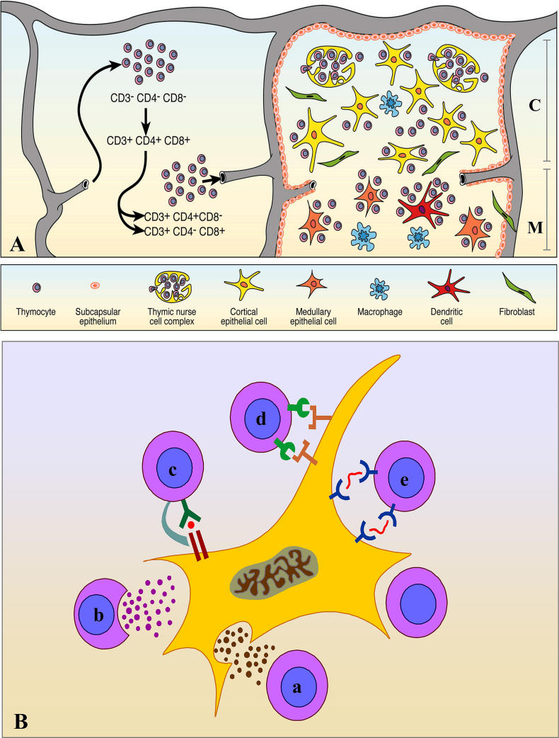
    1. Ritter MA, Palmer DB. The human thymic microenvironment: New approaches to functional analysis. Sem Immunol. 1999;11:13–21. - PubMed
    1. Savino W, Dardenne M. Neuroendocrine control of thymus physiology. Endocrine Rev. 2000;21:412–443. - PubMed
    1. Anderson G, Jenkinson EJ. Lymphostromal interactions in thymic development and function. Nat Rev Immunol. 2001;1:31–40. - PubMed
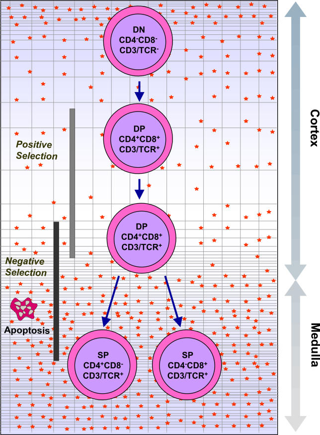
    1. Coutinho A, Caramalho I, Seixas E, Demengeot J. Thymic commitment of regulatory T cells is a pathway of TCR-dependent selection that isolates repertoires undergoing positive or negative selection. Curr Topics Microbiol Immunol. 2005;293:43–71. - PubMed

Publication types