Randomized Controlled Trial
. 2006 Jun;12(6):1441-50.
doi: 10.1089/ten.2006.12.1441.

Effect of rhPDGF-BB delivery on mediators of periodontal wound repair

Affiliations
Randomized Controlled Trial

Effect of rhPDGF-BB delivery on mediators of periodontal wound repair

Jason W Cooke et al. Tissue Eng. 2006 Jun.

Abstract

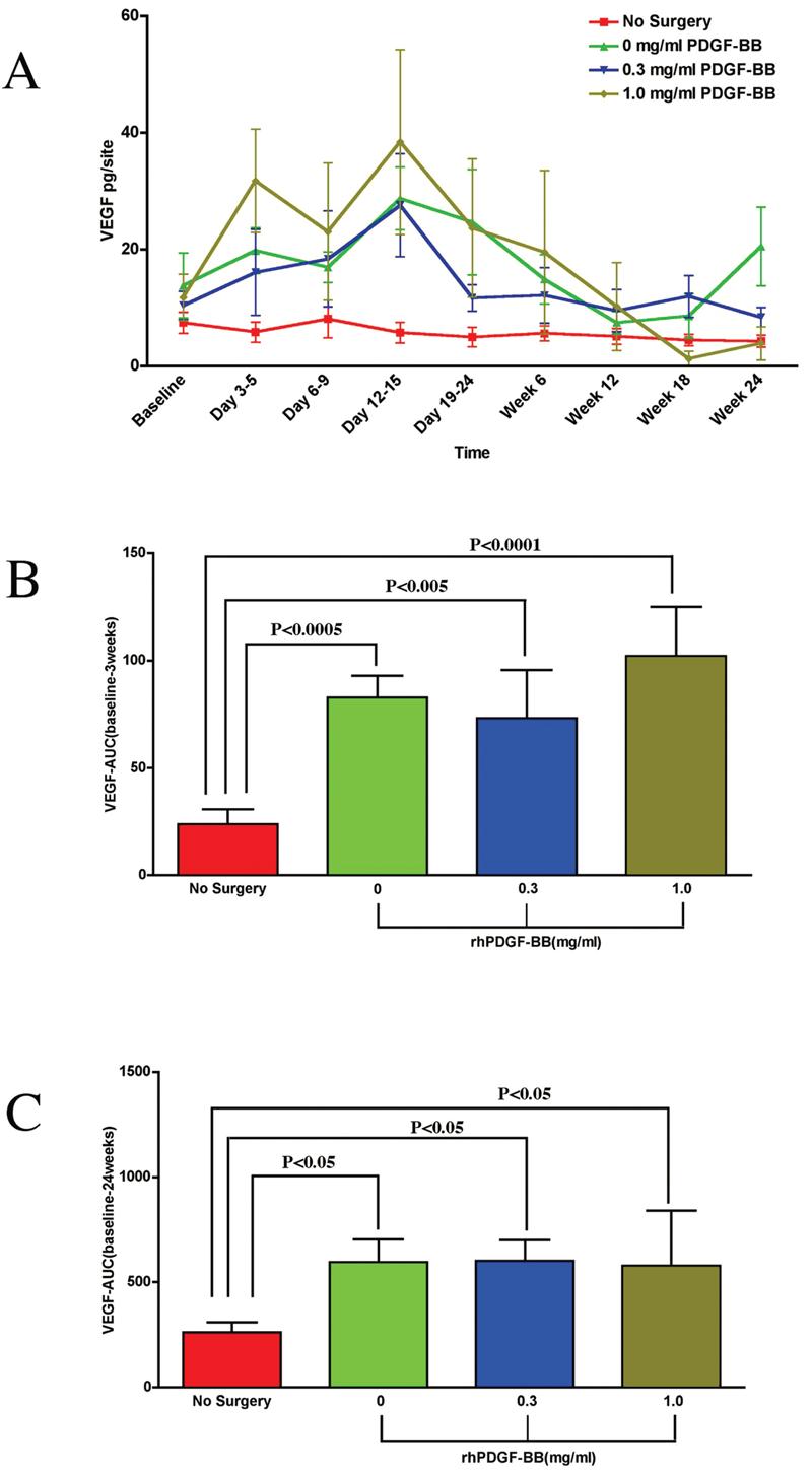
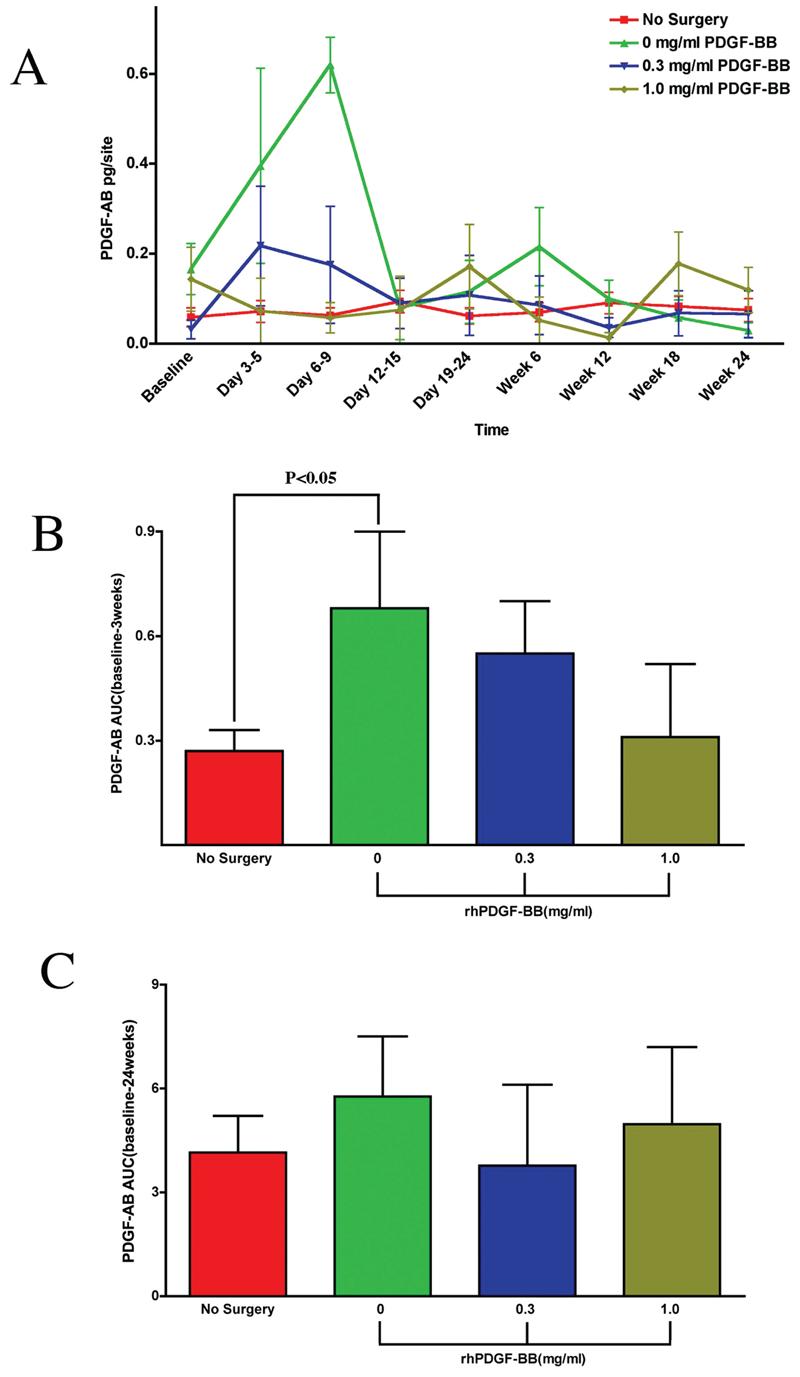
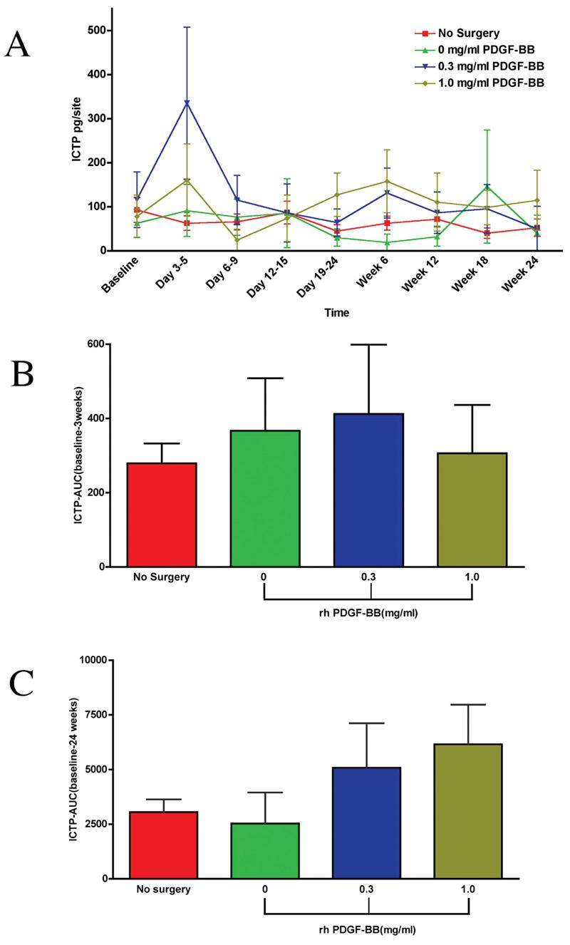
Growth factors such as platelet-derived growth factor (PDGF) exert potent effects on wound healing including the regeneration of tooth-supporting structures. This investigation examined the effect of the local delivery of PDGF-BB when combined with reconstructive periodontal surgery on local wound fluid (WF) levels of PDGF-AB, vascular endothelial growth factor (VEGF), and bone collagen telopeptide (ICTP) in humans with advanced periodontitis. Sixteen patients exhibiting localized periodontal osseous defects were randomized to one of three groups (beta-TCP carrier alone, beta-TCP + 0.3 mg/mL of recombinant human PDGF-BB [rhPDGF-BB], or beta-TCP + 1.0 mg/mL of rhPDGF-BB) and monitored for 6 months. WF was harvested and analyzed for PDGF-AB, VEGF, and ICTP WF levels. Teeth contralateral to the target lesions served as controls. Increased levels of VEGF in the WF was observed for all surgical treatment groups with the 1.0 mg/mL rhPDGF-BB group showing the most pronounced difference at 3 weeks in the AUC analysis versus control (p < 0.0001). PDGF-AB WF levels were increased for the carrier alone group compared to both rhPDGFBB groups. Low-dose rhPDGF-BB application elicited increases in ICTP at days 3-5 in the wound healing process, suggesting a promotion of bone turnover at early stages of the repair process (p < 0.02). These results demonstrate contrasting inducible expression patterns of PDGF-AB, VEGF, and ICTP during periodontal wound healing in humans.


Figures

FIG. 1.
FIG. 1.
Study timeline. GCF, gingival crevicular fluid; WF, periodontal wound fluid; WH, wound healing score.
FIG. 2
FIG. 2
PDGF tissue engineering of periodontal defects promotes local release of vascular endothelial growth factor. (A) Graph of VEGF (pg/site) versus timepoint for all groups: β-TCP carrier alone, 0.3mg/mL PDGF, 1.0 mg/mL PDGF, and the nonsurgical control. Statistically significant differences were found between all treatment groups and the nonsurgical control (p < 0.05) except for Days 6–9 and Week 12. A statistically significant difference was also found between the 0.3 mg/mL PDGF group and the 1.0 mg/mL PDGF group at Days 3–5 (p < 0.05). (B) Area under the curve (AUC) analysis for VEGF up to 3 weeks. The most pronounced statistically significant difference was found between the nonsurgical control and the 1.0 mg/mL PDGF group (p < 0.0001). Statistically significant differences were also observed between the nonsurgical control and the β-TCP carrier alone group (p < 0.0005) and the 0.3 mg/mL PDGF group (p < 0.005). The AUC for VEGF was highest for the 1.0 mg/mL PDGF group compared to the other surgical treatment groups, although there were no statistically significant differences between these treatment groups. (C) Area under the curve (AUC) analysis for VEGF of all time-points. Statistically differences were observed between the nonsurgical control and all treatment groups (p < 0.05). No statistically significant differences were found between treatment groups. Statistical analysis was performed using ANOVA Fisher test at the 5% level. A total of 15 patients were analyzed for VEGF. Bars indicate standard error measurements (SEM).
FIG. 3
FIG. 3
Effect of PDGF-BB-mediated tissue engineering of periodontal defects on local PDGF-AB release. (A) Graph of PDGF-AB release (pg/site) versus time point for all groups: β-TCP carrier alone, 0.3 mg/mL PDGF-BB, 1.0 mg/mL PDGF-BB, and the nonsurgical control. A statistically significant difference was observed between the β-TCP carrier alone group and the nonsurgical control at Days 3–5 (p < 0.04). The 0.3 mg/mL PDGF-BB group and the 1.0 mg/mL PDGF-BB group had similar levels of PDGF-AB released throughout the study. (B) Area under the curve (AUC) analysis for PDGF-AB up to 3 weeks. A statistically significant difference is observed only between the nonsurgical control and the β-TCP carrier alone group (p < 0.05). (C) Area under the curve (AUC) analysis for PDGF-AB release at all time points. Statistically significant differences were not observed when evaluating over 24 weeks for any groups. Statistical analysis was performed using ANOVA Fisher test at the 5% level. A total of 14 patients were analyzed for PDGF-AB (endogenous). Bars indicate standard error measurements (SEM).
FIG. 4
FIG. 4
Effect of PDGF-mediated tissue engineering of periodontal defects on local pyridinoline cross-linked carboxyterminal telopeptide of type I collagen (ICTP) release. (A) Graph of ICTP (pg/site) versus time point for all groups: β-TCP carrier alone, 0.3 mg/mL PDGF-BB, 1.0 mg/mL PDGF-BB, and the nonsurgical control. Statistically significant differences were observed between the 0.3 mg/mL PDGF-BB group and the nonsurgical control (p < 0.003) and between the 0.3 mg/mL PDGFBB group and the β-TCP carrier alone group (p < 0.02) at the Day 3–5 timepoint. At 6 weeks, statistically significant differences were observed for the following groups: β-TCP carrier alone versus the 0.3 mg/mL PDGF-BB (p < 0.03), β-TCP carrier alone versus the 1.0 mg/mL PDGF-BB (p < 0.01), and the 1.0 mg/mL PDGF-BB versus nonsurgical control (p < 0.03). (B) Area under the curve (AUC) analysis for ICTP up to 3 weeks. No statistically significant differences were found between treatment groups. (C) AUC analysis for ICTP of all timepoints. No statistically significant differences were observed between any groups. Statistical analysis was performed using ANOVA Fisher test at the 5% level. A total of 14 patients were analyzed for ICTP. Bars indicate standard error measurements (SEM).

Similar articles

Cited by

References

    1. Williams RC. Periodontal disease. N. Engl. J. Med. 1990;322:373. - PubMed
    1. Heldin CH. Platelet-derived growth factor—an introduction. Cytokine Growth Factor Rev. 2004;15:195. - PubMed
    1. Heldin CH, Ostman A, Ronnstrand L. Signal transduction via platelet-derived growth factor receptors. Biochim. Biophys. Acta. 1998;1378:F79. - PubMed
    1. Rosenkranz S, Kazlauskas A. Evidence for distinct signaling properties and biological responses induced by the PDGF receptor alpha and beta subtypes. Growth Factors. 1999;16:201. - PubMed
    1. Lynch SE, de Castilla GR, Williams RC, Kiritsy CP, Howell TH, Reddy MS, Antoniades HN. The effects of short-term application of a combination of platelet-derived and insulin-like growth factors on periodontal wound healing. J. Periodontol. 1991;62:458. - PubMed

Publication types

MeSH terms