. 2006;8(4):R111.
doi: 10.1186/ar2000.

Autoantibodies against the replication protein A complex in systemic lupus erythematosus and other autoimmune diseases

Affiliations

Autoantibodies against the replication protein A complex in systemic lupus erythematosus and other autoimmune diseases

Yoshioki Yamasaki et al. Arthritis Res Ther. 2006.

Abstract

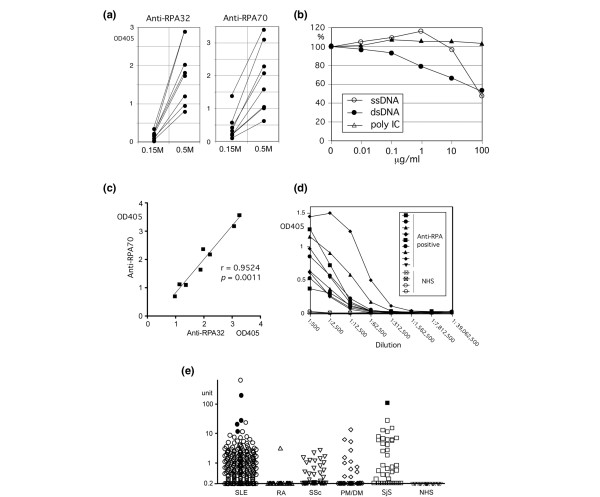
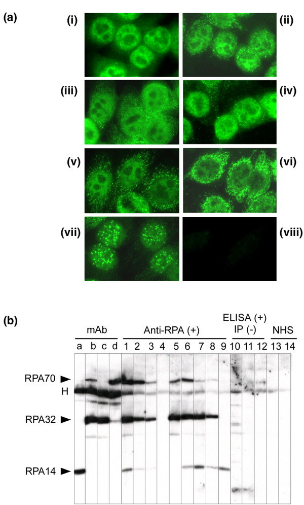
Replication protein A (RPA), a heterotrimer with subunits of molecular masses 70, 32, and 14 kDa, is a single-stranded-DNA-binding factor involved in DNA replication, repair, and recombination. There have been only three reported cases of anti-RPA in systemic lupus erythematosus (SLE) and Sjögren syndrome (SjS). This study sought to clarify the clinical significance of autoantibodies against RPA. Sera from 1,119 patients enrolled during the period 2000 to 2005 were screened by immunoprecipitation (IP) of 35S-labeled K562 cell extract. Antigen-capture ELISA with anti-RPA32 mAb, immunofluorescent antinuclear antibodies (ANA) and western blot analysis with purified RPA were also performed. Our results show that nine sera immunoprecipitated the RPA70-RPA32-RPA14 complex and all were strongly positive by ELISA (titers 1:62,500 to 1:312,500). No additional sera were positive by ELISA and subsequently confirmed by IP or western blotting. All sera showed fine speckled/homogeneous nuclear staining. Anti-RPA was found in 1.4% (4/276) of SLE and 2.5% (1/40) of SjS sera, but not in rheumatoid arthritis (0/35), systemic sclerosis (0/47), or polymyositis/dermatomyositis (0/43). Eight of nine patients were female and there was no racial predilection. Other positive patients had interstitial lung disease, autoimmune thyroiditis/hepatitis C virus/pernicious anemia, or an unknown diagnosis. Autoantibody specificities found in up to 40% of SLE and other diseases, such as anti-nRNP, anti-Sm, anti-Ro, and anti-La, were unusual in anti-RPA-positive sera. Only one of nine had anti-Ro, and zero of nine had anti-nRNP, anti-Sm, anti-La, or anti-ribosomal P antibodies. In summary, high titers of anti-RPA antibodies were found in nine patients (1.4% of SLE and other diseases). Other autoantibodies found in SLE were rare in this subset, suggesting that patients with anti-RPA may form a unique clinical and immunological subset.


Figures

Figure 1
Figure 1
Immunoprecipitation of replication protein A (RPA). (a) Immunoprecipitation of RPA by mAbs and human autoimmune sera. 35S-labeled K562 cell extracts were immunoprecipitated with mAbs against RPA32 (lane 32), human sera with anti-RPA (lanes 1 to 4, SLE; lanes 5 to 8, others) or with normal human serum (NHS). Coexisting anti-Ro (lane 2) and anti-Su (lanes 3 and 8) are indicated by the open arrowheads. (b) Immunoprecipitation of lupus autoantigens that co-migrate or overlap with RPA. [35S]-labeled K562 cell extracts were immunoprecipitated with sera from patients with SLE (lanes labeled Ki to Histones except lane Ku) or PM (lane Ku), or mouse mAbs BM6.5 (anti-histones). These sera or mAbs recognize autoantigens co-migrate with components of RPA. RPA32 co-migrates with Ki (SL, lanes Ki and rP/Ki/his) and U1snRNP-A (U1-A, lanes nRNP and Ku/nRNP), RPA70 co-migrates with Ku p70 (lanes Ku/nRNP and Ku), and RPA14 co-migrates with histone H4 (lanes rP/Ki/his, Histones, and BM6.5). The specificities of human autoimmune sera are indicated. The numbers at the right are the molecular masses of protein standards. his, histones; rP, ribosomal P.
Figure 2
Figure 2
Anti-replication protein A (RPA) antigen-capture ELISA. (a) Effects of NaCl concentration of the cell extracts on the reactivity of anti-RPA human sera. ELISAs were performed as described in the Materials and methods section with mAbs against RPA32 or RPA70 to coat ELISA plates and to capture RPA from K562 cell extracts, which were prepared in buffer containing either 0.15 M or 0.5 M NaCl. Sera diluted to 1:500 in 0.5 M NaCl NET/Nonidet P40 were tested. (b) Effects of single-stranded DNA (ssDNA), double-stranded DNA (dsDNA), or double-stranded RNA (dsRNA) on the reactivity of human anti-RPA autoantibodies. Affinity-purified RPA on a microtiter plate was incubated for 30 minutes with ssDNA, dsDNA, or dsRNA (poly I:C) at concentrations of 0.01 to 100 μg/ml or with buffer alone. Wells were then incubated with 1:2,000 diluted anti-RPA-positive sera followed by ALP mouse mAb anti-human IgG, and developed. The percentage reactivity compared with RPA incubated with buffer alone (100%) is shown. ssDNA or dsDNA, but not dsRNA, inhibited the human anti-RPA binding in a dose-dependent manner. (c) Correlation between levels of anti-RPA by antigen-capture ELISA with mAbs against RPA32 and against RPA70. The reactivity of eight anti-RPA-positive human autoimmune sera in ELISA with mAbs against RPA32 and against RPA70 was compared. Spearman r = 0.9524, p = 0.0011. (d) Titration curves of anti-RPA-positive human sera. Titration curves of nine anti-RPA-positive autoimmune sera and four normal human sera (NHS) were created by ELISA with mAb against RPA32. K562 cell extracts in 0.5 M NaCl NET/Nonidet P40 buffer were used and sera were serially diluted 1:5 starting from 1:500. (e) Screening of anti-RPA antibodies in sera from patients with various systemic rheumatic diseases by ELISA. Sera from SLE (n = 276), rheumatoid arthritis (RA; n = 35), SSc (n = 47), PM/DM (n = 43), SjS (n = 40), and normal control (NHS, n = 30) were tested at 1:2,500 dilutions by ELISA with mAb against RPA32. SLE (p < 0.001 versus RA, p < 0.05 versus SSc, p < 0.01 versus PM/DM, p < 0.001 versus NHS) and SjS (p < 0.001 versus RA or NHS, p < 0.05 versus SSc, p < 0.01 versus PM/DM) showed high reactivity. RA versus SSc, p < 0.05; SSc versus NHS, p < 0.05; all other pairs were not significant. All comparisons were made with the Kruskal–Wallis test with Dunn's multiple comparison test. Open symbols, immunoprecipitation negative; filled symbols, immunoprecipitation positive. SjS, Sjögren syndrome; SSc, systemic sclerosis.
Figure 3
Figure 3
Immumofluorescent ANA and western blot with anti-RPA positive sera (a) Immunofluorescent ANA testing with anti-RPA-positive sera. HEp-2 cells were stained with mAb against RPA32 (i), RPA70 (ii), human autoimmune sera with anti-RPA (1:160 dilution, iii–vii), or normal control (viii). All anti-RPA-positive sera showed nuclear fine speckled/homogeneous staining, similar to the staining by anti-RPA32 or anti-RPA70 mAb. Some sera had an additional immunofluorescent pattern from the other coexisting specificities; mitochondria (vi) and centromere (vii). (b) Western blot analysis of anti-RPA antibodies. RPA was immunoprecipitated from K562 cell extract, fractionated by 12% SDS-PAGE, and transferred to a nitrocellulose filter. Strips of the filter were probed with mAbs against RPA (lanes a to d: a, RPA14; b, RPA32; c, RPA32; d, RPA70), anti-RPA immunoprecipitation-positive sera (lanes 1 to 9), anti-RPA ELISA-positive immunoprecipitation-negative sera (lanes 10 to 12), or control sera (normal human serum (NHS), lanes 13 and 14). H, mouse IgG heavy chain.

Similar articles

Cited by

References

    1. Tan EM. Antinuclear antibodies: diagnostic markers for autoimmune diseases and probes for cell biology. Adv Immunol. 1989;44:93–151. - PubMed
    1. Miyachi K, Tan EM. Antibodies reacting with ribosomal ribonucleoprotein in connective tissue diseases. Arthritis Rheum. 1979;22:87–93. - PubMed
    1. Takasaki Y, Fishwild D, Tan EM. Characterization of proliferating cell nuclear antigen recognized by autoantibodies in lupus sera. J Exp Med. 1984;159:981–992. doi: 10.1084/jem.159.4.981. - DOI - PMC - PubMed
    1. Bravo R, Frank R, Blundell PA, Macdonald-Bravo H. Cyclin/PCNA is the auxiliary protein of DNA polymerase-delta. Nature. 1987;326:515–517. doi: 10.1038/326515a0. - DOI - PubMed
    1. Reeves WH, Narain S, Satoh M. Autoantibodies in systemic lupus erythematosus. In: Koopman WJ, Moreland LW, editor. Arthritis and Allied Conditions. 15. Philadelphia: Lippincott Williams & Wilkins; 2004. pp. 1497–1521.

Publication types

MeSH terms