. 2006 Sep 8;281(36):25903-14.
doi: 10.1074/jbc.M603414200. Epub 2006 Jul 17.

Cross-talk between epidermal growth factor receptor and hypoxia-inducible factor-1alpha signal pathways increases resistance to apoptosis by up-regulating survivin gene expression

Affiliations

Cross-talk between epidermal growth factor receptor and hypoxia-inducible factor-1alpha signal pathways increases resistance to apoptosis by up-regulating survivin gene expression

Xiang-Hong Peng et al. J Biol Chem. .

Abstract

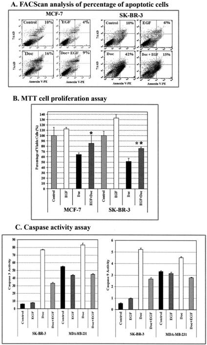
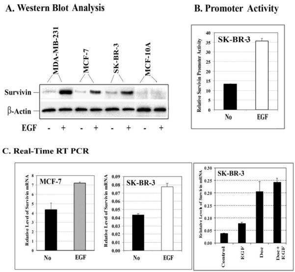
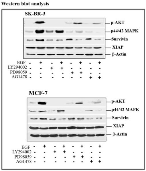
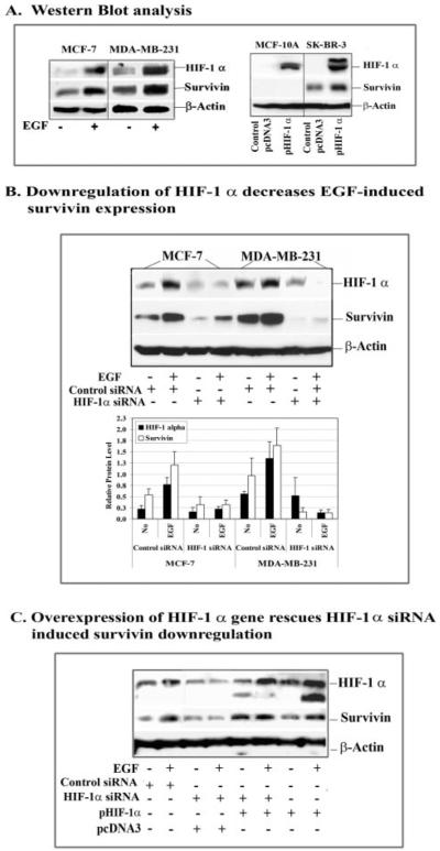
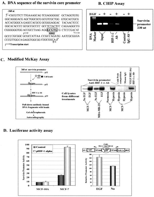
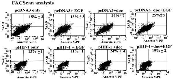
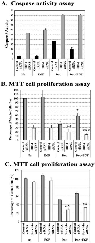
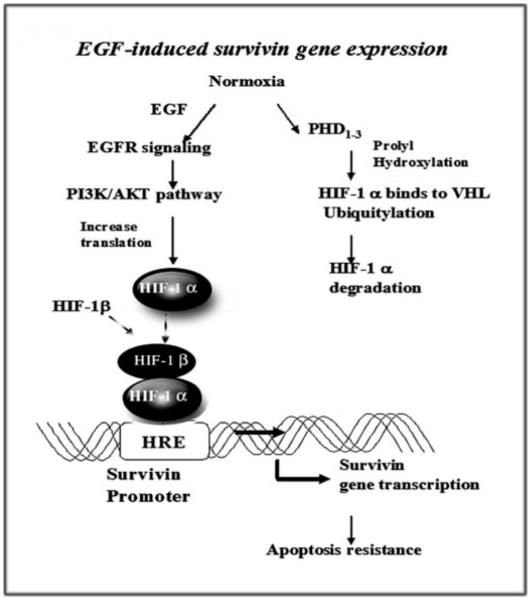
Although increasing evidence supports a link between epidermal growth factor receptor (EGFR) signaling and resistance to apoptosis, the mechanism by which the EGFR signaling pathway inhibits apoptosis is not well understood. In this study, we found that epidermal growth factor (EGF) stimulation increased the level of expression of the inhibitor of apoptosis protein survivin in breast cancer cells but not in normal mammary epithelial cells. We further demonstrated that activation of survivin gene expression is mediated by oxygen-independent hypoxia-inducible factor (HIF)-1alpha up-regulation in EGF-treated cancer cells. EGFR signaling activated the phosphoinositide 3-kinase/AKT pathway, subsequently increasing the level of HIF-1alpha under normoxic conditions. HIF-1alpha then activated survivin gene transcription through direct binding to the survivin promoter. Furthermore, we found that overexpression of HIF-1alpha small interfering RNA blocks EGF-induced survivin gene up-regulation and increases apoptosis induced by the chemotherapy drug docetaxel. However, transfection of a plasmid expressing HIF-1alpha gene activates survivin gene expression and reduces the apoptotic response. Our results demonstrate a novel pathway for EGFR signaling-mediated apoptosis resistance in human cancer cells. Although the role of HIF-1alpha in regulating cell survival under hypoxic conditions has been studied extensively, our results show that normoxic breast cancer cells utilize cross-talk between EGFR signals and HIF-1alpha to up-regulate the anti-apoptotic survivin gene, providing a strong rationale for the targeting of HIF-1alpha as a therapeutic approach for both hypoxic and normoxic tumor cells. Understanding key molecular events in EGFR signaling-induced apoptosis resistance should provide new information for the development of novel therapeutic agents targeting EGFR, HIF-1alpha, and/or survivin.


Figures

FIGURE 1
FIGURE 1. Activation of the EGFR signaling pathway increases resistance to docetaxel-induced apoptosis in human breast cancer cells
A, apoptosis assay using Annexin V staining and FACScan analysis. MCF-7 and SK-BR-3 cells were cultured for 2 days in medium containing 100 ng/ml EGF in the absence or presence of 25 nM (SK-BR-3) or 50 nM (MCF-7) docetaxel and 2% FBS. The cells were then stained with Annexin V-PE and 7-AAD and analyzed by FACScan. The percentages of apoptotic cells are shown in the upper panel, including both early (Annexin-V+, 7-AAD) and late stage (Annexin+, 7-AAD+) apoptotic cells. B, EGF stimulation decreases the sensitivity of human breast cancer cells to docetaxel treatment. Cancer cells cultured in 96-well plates were treated for 3 days with the reagents as described above. The percentage of remaining viable cells was determined using a MTT cell proliferation assay. The results of this assay are expressed relative to the cell density of untreated cells. Each value in the graph represents the mean ± S.D. of five repeat samples. An asterisk indicates a significant difference when compared with the control value (*, p < 0.05; **, p < 0.001). C, activation of the EGFR signal inhibits docetaxel-induced caspase activity. The cells were treated with docetaxel in the absence or presence of EGF for 3 days. 25 μg of proteins from their total cell lysates were examined for caspase-3-like or caspase-9 activity using substrates specific for these caspases. Fluorescence intensity was measured by a fluorescence microplate reader. The numbers in the figure represent mean values ± S.D. from three repeat groups.
FIGURE 2
FIGURE 2. EGF stimulation increases the levels of survivin expression in breast cancer cells
Cells were cultured in 2% FBS medium overnight, followed by treatment with human EGF at 100 ng/ml for 45 min to 4 h. A, examination of the level of survivin protein (16.5 kDa) in breast cancer and normal cell lines following EGF stimulation for 45 min by Western blot analysis. B, EGF stimulation activates survivin promoter activity. SK-BR-3 cells were transfected with a survivin promoter luciferase reporter plasmid (pluc cyc1.2) for 24 h and then treated with EGF for 4 h. The cell lysates were collected for the luciferase assay. C, total RNAs were isolated and analyzed by real time reverse transcription-PCR, as described under “Experimental Procedures,” to determine changes in the level of survivin mRNA after EGF treatment. EGF stimulation increases the levels of survivin mRNA in both MCF-7 and SK-BR-3 cell lines. Additionally, docetaxel treatment could also up-regulate survivin gene expression, and its expression level is further enhanced in the presence of EGF. The relative level of survivin mRNA is a ratio of the quantity of survivin to β-actin PCR products. A mean value of three repeat samples is shown.
FIGURE 3
FIGURE 3. Analysis of signal transduction pathways regulating EGF-induced survivin gene expression
Cells were pretreated with 10 μM LY294002, PD98059, or AG1478 for 24 h in culture medium containing 2% FBS medium. Then 100 ng/ml EGF was added for 45 min. Total cell lysates (50 μg of protein) were examined by Western blot analysis to determine the levels of phosphorylated serine 473 Akt (P-Ser473 Akt), phosphorylated p44/p42 MAPK, survivin, and XIAP. In both breast cancer cell lines, EGF stimulation activated the AKT and MAPK signal pathways, and blocking the PI3K/AKT signal prevented EGF-induced survivin expression. On the other hand, inhibition of the EGFR signal pathway with AG1478 reduced the level of survivin protein. The level of XIAP was not affected by EGF stimulation or the PI3K/AKT, MAPK, and EGFR inhibitors.
FIGURE 4
FIGURE 4. Determination of the role of HIF-1α in survivin up-regulation in normoxic breast cancer cell lines after EGF treatment
A, Western blot analysis showed that EGF stimulation induces high levels of both HIF-1α and survivin expression. Furthermore, transfection of HIF-1α gene-expressing plasmids increases the level of survivin in breast cancer SK-BR-3 cells but not in normal breast epithelial MCF-10A cells. B, overexpression of HIF-1α siRNA by transduction of the cells with a multiplicity of infection of 100 plaque-forming units/cell of adenoviral vectors inhibited EGF-induced as well as basal levels of HIF-1α expression in MCF-7 and MDA-MB-231 cells. The levels of survivin protein were also markedly reduced. The relative levels of HIF-1α and survivin proteins in the cells treated with control and HIF-1α siRNAs were quantified by measuring the density of protein bands using Scion Image (Scion Corp., Frederick, MD). HIF-1α or survivin protein level was calculated from a ratio of the density relative to β-actin for each sample. The number in the figure represents the mean value of three repeat Western blots. C, over-expression of the HIF-1α gene rescues HIF-1α siRNA-induced survivin down-regulation. Breast cancer MCF-7 cells were co-transfected with HIF-1α siRNA and pHIF-1α plasmids for 24 h, and the levels of HIF-1α and survivin proteins were examined by Western blot analysis. In the presence of an excessive amount of exogenous HIF-1α, expression of siRNA to HIF-1α could no longer inhibit the level of EGF-induced survivin expression. Although transfection of pSilencer HIF-1α siRNA plasmids showed less inhibitory effects on the levels of HIF-1α and survivin proteins, compared with the effect produced by using AdHIF-1α siRNA seen in this figure (B), it is appropriate for the design of this study, since the HIF-1α gene is expressed from a plasmid vector.
FIGURE 5
FIGURE 5. Determination of the mechanism of HIF-1α-activated survivin gene expression
A, DNA sequence of the survivin core promoter. A putative HRE site, located at −81 to −85 nt, is marked in the promoter sequence. B, detection of the binding of HIF-1α to the survivin promoter in breast cancer cells using a ChIP assay. SK-BR-3 cells were treated with or without EGF for 4 h. A ChIP assay was then performed. 230-nt PCR products of the survivin promoter were only detectable in the samples pulled down by HIF-1α antibody and not in control IgG samples. A marked higher level of the survivin promoter PCR products was seen in the EGF-treated sample compared with the no treatment sample. C, modified McKay assay for detection of the binding of HIF-1α to the survivin promoter. Cancer cells were treated with or without EGF for 45 min, and nuclear extracts were incubated with 269-nt radiolabeled survivin promoter fragments, and the resulting HIF-1-DNA complexes were pulled down using anti-HIF-1α antibody-conjugated Protein A beads. As shown, a high level of HIF-1α-survivin promoter complexes was found following incubation with nuclear extracts from EGF-treated breast cancer cells. The binding specificity was further demonstrated by the absence of survivin promoter bands in samples with a 10 times excess of unlabeled (cold) survivin promoter fragments or using nonspecific mouse IgG-conjugated Protein A beads. A DNA fragment containing six repeats of the HRE fragments of the human VEGF promoter was used as a positive control. D, luciferase activity assay. Overexpression of the HIF-1α gene after co-transfection of pHIF-1α and pluc cyc1.2 increases survivin promoter activity in MCF-7 tumor cells but fails to induce the promoter activity in normal MCF-10A cells. EGF-induced HIF-1α was able to bind to the HRE and activate firefly luciferase gene expression under normoxic conditions, since luciferase activity was increased in the cell lysates from EGF-treated MDA-MB-231 cells stably transfected with pBI-GL-V6R plasmid, which has six repeats of VEGF HRE fragments.
FIGURE 6
FIGURE 6. Examination of the effect of overexpression of HIF-1α gene on docetaxel-induced apoptosis by FACScan analysis
Breast cancer SK-BR-3 cells, transfected with pHIF-1α or control pcDNA3 plasmids for 24 h, were treated with 25 nM docetaxel in the absence or presence of 100 ng/ml of EGF for an additional 24 h. Both floating and adhesive cells were collected for Annexin V-PE and 7-AAD staining followed by FACScan analysis. Expression of the HIF-1α gene reduced the percentage of docetaxel-induced apoptosis. The combination of EGF activation with HIF-1α gene expression further increased the inhibition effects. Since the cells were cultured in the medium containing 2% of FBS due to EGF treatment, we observed a relatively high level of the basal level of apoptosis in pcDNA3 control plasmid-transfected cells. The number in the figure is the mean value ± S.D. from three repeat samples.
FIGURE 7
FIGURE 7. Down-regulation of HIF-1α or survivin gene expression using siRNAs enhances docetaxel-induced apoptosis and reverses EGF signal-mediated resistance to apoptosis
SK-BR-3 cells were transduced with AdControl siRNA or HIF-1α siRNA vectors for 24 h. The cells were then treated with 25 nM docetaxel, in the absence or presence of 100 ng/ml EGF for an additional 24 h. A, caspase activity was examined in the cell lysates using a caspase-3-like specific substrate. The numbers in the figure represent mean values of three repeat groups. Similar results were obtained from repeat experiments. As shown, inhibition of HIF-1α expression increases caspase-3 activity in tumor cells for all treatment groups. Additionally, EGF-induced inhibition of caspase-3 activity in docetaxel-treated cells was reversed by the down-regulation of HIF-1α with siRNA. B, results from the MTT cell proliferation assay show that down-regulation of HIF-1α significantly increased docetaxel-induced cell death and completely blocked EGF-induced resistance to docetaxel treatment in SK-BR-3 cells. On the other hand, a significant reduction of docetaxel-induced cell death is found in EGF-treated, control siRNA vector-transduced cells. C, transfection of plasmids expressing survivin siRNA significantly increases docetaxel-induced cell death and counteracts the protection effect of EGF stimulation. The absorbance value of cells transduced with AdControl siRNA vector or plasmids expressing control siRNA serves as a relative cell number of 100%. Each value in the bar graph represents a mean value ± S.D. of four repeat samples. *, p < 0.01; **, p < 0.001; ***, p < 0.0001.
FIGURE 8
FIGURE 8. Schematic illustration of a cross-talk between the EGFR and HIF-1α signal pathways showing up-regulation of survivin gene expression and induction of resistance to apoptosis
Activation of EGFR signaling increases HIF-1α protein synthesis under normoxic conditions through the PI3K/AKT signal pathway. HIF-1α then directly interacts with the survivin promoter, up-regulating the level of survivin gene expression, which results in resistance to apoptosis in tumor cells.

Similar articles

Cited by

References

    1. Danielsen AJ, Maihle NJ. Growth Factors. 2002;20:1–15. - PubMed
    1. Chrysogelos SA, Dickson RB. Breast Cancer Res. Treat. 1994;29:29–40. - PubMed
    1. Harris AL. Breast Cancer Res. Treat. 1994;29:1–2. - PubMed
    1. Bucci B, D’Agnano I, Botti C, Mottolese M, Carico E, Zupi G, Vecchione A. Anticancer Res. 1997;17:769–774. - PubMed
    1. Buchholz TA, Tu X, Ang KK, Esteva FJ, Kuerer HM, Pusztai L, Cristofanilli M, Singletary SE, Hortobagyi GN, Sahin AA. Cancer. 2005;104:676–681. - PubMed

Publication types

MeSH terms