. 2006 Jul 17;174(2):245-53.
doi: 10.1083/jcb.200605028.

Entry of muscle satellite cells into the cell cycle requires sphingolipid signaling

Affiliations

Entry of muscle satellite cells into the cell cycle requires sphingolipid signaling

Yosuke Nagata et al. J Cell Biol. .

Abstract

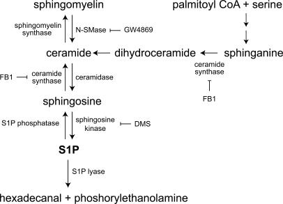
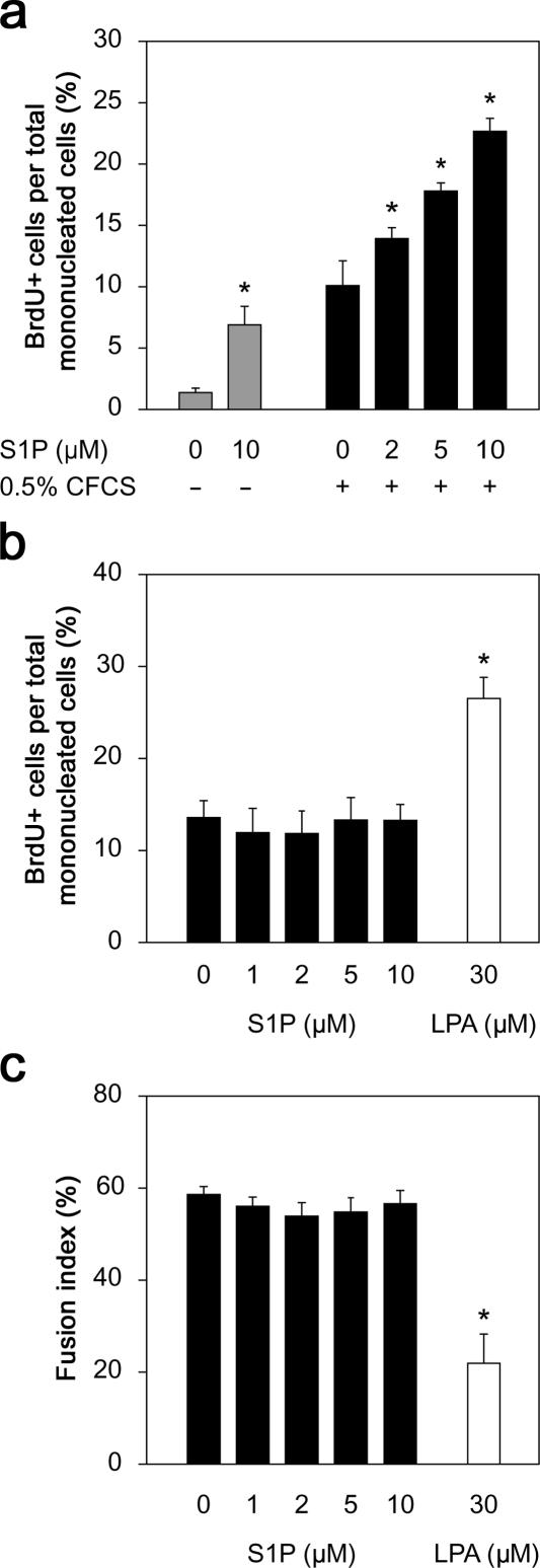
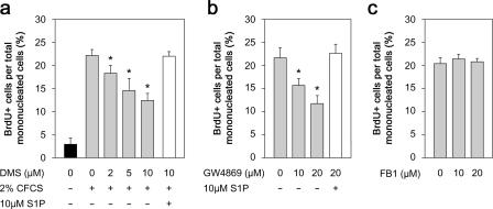
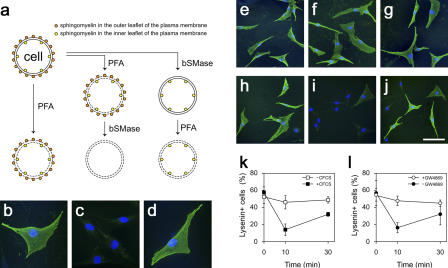
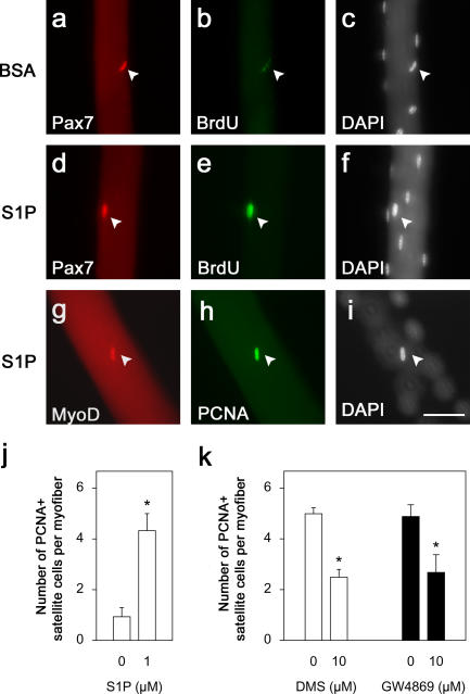
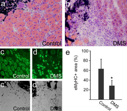
Adult skeletal muscle is able to repeatedly regenerate because of the presence of satellite cells, a population of stem cells resident beneath the basal lamina that surrounds each myofiber. Little is known, however, of the signaling pathways involved in the activation of satellite cells from quiescence to proliferation, a crucial step in muscle regeneration. We show that sphingosine-1-phosphate induces satellite cells to enter the cell cycle. Indeed, inhibiting the sphingolipid-signaling cascade that generates sphingosine-1-phosphate significantly reduces the number of satellite cells able to proliferate in response to mitogen stimulation in vitro and perturbs muscle regeneration in vivo. In addition, metabolism of sphingomyelin located in the inner leaflet of the plasma membrane is probably the main source of sphingosine-1-phosphate used to mediate the mitogenic signal. Together, our observations show that sphingolipid signaling is involved in the induction of proliferation in an adult stem cell and a key component of muscle regeneration.


Figures

Figure 1.
Figure 1.
Simplified overview of sphingolipid metabolism. De novo synthesis of sphingolipids begins with the serine palmitoyltransferase catalyzed condensation of serine with palmitoyl CoA. The resulting 3-ketosphinganine is reduced to sphinganine and then subsequently acylated to form dihydroceramide by ceramide synthase (inhibited by FB1). Ceramide is then produced from dihydroceramide by the insertion of the trans 4,5 double bond by a desaturase. Ceramide can be reversibly converted to either sphingomyelin by sphingomyelin synthase or deacylated to sphingosine by ceramidase. S1P is then generated from sphingosine by the phosphorylation of the primary hydroxyl group by sphingosine kinase (inhibited by DMS). Sphingomyelin can act as a reservoir for sphingolipids and can be cleaved to ceramide by sphingomyelinase (inhibited by GW4869). S1P degradation is mediated by either the reversible dephosphorylation back to sphingosine by specific S1P phosphatases or the irreversible degradation by S1P lyase to hexadecenal and phosphorylethanolamine.
Figure 2.
Figure 2.
Exogenous S1P can directly induce DNA synthesis in quiescent myogenic cells. In the absence of serum stimulation, only ∼1% of reserve cells incorporate BrdU, but the addition of 10 μM S1P increases the number of dividing cells approximately sevenfold (a). The addition of 0.5% CFCS also stimulates cell division, but the proportion of cells incorporating BrdU is significantly increased by S1P in a dose-dependent manner (a). To determine whether S1P also perpetuated proliferation in a manner similar to other lipids, such as lysophosphatidic acid (LPA), proliferating C2C12 cells were switched to differentiation medium containing 0–10 μM S1P or 30 μM LPA and cultured with daily medium changes for 3 d. After a 3-h BrdU pulse, either cells were immunostained for BrdU (b) or the percentage of nuclei incorporated in myotubes was determined (fusion index; c). S1P was unable to perpetuate division after mitogen withdrawal or prevent differentiation, in contrast to LPA. Data presented are the mean percentage ± SEM from four independent experiments. Asterisks indicate that data are statistically significant using a t test (P < 0.01).
Figure 3.
Figure 3.
S1P is generated from sphingomyelin. When reserve cells were pretreated with DMS to inhibit sphingosine kinase activity before stimulation with 2% CFCS and pulsed with BrdU, the proportion of proliferating cells was significantly reduced, indicating that S1P was being produced from sphingosine during entry to the cell cycle (a). 10 μM S1P overcame the DMS-induced block on S1P synthesis and restored proliferation, showing that the DMS was specifically inhibiting sphingosine kinases (a). Sphingosine is derived from ceramide, of which there are two main sources, sphingomyelin cleavage by SMases or de novo formation (Fig. 1). Reserve cells were treated with GW4869 for 45 min to inhibit the N-SMase catalyzed synthesis from sphingomyelin and then stimulated with 2% CFCS and pulsed with BrdU. Immunostaining for BrdU showed that the number of proliferating cells was significantly reduced after GW4869 treatment (b). The inhibition by GW4869 was specific to the generation of S1P, as S1P overcame the block (b). In contrast, blocking de novo synthesis of ceramide by inhibiting ceramide synthase with FB1 had no effect on reserve cell activation (c). Mean percentage ± SEM of BrdU-positive cells per total mononucleated cells from four independent experiments. Asterisks indicate that data are statistically significant using a t test (P < 0.05).
Figure 4.
Figure 4.
Sphingomyelin in the inner leaflet of the plasma membrane is the source of S1P. The dynamics of sphingomyelin during reserve cell activation was examined using bSMase to selectively remove sphingomyelin from either the outer or inner leaflet while monitoring sphingomyelin levels with lysenin (a). Reserve cells were fixed/permeabilized with 4% PFA, probed with 0.2 μg/ml lysenin, and immunostained (green), and all nuclei were identified using DAPI (blue). The vast majority of reserve cells bound lysenin, showing sphingomyelin on their surface (b). When reserve cells were fixed/permeabilized and then treated with 100 mU/ml bSMase to digest sphingomyelin on both the outer and inner leaflet, sphingomyelin was no longer detectable (c). To distinguish between sphingomyelin on the inner and outer surfaces of the cell membrane, live reserve cells were treated with bSMase so that only external sphingomyelin was accessible to digestion. Lysenin (10 μg/ml) binding then revealed sphingomyelin located only in the inner leaflet (d). To determine whether sphingomyelin in the outer leaflet was metabolized to generate S1P, reserve cells were activated with 2% CFCS and either fixed/permeabilized immediately (e) or after 10 (f) or 30 min (g). No change in lysenin immunostaining was observed (e–g). To specifically assay sphingomyelin in the inner leaflet, live reserve cells were incubated with bSMase for 2 h to digest sphingomyelin in the outer leaflet before activation with 2% CFCS. Cells were then fixed/permeabilized, probed with lysenin, and immunostained immediately (h) or after 10 (i) or 30 min (j) of stimulation. There was a transient drop in the number of reserve cells with lysenin immunostaining after 10 min of stimulation (i, quantified in k [closed squares]), whereas unstimulated reserve cells exhibited no such drop (k, open squares). Live reserve cells treated with bSMase for 2 h and then incubated with 20 μM GW4869 before activation with 2% CFCS (l) showed that inhibition of N-SMase prevented the transient loss of sphingomyelin in the inner leaflet by blocking its cleavage (l, open circles). Data were obtained from three independent experiments and are expressed as mean percentage ± SEM of lysenin-positive cells per total mononucleated cells. Bar: (b–d) 50 μm; (e–j) 100 μm.
Figure 5.
Figure 5.
S1P is required for inducing entry of satellite cells into the cell cycle. Isolated myofibers were cultured in DME/BSA (a–c) or DME/BSA/ 1 μM S1P (d–f) in the presence of BrdU. Myofibers were fixed 48 h later, and their associated satellite cells were coimmunostained for Pax7 (red) and BrdU (green; arrowheads in a–f). Similarly, immunostaining for MyoD (red) and PCNA (green; arrowheads in g–i) showed that there were significantly more satellite cells entering the cell cycle in the presence of S1P than in DME/BSA (j). All nuclei present were identified with DAPI (c, f, and i). Bar, 50 μm. To determine whether S1P is generated in response to mitogen stimulation during satellite cell activation, isolated myofibers were treated with 10 μM DMS in DME for 45 min before their associated satellite cells were stimulated with CEE/serum and analyzed for the expression of PCNA (k). In the absence of DMS, satellite cells were readily activated and practically all entered the cell cycle, but after exposure to the inhibitor, significantly less did so (k). To determine whether sphingomyelin was the source of S1P, isolated myofibers were incubated with 10 μM GW4869 to inhibit N-SMase activity before mitogen stimulation and then immunostained. Again, there was a significant reduction in the number of satellite cells entering the cell cycle (k), showing that S1P is derived from sphingomyelin. Data represent the mean ± SEM of immunostained cells per myofiber from three independent experiments (20 myofibers per experiment). Asterisks indicate that data are statistically significant using a t test (P < 0.05).
Figure 6.
Figure 6.
Inhibition of S1P synthesis perturbs muscle regeneration. TA muscles were induced to regenerate by injection of cardiotoxin together with India ink to mark the injection site. At the same time, the right TA muscle was also injected with DMS to inhibit sphingosine kinase activity, thus preventing S1P synthesis. The left TA muscle served as a control, and only carrier DMSO was injected. 7 d later, both muscles were removed, cryosectioned, and analyzed. Hematoxylin and eosin staining showed that control left TA muscles had regenerated successfully (a), with many large new myofibers identified by the central location of their myonuclei, in the vicinity of the injection site, as shown by the presence of the black India ink (a, top right). In contrast, even 7 d after injection, right TA muscles in which DMS had been administered exhibited severely retarded regeneration (b). To quantify regenerated muscle, sections were immunostained for eMyHC to identify new myofibers (c and d) in the vicinity of the injection site (c′ and d′ bright field image included to show location of India ink). The area occupied by regenerating myofibers was determined from several such experiments, and data were pooled and expressed as a percentage ± SEM of the total area assayed (e). This showed that administration of DMS resulted in significantly less regenerating myofibers. Asterisks indicate that data are statistically significant using a t test (P < 0.05).

References

    1. Andrieu, N., R. Salvayre, and T. Levade. 1996. Comparative study of the metabolic pools of sphingomyelin and phosphatidylcholine sensitive to tumor necrosis factor. Eur. J. Biochem. 236:738–745. - PubMed
    1. Andrieu-Abadie, N., and T. Levade. 2002. Sphingomyelin hydrolysis during apoptosis. Biochim. Biophys. Acta. 1585:126–134. - PubMed
    1. Beauchamp, J.R., L. Heslop, D.S. Yu, S. Tajbakhsh, R.G. Kelly, A. Wernig, M.E. Buckingham, T.A. Partridge, and P.S. Zammit. 2000. Expression of CD34 and Myf5 defines the majority of quiescent adult skeletal muscle satellite cells. J. Cell Biol. 151:1221–1234. - PMC - PubMed
    1. Bischoff, R. 1986. Proliferation of muscle satellite cells on intact myofibers in culture. Dev. Biol. 115:129–139. - PubMed
    1. Chargé, S.B., and M.A. Rudnicki. 2004. Cellular and molecular regulation of muscle regeneration. Physiol. Rev. 84:209–238. - PubMed

Publication types