. 2006 Aug;26(15):5621-35.
doi: 10.1128/MCB.02357-05.

The normal phenotype of Pmm1-deficient mice suggests that Pmm1 is not essential for normal mouse development

Affiliations

The normal phenotype of Pmm1-deficient mice suggests that Pmm1 is not essential for normal mouse development

K Cromphout et al. Mol Cell Biol. 2006 Aug.

Abstract

Phosphomannomutases (PMMs) are crucial for the glycosylation of glycoproteins. In humans, two highly conserved PMMs exist: PMM1 and PMM2. In vitro both enzymes are able to convert mannose-6-phosphate (mannose-6-P) into mannose-1-P, the key starting compound for glycan biosynthesis. However, only mutations causing a deficiency in PMM2 cause hypoglycosylation, leading to the most frequent type of the congenital disorders of glycosylation (CDG): CDG-Ia. PMM1 is as yet not associated with any disease, and its physiological role has remained unclear. We generated a mouse deficient in Pmm1 activity and documented the expression pattern of murine Pmm1 to unravel its biological role. The expression pattern suggested an involvement of Pmm1 in (neural) development and endocrine regulation. Surprisingly, Pmm1 knockout mice were viable, developed normally, and did not reveal any obvious phenotypic alteration up to adulthood. The macroscopic and microscopic anatomy of all major organs, as well as animal behavior, appeared to be normal. Likewise, lectin histochemistry did not demonstrate an altered glycosylation pattern in tissues. It is especially striking that Pmm1, despite an almost complete overlap of its expression with Pmm2, e.g., in the developing brain, is apparently unable to compensate for deficient Pmm2 activity in CDG-Ia patients. Together, these data point to a (developmental) function independent of mannose-1-P synthesis, whereby the normal knockout phenotype, despite the stringent conservation in phylogeny, could be explained by a critical function under as-yet-unidentified challenge conditions.


Figures
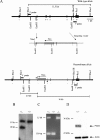
FIG. 1.
FIG. 1.
Generation of Pmm1 knockout mice. (A) Generation of the targeting vector. A partial restriction map of the wild-type allele of Pmm1 is shown. To disrupt exon 3 in the mouse genome, a 6-kb EcoRI fragment, including exons 2 to 6 was subcloned into pBSK targeting vector. The Neo gene contains stop codons in all three reading frames. Homologous recombination between genomic DNA and the targeting vector results in the insertion of a 1.25-kb Neo cassette in exon 3. (B) Southern blot analysis of Pmm1+/− ES cells. Southern blot analysis was carried out on XhoI-digested genomic DNA from G418-resistant ES clones with the 3′ external probe, a HindIII/PstI fragment 3′ of the homologous region (see panel A). The wild-type allele results in a 11.5-kb band and the knockout allele results in a 10-kb band. (C) PCR genotyping of Pmm1 knockout mice. PCR was done on genomic tail DNA with two primers: one 3′ primer and 5′ primer (arrowheads in panel A). The wild-type allele results in a 330-bp band, and the knockout allele results in a 1.5-kb band. (D) Western blot analysis of brain extracts of Pmm1+/+ and Pmm1−/− mice using the anti-Pmm1 antibody. Pmm1 knockout mice show no detectable Pmm1 and equal intensity of the actin signal as a loading control.
FIG. 2.
FIG. 2.
Western blot analysis of embryonic (E17) (A) and adult (B) tissues and endocrine glands (C). Blots of tissue extracts (15 μg) and recombinant Pmm1 (10 ng) were probed with affinity-purified anti-Pmm1 antibody (1 in 10,000 for brain extracts; 1 in 500 for nonneural tissues) or with anti-β-actin antibody (1 in 5,000).The sizes of the molecular mass markers are indicated in kilodaltons.
FIG. 3.
FIG. 3.
Pmm1 immunoreactivity in the E17 head region. (A) Pmm1 signals have a widespread distribution throughout the developing brain, and particular intense signals are found in the sensory ganglia. (B) Peripheral processes from these ganglia are intensely labeled for Pmm1 in contrast to the central processes (panel C). Abbreviations: C, central branches; Cb, cerebellum; Cer, cerebrum; H, hippocampus; P, peripheral processes; T, trigeminal ganglion.
FIG. 4.
FIG. 4.
Localization of the Pmm1 enzyme in developing visceral organs of the E17 mouse embryo. Intense staining in the mucosal lining of the stomach (A) and intestine (B). The surrounding submucosa is almost devoid of signal. (C) The liver parenchyma shows intense signals in hepatocytes. (D) Secretory cells of the exocrine pancreas are negative for Pmm1. The endocrine component is intensely stained with the anti-Pmm1 antibody. (E) In the adrenal gland the differentiating cells are Pmm1 positive, whereas in the developing kidney the glomeruli in the cortical region are highly immunoreactive. (F) The lung parenchyma is moderately stained, with intense signals in the epithelial cells lining the alveolar ducts. Abbreviations: A, alveolus; AG, adrenal gland; C, cortex; CT proximal or distal convoluted tubule; End, endocrine pancreas; Ex, exocrine pancreas; G, glomerulus; M, mucosa; Me, medulla; P, pelvic region; SM, submucosa; TB, terminal bronchus.
FIG. 5.
FIG. 5.
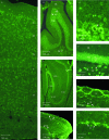
Localization of Pmm1 signals in adult mouse brain. (A) The expression of Pmm1 in the adult cerebral cortex was uniformly distributed over the six cortical layers, with the exception of layer I showing only faint neuropil staining. Ependymal and glial cells of the white matter were mostly negative. (B) In adult cerebellum, striking Pmm1-positive signals are seen in the Purkinje cell layer, whereas the granule cell layer has fainter signals, and the molecular layer is almost devoid of staining. Staining of the molecular layer was confined to faint neuropil staining. In the Purkinje cells (panel C, arrowhead) intense signals were observed in the cell body cytoplasm. (D) In the hippocampus, Pmm1 is expressed in adult CA regions and dentate gyrus. (E) Detailed photographs show high Pmm1 reactivity in dentate gyrus granular cell bodies and polymorphic neurons and faint neuropil staining in the molecular layer. (F) In the olfactory bulb intense signals are observed in the glomerular layer and the layer of mitral cells. (G) In the glomerular layer, the body of the glomeruli is devoid of Pmm1 signal, whereas surrounding nerve endings are intensely labeled. (H) In the mitral cell layer especially, the cell bodies of the mitral cells are immunoreactive for the Pmm1 antibody. Abbreviations: CA, cornus ammonis; DCN, deep cerebellar nuclei; DG, dentate gyrus; EGL, external granular layer; G, glomerulus; GC, granular cells; IGL, internal granular layer; M, mitral cell; ML, molecular layer; PC, Purkinje cell; PN, polymorphic neuron. (Panels A to E were reprinted from the European Journal of Neuroscience, Fig. 2C and Fig. 3C, E, I, and K [7], with the permission of the publisher.)
FIG. 6.
FIG. 6.
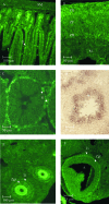
Pmm1 immunoreactivity in adult mouse organs. (A) In the adult intestine Pmm1 signals are found in the mucosa, whereas the submucosa is almost devoid of Pmm1 signal. The Pmm1 signals are concentrated in the crypts, whereas in the villi Pmm1 the immunoreactivity is much less intense. (B) In the adrenal gland, especially the zona glomerulosa, cells show high immunoreactivity, whereas cells of the other layers are less intensely labeled. (C) In the seminiferous tubuli, spermatogonia are Pmm1 positive and not so much for the Sertoli and Leydig cells. (D) mRNA analysis reveals high signals in postmeiotic cells. (E and F) Strong cytoplasmic staining is seen in secondary follicles (E), and strong staining is obvious in the theca interna cells of the graafian follicles (F). Abbreviations: A, antrum; C, crypt; G, goblet cell; M, medulla; S, spermatogonium; SM, submucosa; St, spermatid; Ti, theca interna; ZF, zona fasciculate; ZG, zona glomerulosa; Zgr, zona granulosa; ZR, zona reticularis.
FIG. 7.
FIG. 7.
Pmm1 localization in pancreatic cell populations. (A) Pmm1 shows a high expression level in pancreatic islets. (B) Staining of the same section with antiglucagon reveals a partial overlap in the signals. (C and D) Double stainings with an anti-insulin antibody also reveals a colocalization with pancreatic β cells.
FIG. 8.
FIG. 8.
Hematoxylin-and-eosin-stained sagittal sections of brain structures from wild-type and Pmm1 knockout embryonic (A to F) or adult mice (G to L). The results indicate that the Pmm1 deficiency does not affect cerebral cortex (A, B, G, and H), cerebellar (C, D, I, and J), or hippocampal development (E, F, K, and L).
FIG. 9.
FIG. 9.
Western blot analysis of Pmm2 in adult wild-type and knockout brain. Blots of brain extracts (10 and 15 μg) were probed with affinity-purified anti-Pmm2 antibody (1:10,000) or with anti-β-actin antibody (1:5,000), showing identical levels of Pmm2 in wild-type and knockout brain.

References

    1. Aebi, M., and T. Hennet. 2001. Congenital disorders of glycosylation: genetic model systems lead the way. Trends Cell Biol. 11:136-141. - PubMed
    1. Akaboshi, S., K. Ohno, and K. Takeshita. 1995. Neuroradiological findings in the carbohydrate-deficient glycoprotein syndrome. Neuroradiology 37:491-495. - PubMed
    1. Antoun, H., N. Villeneuve, A. Gelot, S. Panisset, and C. Adamsbaum. 1999. Cerebellar atrophy: an important feature of carbohydrate deficient glycoprotein syndrome type 1. Pediatr. Radiol. 29:194-198. - PubMed
    1. Artuch, R., I. Ferrer, J. Pineda, J. Moreno, C. Busquets, P. Briones, and M. A. Vilaseca. 2003. Western blotting with diaminobenzidine detection for the diagnosis of congenital disorders of glycosylation. J. Neurosci. Methods 125:167-171. - PubMed
    1. Barone, R., L. Pavone, A. Fiumara, R. Bianchini, and J. Jaeken. 1999. Developmental patterns and neuropsychological assessment in patients with carbohydrate-deficient glycoconjugate syndrome type IA (phosphomannomutase deficiency). Brain Dev. 21:260-263. - PubMed

Publication types

MeSH terms