. 2006 Aug;26(15):5650-62.
doi: 10.1128/MCB.00313-06.

Hypomorphic mutation of the TALE gene Prep1 (pKnox1) causes a major reduction of Pbx and Meis proteins and a pleiotropic embryonic phenotype

Affiliations

Hypomorphic mutation of the TALE gene Prep1 (pKnox1) causes a major reduction of Pbx and Meis proteins and a pleiotropic embryonic phenotype

Elisabetta Ferretti et al. Mol Cell Biol. 2006 Aug.

Abstract

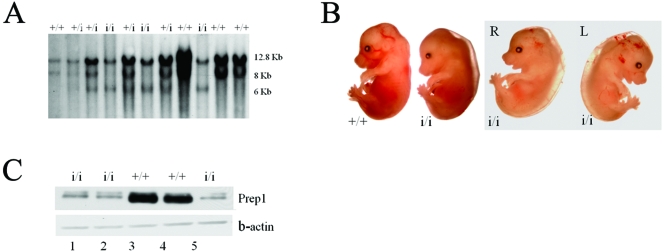
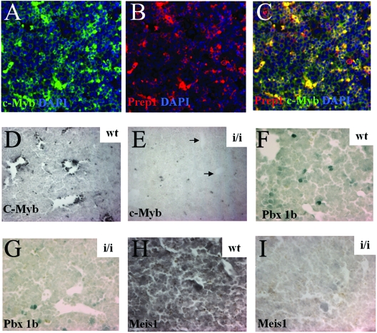
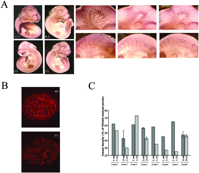
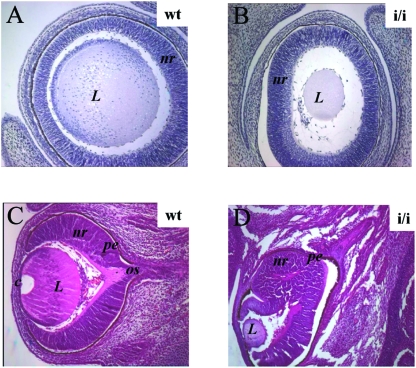
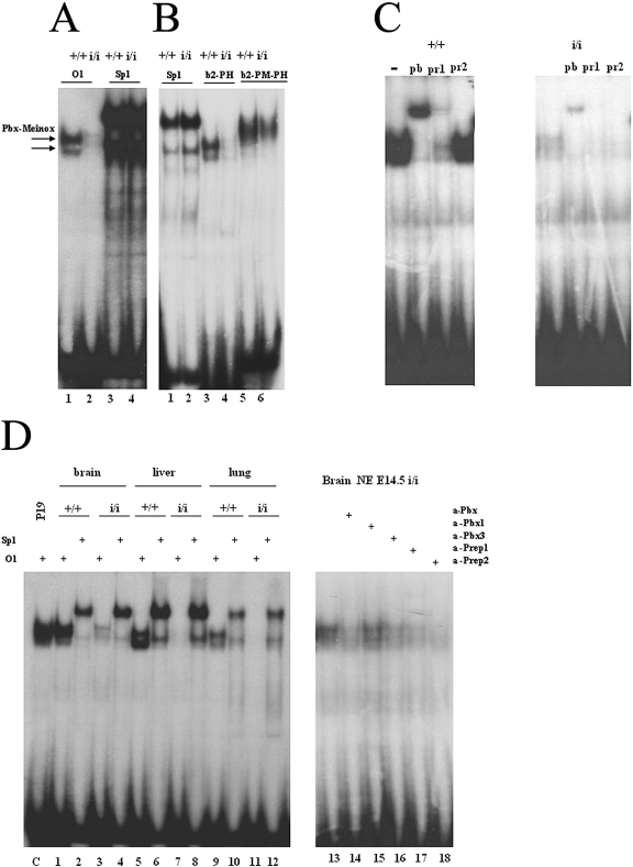
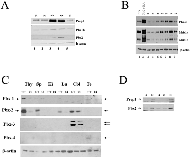
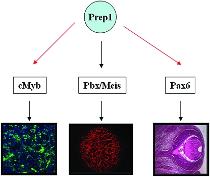
The interaction of Prep1 and Pbx homeodomain transcription factors regulates their activity, nuclear localization, and likely, function in development. To understand the in vivo role of Prep1, we have analyzed an embryonic lethal hypomorphic mutant mouse (Prep1(i/i)). Prep1(i/i) embryos die at embryonic day 17.5 (E17.5) to birth with an overall organ hypoplasia, severe anemia, impaired angiogenesis, and eye anomalies, particularly in the lens and retina. The anemia correlates with delayed differentiation of erythroid progenitors and may be, at least in part, responsible for intrauterine death. At E14.5, Prep1 is present in fetal liver (FL) cMyb-positive cells, whose deficiency causes a marked hematopoietic phenotype. Prep1 is also localized to FL endothelial progenitors, consistent with the observed angiogenic phenotype. Likewise, at the same gestational day, Prep1 is present in the eye cells that bear Pax6, implicated in eye development. The levels of cMyb and Pax6 in FL and in the retina, respectively, are significantly decreased in Prep1(i/i) embryos, consistent with the hematopoietic and eye phenotypes. Concomitantly, Prep1 deficiency results in the overall decrease of protein levels of its related family member Meis1 and its partners Pbx1 and Pbx2. As both Prep1 and Meis interact with Pbx, the overall Prep1/Meis-Pbx DNA-binding activity is strongly reduced in whole Prep1(i/i) embryos and their organs. Our data indicate that Prep1 is an essential gene that acts upstream of and within a Pbx-Meis network that regulates multiple aspects of embryonic development.


Figures

FIG. 1.
FIG. 1.
Prep1i/i phenotype. (A) Southern blotting analysis of EcoRI-digested DNAs from the progeny obtained by crossing F1 Prep1i/+ × Prep1i/+. (B) Gross morphology of Prep1i/i embryos. The two rightmost panels show the same embryo viewed from both sides (R and L), exhibiting edema, pallor, smaller size, small liver spot and hemorrhaging. (C) Nuclear extracts prepared from E14.5 embryonic brains of 5 littermate embryos (2 wt and 3 Prep1i/i, as indicated) were immunoblotted with monoclonal anti-Prep1 and anti-beta-actin antibodies. Lane 1, 2, and 5 are extracts from Prep1i/i embryos; lanes 3 and 4 are extracts from wt embryos.
FIG. 2.
FIG. 2.
Presence of Prep1, Pbx1b, Meis1, and cMyb in wt and Prep1i/i FL. (A to C) Immunofluorescence analysis of E14.5 wt FL sections. Triple staining with anti-cMyb and anti-Prep antibodies and 4′,6′-diamidino-2-phenylindole (DAPI) to stain nuclei is shown. The antibodies used are indicated. Magnification (A to C), ×20. In panel C, colocalization of cMyb and Prep1 appears as a yellow color. (D to I) Immunohistochemistry of E14.5 wt and Prep1i/i FL, developed with the DAB kit. (D and E) Anti-cMyb antibodies; (F and G) anti-Pbx1b antibodies; (H and I) anti-Meis1 antibodies. Magnification, ×20.
FIG. 3.
FIG. 3.
Prep1i/i embryos exhibit angiogenesis defects. (A) Whole-mount CD31 (PECAM) immunofluorescence on E10.5 embryos. The genotype is shown in each panel. A nonimmune (n.i.) serum gave essentially no staining (not shown). A set of close-up pictures is inserted. The top row shows details of the head region; the bottom row the intersomitic area. (B) Immunofluorescence on E7.5 wt and Prep1i/i allantois cultured for 18 h and stained with anti-CD31 antibodies. (C) Vessel density (percentage of CD31-stained pixels) in 11 different litters containing wt and Prep1i/i embryos. At the bottom of each histogram, the symbols indicate the numbers of wt and Prep1i/i littermates from different crosses.
FIG. 4.
FIG. 4.
Eye defects in E14.5 Prep1i/i embryos. Comparison of hematoxylin (A and B) or hematoxylin-eosin (C and D) staining of wt and Prep1i/i embryonic eyes (indicated). Notice the reduction of the lens size (A and B) and anomalies and duplication of the retinal epithelium (D). L, lens; nr, neural retina; pe, pigmented retinal epithelium; c, cornea; os, optic stalk. Magnification, ×10 (A and B); ×4 (C and D).
FIG. 5.
FIG. 5.
Prep1 and Pax6 are colocalized in wt embryonic eye structures, and Pax6 is present at lower levels in Prep1i/i embryos. Immunohistochemistry with anti-Prep1 (A, B, D, F, G, and I), antiactin (C and E), anti-Pax6 (H, J, and K), and anti-Meis1 (L to O) antibodies in wt and Prep1i/i embryonic eye sections (genotype and antibody used are indicated). Comparison of plates H and I shows that Prep1 and Pax6 colocalize, while plates J and K show lower levels of Pax6 in Prep1i/i eye structures. ch, choroids; c, cornea; ce, corneal epithelium; gc, ganglion cell layer; in, inner nuclear layer; i, iris/ciliar body; L, lens; le, lens epithelium; lf, lens fiber cells; nr, neural retina; os, optic stalk; on, outer nuclear layer; pe, pigmented retinal epithelium; rc, rod and cone photoreceptor cell layer; s, sclera. All panels were developed by the alkaline phosphatase reaction, except panels H and L to O. Magnification, ×60 (A); ×20 (B to E and L to O); ×40 (F, G); ×10 (H to K).
FIG. 6.
FIG. 6.
Decreased Pbx DNA-binding activity in Prep1i/i embryos. (A) Electrophoretic mobility shift analysis of the DNA-binding activity of nuclear extracts from an E10.5 total embryo with labeled O1 (specific for Prep1/Meis-Pbx dimers) and Sp1 (control) oligonucleotides. On the top of the panels, the genotype of the embryo is indicated. The two arrows to the left indicate the migration of Pbxa and Pbxb-Prep/Meis complexes. (B) Same as in panel A, with labeled oligonucleotide Sp1 (control), b2-PH (binding both Pbx-Hoxb1 and Prep1/Meis-Pbx dimeric complexes), and b2-PM-PH (binding also Meis/Prep1-Pbx-Hoxb1 ternary complexes). TC indicates the migration of the ternary complex. (C) Identification of the nature of the binding activity by EMSA in the presence of anti-Prep1 (pr1), anti-panPbx (pb), and anti-Prep2 (pr2) antibodies. Comparison of nuclear extracts from E10.5 wt and Prep1i/i embryos. The oligonucleotide used was O1. (D) Left panel, analysis by EMSA on nuclear extracts obtained from embryonic brain (lanes 1 to 4), liver (lanes 5 to 8), and lung (lanes 9 to 12). The genotype is indicated on the top of the panel. Right panel, analysis by EMSA of the residual binding activity of nuclear extracts obtained from a Prep1i/i brain in the presence of different antibodies (+), as indicated on the top of the panel. α, anti.
FIG. 7.
FIG. 7.
Prep1i/i embryonic and adult organs show a decrease of Pbx1b, Pbx2, and Meis1 proteins. (A) Immunoblotting analysis of the same filter shown in Fig. 1C: nuclear extracts from the E14.5 embryonic brain of 5 littermate embryos (2 wt and 3 Prep1i/i, as indicated) tested with monoclonal anti-Pbx1b, anti-Pbx2, and anti-beta-actin antibodies. Lanes 1, 2, and 5 contain extracts from Prep1i/i embryos; lanes 3 and 4 contain extracts from wt embryos. (B) Immunoblotting analysis of brain extracts from 1 wt, 3 heterozygous, and 3 Prep1i/i embryos, using anti-Pbx2 and anti-Meis1 antibodies. (C) Immunoblotting analysis of nuclear extracts obtained from organs, indicated at the top of the panel, of an adult Prep1i/i mouse, tested with anti-Pbx1, anti-Pbx2, anti-Pbx3, anti-Pbx4, and antiactin antibodies. Cbl, cerebellum; Lu, lung; Ki, kidney; Sp, spleen; Te, testis; Thy, thymus. (D) Immunoblotting analysis of E14.5 liver extracts from 1 wt, 1 heterozygous, and 3 Prep1i/i embryos, using anti-Pbx2 (α-Pbx2) and anti-Prep1 (α-Prep1) antibodies.
FIG. 8.
FIG. 8.
Establishment of a hierarchical role for Prep1 within the TALE protein network in embryogenesis. Prep1 is required for normal hematopoiesis, angiogenesis, and oculogenesis, as illustrated. The scheme depicts the upstream role of Prep1 as it controls Pbx and Meis TALE class homeoproteins and their target genes. Such genes become “effectors” within specific developmental processes: cMyb for erythropoiesis and Pax6 for eye development. Black arrows indicate direct control; red arrows indicate hierarchical control, whose direct or indirect nature remains to be established.

References

    1. Azcoitia, V., M. Aracil, A. Carlos-Martinez, and M. Torres. 2005. The homeodomain protein Meis1 is essential for definitive hematopoiesis and vascular patterning in the mouse embryo. Dev. Biol. 280:307-320. - PubMed
    1. Berkes, C., D. A. Bergstrom, B. H. Penn, K. J. Seaver, P. S. Knoepfler, and S. J. Tapscott. 2004. Pbx marks genes for activation by MyoD indicating a role for a homeodomain protein in establishing myogenic potential. Mol. Cell 14:465-477. - PubMed
    1. Berthelsen, J., J. Vandekerkhove, and F. Blasi. 1996. Purification and characterization of UEF3, a novel factor involved in the regulation of the urokinase and other AP-1 controlled promoters. J. Biol. Chem. 271:822-3830. - PubMed
    1. Berthelsen, J., V. Zappavigna, E. Ferretti, F. Mavilio, and F. Blasi. 1998. Prep1, a novel partner of Pbx proteins, modifies Pbx-Hox protein cooperativity. EMBO J. 17:1434-1445. - PMC - PubMed
    1. Berthelsen, J., V. Zappavigna, F. Mavilio, and F. Blasi. 1998. Prep1, a novel functional partner of Pbx proteins. EMBO J. 17:1423-1433. - PMC - PubMed

Publication types

MeSH terms

Substances