. 2006 Aug;26(15):5675-87.
doi: 10.1128/MCB.00112-06.

Role of human mitochondrial Nfs1 in cytosolic iron-sulfur protein biogenesis and iron regulation

Affiliations

Role of human mitochondrial Nfs1 in cytosolic iron-sulfur protein biogenesis and iron regulation

Annette Biederbick et al. Mol Cell Biol. 2006 Aug.

Abstract

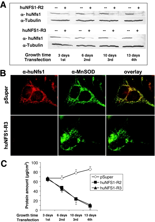
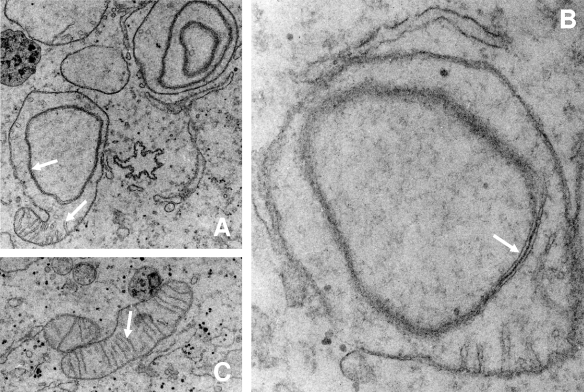
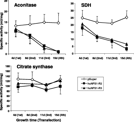
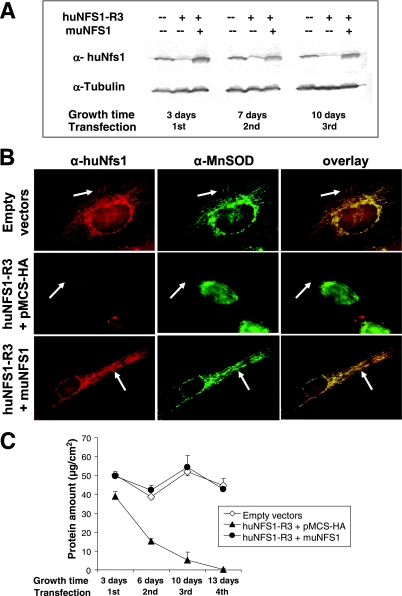
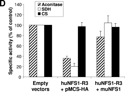
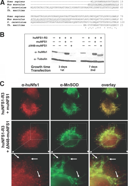
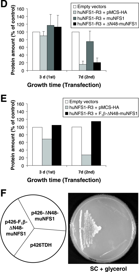
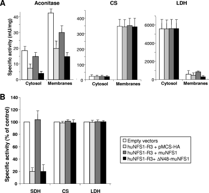
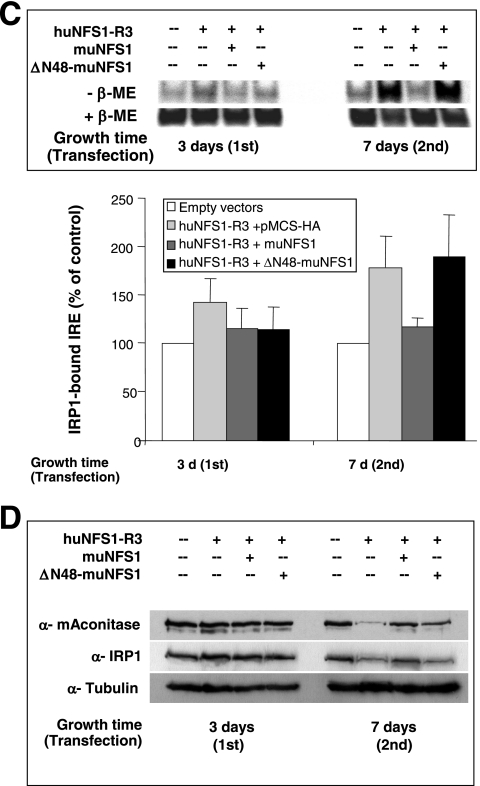
The biogenesis of iron-sulfur (Fe/S) proteins in eukaryotes is a complex process involving more than 20 components. So far, functional investigations have mainly been performed in Saccharomyces cerevisiae. Here, we have analyzed the role of the human cysteine desulfurase Nfs1 (huNfs1), which serves as a sulfur donor in biogenesis. The protein is located predominantly in mitochondria, but small amounts are present in the cytosol/nucleus. huNfs1 was depleted efficiently in HeLa cells by a small interfering RNA (siRNA) approach, resulting in a drastic growth retardation and striking morphological changes of mitochondria. The activities of both mitochondrial and cytosolic Fe/S proteins were strongly impaired, demonstrating that huNfs1 performs an essential function in Fe/S protein biogenesis in human cells. Expression of murine Nfs1 (muNfs1) in huNfs1-depleted cells restored both growth and Fe/S protein activities to wild-type levels, indicating the specificity of the siRNA depletion approach. No complementation of the growth retardation was observed, when muNfs1 was synthesized without its mitochondrial presequence. This extramitochondrial muNfs1 did not support maintenance of Fe/S protein activities, neither in the cytosol nor in mitochondria. In conclusion, our study shows that the essential huNfs1 is required inside mitochondria for efficient maturation of cellular Fe/S proteins. The results have implications for the regulation of iron homeostasis by cytosolic iron regulatory protein 1.


Figures

FIG. 1.
FIG. 1.
Depletion of huNfs1 by RNAi leads to impaired cell growth. HeLa cells were transfected by electroporation with RNAi vectors pSuper, huNFS1-R2, or huNFS1-R3. After the time points indicated cells were harvested, and a fraction was retransfected. (A) Depletion of huNfs1 was analyzed by Western blotting of cell lysates using antibodies against huNfs1 and tubulin as a control. (B) Immunofluorescence detection of huNfs1 and mitochondrial matrix protein MnSOD in HeLa cells 12 days after first transfection. (C) At each time point the protein content of the cells was determined as a measure of cell growth (mean ± standard deviation; n = 4).
FIG. 2.
FIG. 2.
Alteration of mitochondrial inner membrane structure after huNfs1 depletion. HeLa cells were repeatedly transfected with huNFS1-R3 (A, B) or pSuper (C) as described in the legend for Fig. 1 and analyzed by electron microscopy (total growth time, 10 days). (A) Some regions of huNFS1-depleted mitochondria still contained cristae (lower arrow), while other regions had an altered mitochondrial inner membrane structure (upper arrow). (B) huNFS1-depleted mitochondria with altered mitochondrial inner membrane structure (arrow) in higher magnification than the one in panel A. (C) Mitochondrion of a pSuper-treated control cell with intact cristae (arrow). Magnifications: panels A and C, ×7,000; panel B, ×20,000.
FIG. 3.
FIG. 3.
Defects in mitochondrial Fe/S proteins upon huNfs1 depletion. HeLa cells were repeatedly transfected with RNAi vectors pSuper, huNFS1-R2, or huNFS1-R3 and harvested at the time points indicated. Specific activities of mitochondrial Fe/S proteins aconitase and SDH and of the non-Fe/S cluster-containing enzyme citrate synthase were measured in whole cell lysates (mean ± standard deviation; n = 3).
FIG. 4.
FIG. 4.
Expression of muNfs1 in huNfs1-depleted HeLa cells restores cell growth and mitochondrial Fe/S protein activities. (A) HeLa cells were cotransfected with an expression vector containing full-length murine NFS1 cDNA (muNFS1) and the RNAi vector huNFS1-R3, or the corresponding empty vectors. After the time points indicated cells were harvested, and a fraction was retransfected. Expression of muNfs1 and depletion of huNfs1 were analyzed by Western blotting of cell lysates. Blots were probed for Nfs1 and tubulin. The antiserum against huNfs1 also recognizes muNfs1. (B) Immunofluorescence detection of Nfs1 and mitochondrial matrix protein MnSOD 10 days after the first transfection. Arrows indicate areas of mitochondrial staining. (C) Cellular protein content was determined at each harvest (mean ± standard deviation; n = 4). (D) The specific activities of mitochondrial Fe/S proteins aconitase and SDH and of the non-Fe/S cluster-containing enzyme CS were determined in lysates of cells harvested 7 days after the first transfection (mean + standard deviation; n = 6).
FIG. 4.
FIG. 4.
Expression of muNfs1 in huNfs1-depleted HeLa cells restores cell growth and mitochondrial Fe/S protein activities. (A) HeLa cells were cotransfected with an expression vector containing full-length murine NFS1 cDNA (muNFS1) and the RNAi vector huNFS1-R3, or the corresponding empty vectors. After the time points indicated cells were harvested, and a fraction was retransfected. Expression of muNfs1 and depletion of huNfs1 were analyzed by Western blotting of cell lysates. Blots were probed for Nfs1 and tubulin. The antiserum against huNfs1 also recognizes muNfs1. (B) Immunofluorescence detection of Nfs1 and mitochondrial matrix protein MnSOD 10 days after the first transfection. Arrows indicate areas of mitochondrial staining. (C) Cellular protein content was determined at each harvest (mean ± standard deviation; n = 4). (D) The specific activities of mitochondrial Fe/S proteins aconitase and SDH and of the non-Fe/S cluster-containing enzyme CS were determined in lysates of cells harvested 7 days after the first transfection (mean + standard deviation; n = 6).
FIG. 5.
FIG. 5.
Expression and localization of Δ48-muNfs1 lacking a mitochondrial presequence. (A) Sequence alignment of the N-terminal regions of Nfs1-like proteins from the indicated species. The segment deleted in ΔN48-muNfs1 is underlined. Asterisks indicate conserved amino acid residues within the N-terminal part of Nfs1-like proteins including the NifS-like protein of the bacterium Thermotoga maritima (23) which lacks the typical eukaryotic mitochondrial targeting sequence. (B) HeLa cells were cotransfected as indicated with the RNAi vector huNFS1-R3, an expression vector encoding full-length muNfs1 (muNFS1) or N-terminally truncated muNfs1 (ΔN48-muNFS1), or the corresponding empty vectors. After 3 or 7 days, cells were analyzed by Western blotting of cell lysates for Nfs1 and tubulin (see Fig. 4A). (C) Immunofluorescence detection of muNfs1 and MnSOD 7 days after the first transfection. Arrows indicate the absence (left column) or presence (middle column) of mitochondrial staining. The lower panel shows cellular sections magnified from the panel in the middle. In contrast to the full-length muNfs1, ΔN48-muNfs1 does not colocalize with mitochondrial MnSOD. (D) HeLa cells cotransfected with the vectors as indicated were harvested 3 or 7 days after the first transfection, and their protein content was determined as a measure for cell growth (mean + standard deviation; n = 4). (E) Mitochondria-localized ΔN48-muNfs1 can restore growth of huNfs1-depleted HeLa cells. To verify the functionality of ΔN48-muNfs1, HeLa cells were cotransfected as indicated with the RNAi vector huNFS1-R3 and with an expression vector containing the coding information of mitochondria-targeted ΔN48-muNfs1 (F1β-ΔN48-muNFS1), or the corresponding empty vectors. Cells were harvested 3 or 7 days after the first transfection, and their protein content was determined as a measure of cell growth. One representative experiment is shown. (F) Mitochondria-localized ΔN48-muNfs1 can restore growth of huNfs1p-depleted yeast cells. To verify the enzymatic functionality of ΔN48-muNfs1, yeast Gal-NFS1 cells were transformed with expression vectors containing the coding information of ΔN48-muNfs1 either with (p426-F1β-ΔN48-muNFS1) or without (p426-ΔN48-muNFS1) an additional mitochondrial presequence, or the vector alone (p426TDH). To deplete endogenous yeast Nfs1p, cells were first cultured on SC supplemented with dextrose and further cultured on SC supplemented with glycerol at 30°C.
FIG. 5.
FIG. 5.
Expression and localization of Δ48-muNfs1 lacking a mitochondrial presequence. (A) Sequence alignment of the N-terminal regions of Nfs1-like proteins from the indicated species. The segment deleted in ΔN48-muNfs1 is underlined. Asterisks indicate conserved amino acid residues within the N-terminal part of Nfs1-like proteins including the NifS-like protein of the bacterium Thermotoga maritima (23) which lacks the typical eukaryotic mitochondrial targeting sequence. (B) HeLa cells were cotransfected as indicated with the RNAi vector huNFS1-R3, an expression vector encoding full-length muNfs1 (muNFS1) or N-terminally truncated muNfs1 (ΔN48-muNFS1), or the corresponding empty vectors. After 3 or 7 days, cells were analyzed by Western blotting of cell lysates for Nfs1 and tubulin (see Fig. 4A). (C) Immunofluorescence detection of muNfs1 and MnSOD 7 days after the first transfection. Arrows indicate the absence (left column) or presence (middle column) of mitochondrial staining. The lower panel shows cellular sections magnified from the panel in the middle. In contrast to the full-length muNfs1, ΔN48-muNfs1 does not colocalize with mitochondrial MnSOD. (D) HeLa cells cotransfected with the vectors as indicated were harvested 3 or 7 days after the first transfection, and their protein content was determined as a measure for cell growth (mean + standard deviation; n = 4). (E) Mitochondria-localized ΔN48-muNfs1 can restore growth of huNfs1-depleted HeLa cells. To verify the functionality of ΔN48-muNfs1, HeLa cells were cotransfected as indicated with the RNAi vector huNFS1-R3 and with an expression vector containing the coding information of mitochondria-targeted ΔN48-muNfs1 (F1β-ΔN48-muNFS1), or the corresponding empty vectors. Cells were harvested 3 or 7 days after the first transfection, and their protein content was determined as a measure of cell growth. One representative experiment is shown. (F) Mitochondria-localized ΔN48-muNfs1 can restore growth of huNfs1p-depleted yeast cells. To verify the enzymatic functionality of ΔN48-muNfs1, yeast Gal-NFS1 cells were transformed with expression vectors containing the coding information of ΔN48-muNfs1 either with (p426-F1β-ΔN48-muNFS1) or without (p426-ΔN48-muNFS1) an additional mitochondrial presequence, or the vector alone (p426TDH). To deplete endogenous yeast Nfs1p, cells were first cultured on SC supplemented with dextrose and further cultured on SC supplemented with glycerol at 30°C.
FIG.6.
FIG.6.
Extramitochondrial ΔN48-muNfs1 cannot reverse the defects in cytosolic and mitochondrial Fe/S proteins. (A) HeLa cells were transfected as described in the legend for Fig. 5 and harvested 7 days after the first transfection. Soluble cytosolic and membrane fractions were prepared and used to measure the specific activities (mean + standard deviation; n = 5) of aconitase and two non-Fe/S cluster-containing enzymes (CS and LDH). Aconitase activity is composed of mitochondrial aconitase and cytosolic IRP1. (B) Whole-cell lysates were used to determine the specific activities of the mitochondrial Fe/S cluster-containing enzyme SDH and of CS and LDH. Data were normalized to the control vector-transfected cells (mean + standard deviation; n = 3). (C) Transfected cells were harvested 3 or 7 days after the first transfection and analyzed for total binding activity of IRP1 to 32P-labeled IRE of human ferritin mRNA. After native gel electrophoresis, binding of IREs to IRP1 was visualized by autoradiography in the absence (−β-ME) or presence (+β-ME) of β-mercaptoethanol (upper panel) and quantified using a phosphorimager. The amounts of IRP1-bound IRE in the various transfected cells were normalized to the maximal binding capacity of β-ME-treated samples and expressed relative to the amount of IRP1-bound IRE in control vector-transfected cells (mean + standard deviation; n = 3) (lower panel). (D) Cell lysates from a representative experiment were analyzed by immunostaining for mitochondrial aconitase (mAconitase), cytosolic IRP1, and tubulin (see also Fig. 4A).
FIG.6.
FIG.6.
Extramitochondrial ΔN48-muNfs1 cannot reverse the defects in cytosolic and mitochondrial Fe/S proteins. (A) HeLa cells were transfected as described in the legend for Fig. 5 and harvested 7 days after the first transfection. Soluble cytosolic and membrane fractions were prepared and used to measure the specific activities (mean + standard deviation; n = 5) of aconitase and two non-Fe/S cluster-containing enzymes (CS and LDH). Aconitase activity is composed of mitochondrial aconitase and cytosolic IRP1. (B) Whole-cell lysates were used to determine the specific activities of the mitochondrial Fe/S cluster-containing enzyme SDH and of CS and LDH. Data were normalized to the control vector-transfected cells (mean + standard deviation; n = 3). (C) Transfected cells were harvested 3 or 7 days after the first transfection and analyzed for total binding activity of IRP1 to 32P-labeled IRE of human ferritin mRNA. After native gel electrophoresis, binding of IREs to IRP1 was visualized by autoradiography in the absence (−β-ME) or presence (+β-ME) of β-mercaptoethanol (upper panel) and quantified using a phosphorimager. The amounts of IRP1-bound IRE in the various transfected cells were normalized to the maximal binding capacity of β-ME-treated samples and expressed relative to the amount of IRP1-bound IRE in control vector-transfected cells (mean + standard deviation; n = 3) (lower panel). (D) Cell lysates from a representative experiment were analyzed by immunostaining for mitochondrial aconitase (mAconitase), cytosolic IRP1, and tubulin (see also Fig. 4A).

Similar articles

Cited by

References

    1. Acquaviva, F., I. De Biase, L. Nezi, G. Ruggiero, F. Tatangelo, C. Pisano, A. Monticelli, C. Garbi, A. M. Acquaviva, and S. Cocozza. 2005. Extra-mitochondrial localisation of frataxin and its association with IscU1 during enterocyte-like differentiation of the human colon adenocarcinoma cell line Caco-2. J. Cell Sci. 118:3917-3924. - PubMed
    1. Adam, A. C., C. Bornhövd, H. Prokisch, W. Neupert, and K. Hell. 2006. The Nfs1 interacting protein Isd11 has an essential role in Fe/S cluster biogenesis in mitochondria. EMBO J. 25:174-183. - PMC - PubMed
    1. Amarzguioui, M., J. J. Rossi, and D. Kim. 2005. Approaches for chemically synthesized siRNA and vector-mediated RNAi. FEBS Lett. 579:5974-5981. - PubMed
    1. Balk, J., A. J. Pierik, D. Aguilar Netz, U. Mühlenhoff, and R. Lill. 2004. The hydrogenase-like Nar1p is essential for maturation of cytosolic and nuclear iron-sulphur proteins. EMBO J. 23:2105-2115. - PMC - PubMed
    1. Balk, J., D. J. Aguilar Netz, K. Tepper, A. J. Pierik, and R. Lill. 2005. The essential WD40 protein Cia1 is involved in a late step of cytosolic and nuclear iron-sulfur protein assembly. Mol. Cell. Biol. 25:10833-10841. - PMC - PubMed

Publication types

MeSH terms