. 2006 Aug;26(15):5715-27.
doi: 10.1128/MCB.00001-06.

MafB is essential for renal development and F4/80 expression in macrophages

Affiliations

MafB is essential for renal development and F4/80 expression in macrophages

Takashi Moriguchi et al. Mol Cell Biol. 2006 Aug.

Abstract

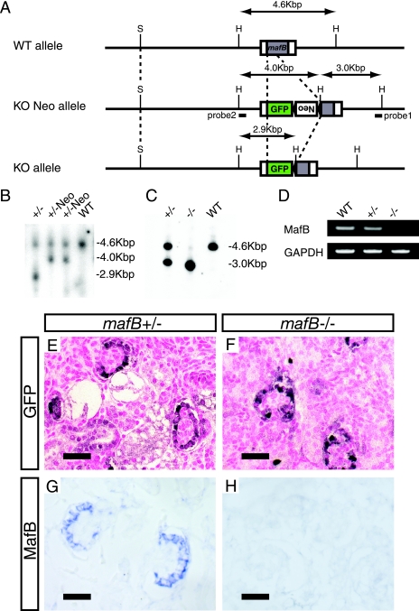
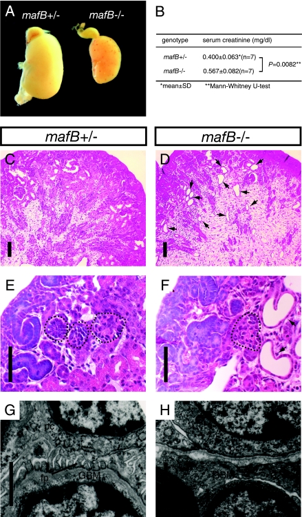
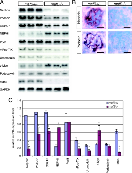
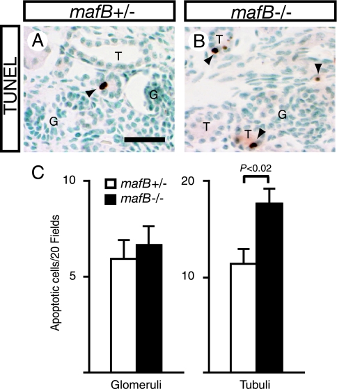
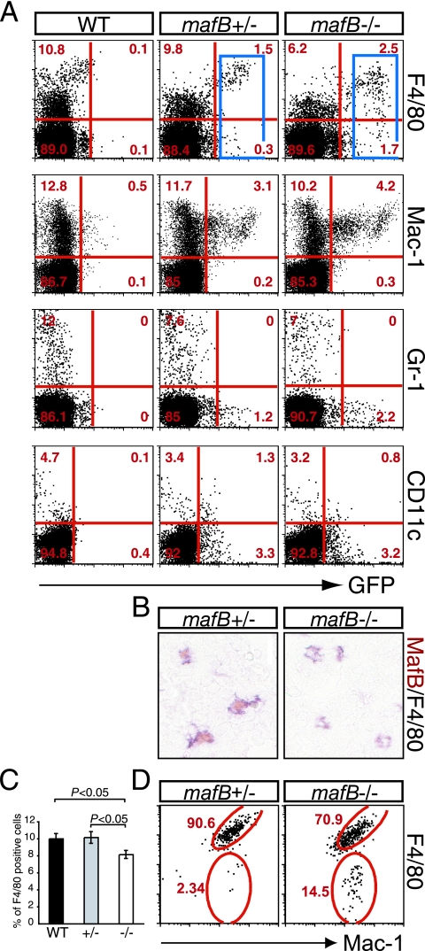
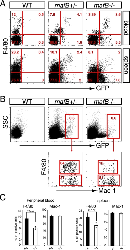
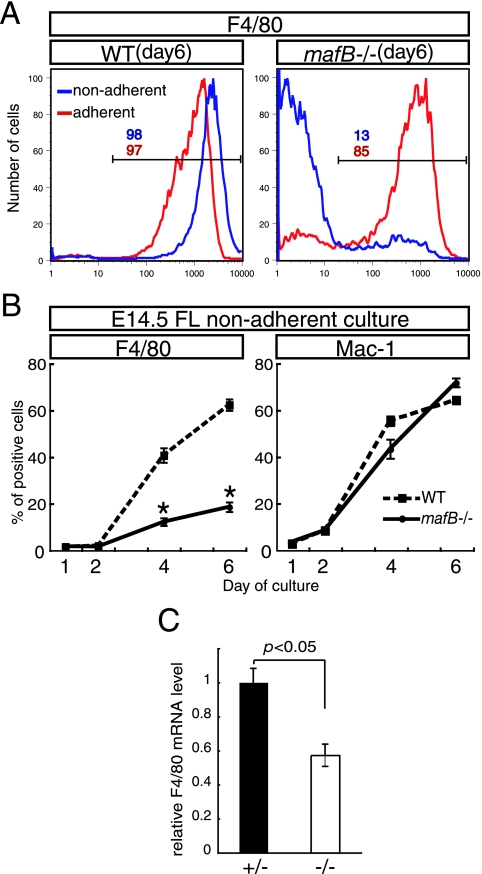
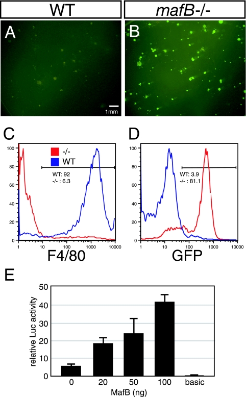
MafB is a member of the large Maf family of transcription factors that share similar basic region/leucine zipper DNA binding motifs and N-terminal activation domains. Although it is well known that MafB is specifically expressed in glomerular epithelial cells (podocytes) and macrophages, characterization of the null mutant phenotype in these tissues has not been previously reported. To investigate suspected MafB functions in the kidney and in macrophages, we generated mafB/green fluorescent protein (GFP) knock-in null mutant mice. MafB homozygous mutants displayed renal dysgenesis with abnormal podocyte differentiation as well as tubular apoptosis. Interestingly, these kidney phenotypes were associated with diminished expression of several kidney disease-related genes. In hematopoietic cells, GFP fluorescence was observed in both Mac-1- and F4/80-expressing macrophages in the fetal liver. Interestingly, F4/80 expression in macrophages was suppressed in the homozygous mutant, although development of the Mac-1-positive macrophage population was unaffected. In primary cultures of fetal liver hematopoietic cells, MafB deficiency was found to dramatically suppress F4/80 expression in nonadherent macrophages, whereas the Mac-1-positive macrophage population developed normally. These results demonstrate that MafB is essential for podocyte differentiation, renal tubule survival, and F4/80 maturation in a distinct subpopulation of nonadherent mature macrophages.


Figures

FIG. 1.
FIG. 1.
Generation of mafB null mutant mice. (A) Strategy for GFP reporter gene knock-in to the mafB locus by homologous recombination. H and S are abbreviations for restriction enzyme cleavage sites for HindIII and SacI, respectively. Neo indicates the neomycin resistance gene flanked by loxP elements. The positions of the 3′ genomic probe 1 and internal probe 2 used for Southern blot analysis are indicated by the horizontal bar. (B) The HindIII restriction fragments detected with internal probe 2 in the wild type (WT) (4.6 kbp), knockout (KO) Neo allele (4.0 kbp), and KO allele (2.9 kbp) are indicated. (C) Genotyping by Southern blotting. HindIII digestion of genomic DNA extracted from the tails of sibling pups of each genotype generated 4.6-kbp and 3.0-kbp bands for the wild-type and KO alleles, respectively, using 3′ genomic probe 1. (D) RT-PCR analysis of MafB mRNA. RNA was prepared from the kidneys of wild-type and heterozygous and homozygous mutant mafB neonates. (E and F) GFP immunoreactivity in podocytes of mafB+/− and mafB−/− neonates. (G and H) Endogenous MafB mRNA expression was specifically detected in the podocytes of mafB+/ kidneys, whereas mafB−/− neonatal kidneys had no MafB mRNA accumulation. Scale bars represent 20 μm in all panels.
FIG. 2.
FIG. 2.
Kidney abnormalities in mafB−/− neonatal mice. (A) Gross appearance of a neonatal kidney from a mafB−/− mouse. The mafB−/− kidney (right) has a punctate surface hemorrhage and a dystrophic general appearance, unlike kidneys recovered from a mafB+/ littermate (left). (B) mafB−/− mice display a significant difference from mafB+/ littermates in mean serum creatinine concentration (P = 0.0082, Mann-Whitney U test). (C and D) periodic acid-Schiff staining of mafB+/ and mafB−/− neonatal kidneys. (C) Tissue sections of mafB+/ kidneys showed numerous proximal and distal tubules. (D) Kidneys from mafB−/− mice have a reduced number of proximal and distal tubules accompanied by numerous cysts, primarily in the area of proximal tubuli (arrows in panels D and F). (E and F) mafB+/ kidneys had mature glomeruli, whereas kidneys from mafB−/− animals had fewer mature glomeruli (outlined by dotted lines). (G and H) Lack of normal podocyte foot processes in mafB−/− glomeruli. Electron microscopy revealed that mafB−/− podocyte foot processes were fused and did not interdigitate (H), whereas normal discrete foot processes were observed in the glomeruli of mafB+/ kidneys (G). pc, podocyte; fp, foot process; GBM, glomerular basement membrane. The scale bars in panels C, D, E, and F are 60 μm, whereas those in panels G and H represent 1 μm.
FIG. 3.
FIG. 3.
Expression of kidney disease-related genes in mafB mutant mice. (A) mafB homozygous mutant kidneys displayed a significant reduction in the accumulation of kidney disease-related mRNAs (nephrin, podocin, and CD2AP) on Northern blots. The proximal/distal tubular marker genes (mFuc-TIX and uromodulin) were also reduced. In contrast, expression of NEPH1 and c-Myc was significantly elevated in the mafB−/− kidney. Pod1, a putative upstream regulator of mafB, was unchanged. The podocalyxin gene was unaffected (see Discussion). (B) Nephrin and podocin immunoreactivities were significantly reduced in the glomeruli of mafB homozygous mutant kidneys in comparison to their heterozygous littermates. The scale bars represent 20 μm. (C) Quantification of the RNA blotting analysis in panel A. The band intensities were quantified with ImageQuant version 5.2 (Molecular Dynamics) software and then normalized to GAPDH mRNA abundance. Three independent animals of each genotype were analyzed. Data are presented as the means ± SD. The statistical significances of the differences are indicated by asterisks (*, P < 0.05; **, P < 0.01 [Student's t test]).
FIG. 4.
FIG. 4.
Increased apoptosis in the kidneys of mafB−/− mice. (A and B) TUNEL-positive cells in mafB+/ and mafB−/− kidneys (indicated by arrowheads), respectively, were predominantly observed in the tubular cells of mafB−/− kidneys, whereas significantly fewer tubule cells are TUNEL positive in mafB+/ kidneys (T, tubule; G, glomerulus). The scale bar is 20 μm. (C) TUNEL-positive cells were counted in 20 randomly selected fields within the periglomerular area or cortical interstitium and were then recorded as TUNEL-reactive cells/20 fields. For each genotype, one central section from each of four kidneys from two different pups was analyzed. The data were recorded as means ± SD. The statistical significance of differences was determined by Student's t test.
FIG. 5.
FIG. 5.
Flow cytometric analysis of e14.5 fetal liver hematopoietic cells. (A) Single-cell suspensions were prepared from the livers of wild-type (WT) mice (+/+) or heterozygous (mafB+/) or homozygous (mafB−/−) mutant e14.5 embryos. The cells were stained with PE- or APC-conjugated primary antibodies (indicated on the right) and then analyzed by flow cytometry. The profiles display GFP fluorescence on the horizontal axis and antibody-mediated PE or APC fluorescence on the vertical axis. The percentage of cells in each of the four quadrants is indicated. Specificities of the antibodies are as follows: F4/80, differentiated macrophage; Mac-1, monocyte-macrophage lineage; Gr-1, granulocytes; CD11c, dendritic cells. (B) MafB and F4/80 double immunostaining on frozen sections of e14.5 fetal liver from each genotype. In the mafB+/ control, MafB and F4/80 expression is nicely colocalized, whereas MafB immunoreactivity was absent in mafB−/− fetal livers. (C) In e14.5 fetal liver cells from mafB−/− mice, the total population of F4/80-positive cells was slightly reduced in comparison to the number recovered in either mafB+/ or wild-type mice. (D) F4/80 and Mac-1 expression profile in GFP-positive (MafB-expressing) macrophages. A total of 1.1% of GFP-positive cells (blue gates in A) from each genotype were analyzed. In the mafB−/− cells, an increase of F4/80 Mac-1+ premature macrophages and a reduction of F4/80+ Mac-1+ mature macrophages were observed.
FIG. 6.
FIG. 6.
F4/80 expression is suppressed in MafB-deficient macrophages. (A and C) Reduction of the F4/80-positive population and the increase in the GFP+ F4/80 population in neonatal peripheral blood and spleen cells. The Mac-1-positive macrophage population was unaffected (C). (B) F4/80 and Mac-1 expression profiles in GFP-positive (MafB-expressing) macrophages. A total of 0.6% of GFP-positive cells (red gates in B) from each genotype were analyzed. In the mafB−/− cells, an increase of F4/80 Mac-1+ premature macrophages and a reduction of F4/80+ Mac-1+ mature macrophages were observed. Seven independent animals of each genotype were analyzed. The data were recorded as means ± SD. The statistical significance of differences was determined by Student's t test. WT, wild type.
FIG. 7.
FIG. 7.
Failure to induce F4/80 in nonadherent mafB−/− macrophages. (A) The mafB mutant fetal liver-derived macrophages (right panel, blue line) failed to express F4/80 immunoreactivity when cultured under nonadherent conditions in the presence of M-CSF for 6 days, although 98% of wild-type (WT) fetal liver cells generated F4/80-positive mature macrophages (left panel, blue line). When wild-type or mafB−/− cells were cultured on adherent substrates (red lines), more than 85% of the hematopoietic cells of both genotypes differentiated into F4/80-positive cells. (B) When cultured under nonadherent conditions, MafB deficiency suppressed F4/80 expression from day 2 onward, although Mac-1 expression was not affected. FL, fetal liver. (C) Real-time PCR analysis of nonadherent macrophages showed a 50% reduction of F4/80 mRNA expression in the homozygous mutant macrophages. Seven independent animals of each genotype were analyzed. The data were recorded as means ± SD. The statistical significance of differences was determined by Student's t test.
FIG. 8.
FIG. 8.
mafB−/− macrophages derived from methylcellulose culture fail to induce F4/80 expression. In methylcellulose medium supplemented with M-CSF, 81.1% of mafB−/− e14.5 fetal liver cells gave rise to GFP-positive macrophages (A, B, and D). A total of 92% of e14.5 wild-type (WT) fetal liver cells gave rise to F4/80-positive macrophages, whereas most mafB−/− fetal liver cells failed to induce F4/80 expression (C). (E) Cotransfection reporter assay using the 668-bp F4/80 promoter reporter and the MafB expression vector. Luciferase (Luc) activity was significantly activated by cotransfection of the MafB expression vector in a dose-dependent manner in the murine macrophage cell line RAW264.7.

References

    1. Abrahamson, D. 1991. Glomerulogenesis in the developing kidney. Semin. Nephrol. 11:375-389. - PubMed
    1. Austyn, J. M., and S. Gordon. 1981. F4/80, a monoclonal antibody directed specifically against the mouse macrophage. Eur. J. Immunol. 11:805-815. - PubMed
    1. Barletta, G., I. A. Kovari, R. K. Verma, D. Kerjaschkil, and L. B. Holzman. 2003. Nephrin and Neph1 co-localize at the podocyte foot process intercellular junction and form cis hetero-oligomers. J. Biol. Chem. 278:19266-19271. - PubMed
    1. Blanchi. B., L. M. Kelly, J. C. Viemari, I. Lafon, H. Burnet, M. Bevengut, S. Tillmanns, L. Daniel, T. Graf, G. Hilaire, and M. H. Sieweke. 2003. MafB deficiency causes defective respiratory rhythmogenesis and fatal central apnea at birth. Nat. Neurosci. 10:1091-1100. - PubMed
    1. Boute, N., O. Gribouval, S. Roselli, F. Benessy, H. Lee, A. Fuchshuber, K. Dahan, M. C. Gubler, P. Niaudet, and C. Antignac. 2000. NPHS2, encoding the glomerular protein podocin, is mutated in autosomal recessive steroid-resistant nephrotic syndrome. Nat. Genet. 24:349-354. - PubMed

Publication types

MeSH terms

Substances