. 2006 Aug;26(15):5759-70.
doi: 10.1128/MCB.00271-06.

Mechanism of action of a distal NF-kappaB-dependent enhancer

Affiliations

Mechanism of action of a distal NF-kappaB-dependent enhancer

Belete Teferedegne et al. Mol Cell Biol. 2006 Aug.

Abstract

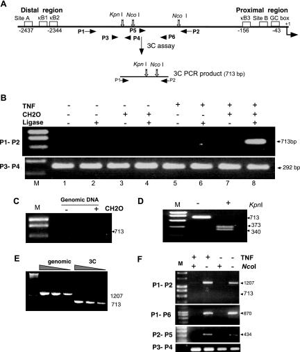
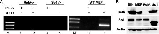
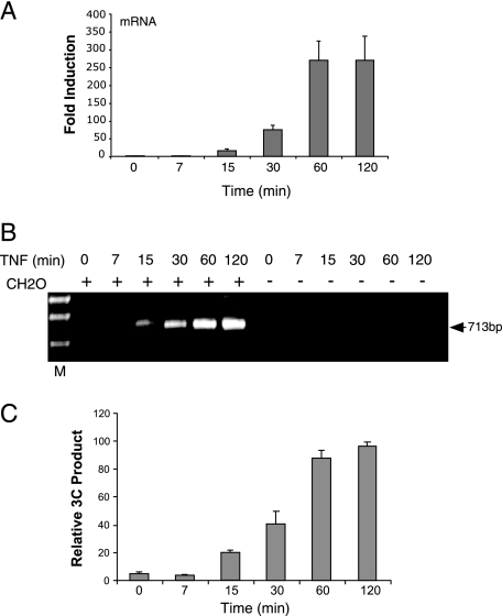
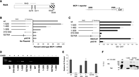
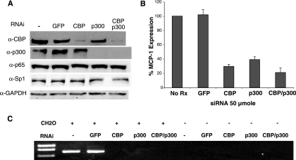
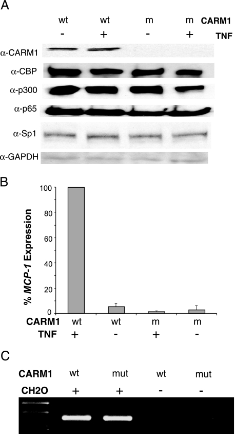
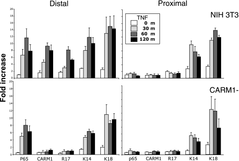
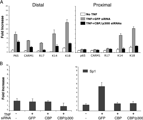
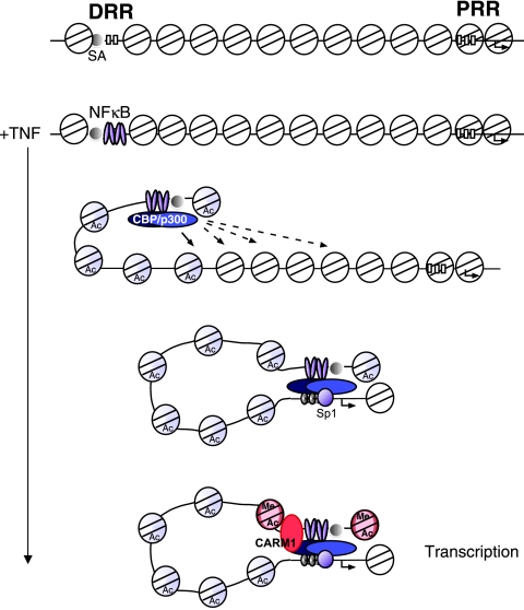
The monocyte chemoattractant protein 1 gene (MCP-1) is regulated by TNF through an NF-kappaB-dependent distal enhancer and an Sp1-dependent promoter-proximal regulatory region. In the silent state, only the distal regulatory region is accessible to transcription factors. Upon activation by tumor necrosis factor, NF-kappaB binds to the distal regulatory region and recruits CBP and p300. CBP and p300 recruitment led to specific histone modifications that ultimately enabled the binding of Sp1 to the proximal regulatory region. During this process, a direct interaction between the distal and proximal regulatory regions occurred. Sp1, NF-kappaB, CBP, and p300 were required for this interaction. CBP/p300-mediated histone modifications enhanced the binding of the coactivator CARM1 to the distal regulatory region. CARM1, which is necessary for MCP-1 expression, was not required for distal-proximal region interactions, suggesting that it plays a later downstream activation event. The results describe a model in which the separation of the distal enhancer from the promoter-proximal region allows for two independent chromatin states to exist, preventing inappropriate gene activation at the promoter while at the same time allowing rapid induction through the distal regulatory region.


Figures

FIG. 1.
FIG. 1.
An interaction between the distal and proximal regulatory regions of the MCP-1 gene forms following TNF stimulation. (A) A schematic representation of the 3C assay and the architecture of the MCP-1 promoter with cis regulatory sites with selected restriction sites and primer positions (P1 through P4) is shown. In the 3C assay, cells were fixed with formaldehyde and chromatin was isolated and digested with NcoI. Following inactivation of the restriction enzyme, the sample was diluted and subjected to DNA ligation such that only intramolecular ligations would be preferred. The cross-links were then reversed, and the DNA was purified. PCR primers P1 and P2 were used to detect the formation of the novel ligated 3C product. Primers P3 and P4 were used to verify that similar levels of MCP-1 DNA were present in the assay and that the DNA was amplifiable. Products from the 3C assay were examined on agarose gels stained with ethidium bromide. (B) NIH 3T3 cells treated in the absence (−) (lanes 1 to 4) or presence (+) (lanes 5 to 8) of TNF for 2 h were processed in the above 3C assay with and without formaldehyde (CH2O) or DNA ligase, as indicated, to control for specificity of the protocol. M, DNA marker. (C) Purified mouse genomic DNA was processed in the 3C assay and demonstrates that the unique PCR product cannot form with naked DNA templates. (D) KpnI cleaves the PCR product generated by the 3C assay, producing the anticipated DNA fragments as shown on the agarose gel. (E) PCR amplification of the 3C product and the non-3C product display similar relative efficiencies. PCR products generated from undigested genomic DNA (1,207 bp) and DNA from a 3C assay (713 bp) were quantitated by fluorometry and used as templates in PCRs to determine if there were major differences in their PCR efficiency. DNA templates (40 pg, 8 pg, and 1.6 pg) were amplified by primers P1 and P2 under identical PCR conditions. (F) Chromatin isolated from TNF-treated or control cells fixed with formaldehyde was subjected to NcoI digestion as indicated. Following deproteination, the DNA was purified and analyzed by PCR using the indicated primers. Because the ligation step of the 3C assay was not performed, P1/P2, P1/P6, and P2/P5 PCR products can only be observed when NcoI was omitted from the reaction, indicating that the chromatin is accessible to restriction digestion even in the absence of TNF.
FIG. 2.
FIG. 2.
RelA and Sp1 are required for generation of the 3C product. (A) Murine cell lines deficient for RelA or Sp1 were compared to a wild-type MEF line for their ability to form the MCP-1 3C product. Cells were treated with TNF as indicated (+, present; −, absent), and formaldehyde was excluded in some experiments to control for nonspecific events. Only the wild-type (WT MEF) cells were able to generate a 3C assay product, suggesting that the 3C product is mediated by the factors bound to the DRR and PRR and that interactions between these regions occur in response to TNF. (B) Western blots for the presence of RelA, Sp1, or actin were conducted on the indicated cell lines. The lower migrating Sp1 band in the Sp1−/− cells represents the N-terminal portion of Sp1 that is expressed in the knockout line. As described by Marin et al., this mutant is inactive in DNA binding and transactivation (38).
FIG. 3.
FIG. 3.
MCP-1 mRNA accumulation and distal/proximal region interactions are coincident events. (A) MCP-1 mRNA is detectable in the cytoplasm after 15 min of TNF stimulation and increases over the first hour of the time course. Real-time RT-PCR was carried out on RNA isolated from NIH 3T3 cells treated with 500 units of TNF for the indicated time. The results from three independent assays were averaged following normalization to the levels of GAPDH mRNA detected by real-time RT-PCR. (B) The 3C product was not detected until 15 min of TNF stimulation. NIH 3T3 cells treated with TNF for the indicated time were assayed using the 3C assay described above. The presence (+) of the formaldehyde (CH2O) was required for generation of the 3C product. (C) The 3C product increased in intensity over the first hour of the time course and was present at 2 h. Images from stained agarose gels, representing data from three independent sample sets, were quantitated and averaged as described in Materials and Methods. The mean data are plotted with their standard deviations.
FIG. 4.
FIG. 4.
The Rel homology domain and serine 276 of RelA are required for expression and distal/proximal region interactions. A series of RelA mutants (A) were transfected into RelA-deficient cells and tested for their ability to drive expression of the endogenous gene (B) or an MCP-1 promoter CAT reporter gene (C) and for their ability to form the 3C product on the endogenous MCP-1 locus (D). TNF was not added to these assays, as overexpression of RelA is sufficient to bypass sequestration in the cytoplasm (48). All RelA expression constructions that contained a wild-type (WT) Rel homology domain were able to drive MCP-1 expression and generate a 3C product. (A) A schematic of the MCP-1 reporter gene construct (pJECAT2.6) and of the RelA p65 domain structure is shown. RHD, rel homology domain; TAD, transcriptional activation domain. Phosphorylatable serine residues shown to be important for RelA activity are indicated. (B) Real-time RT-PCR of MCP-1 mRNA levels from RelA-deficient cells transiently transfected as above with the indicated plasmids were normalized to the amount of GAPDH mRNA and plotted as the percentage of the wild-type RelA transfectant. The averages of results from three independent transfections are shown. (C) RelA-deficient cells transfected with the indicated RelA expression vector and the MCP-1 reporter. CAT values, derived from an enzyme-linked immunosorbent assay for CAT protein, normalized to the expression of an alkaline phosphatase expression vector were averaged from three experiments. (D) The 3C assay was carried out on the indicated plasmids transfected into the RelA-deficient cells as above. (E) Images from stained agarose gels representing three independent assays of the experiment shown in panel D were quantitated and plotted as described in Materials and Methods. (F) Western blots performed on cells transfected with the RelA mutant series show similar levels of expression between wild-type and mutant proteins. Different antibodies were used due to the loss of epitopes in some of the mutants (left blot, sc-372 [Santa Cruz, Inc.]; right blot, 12145-30 [Abcam, Inc.]).
FIG. 5.
FIG. 5.
CBP and p300 are required for MCP-1 expression and distal/proximal region interaction. siRNAs to CBP, p300, or both were transfected into NIH 3T3 cells. Cells were incubated for 48 h and then treated with TNF for 2 h. (A) Western blot analysis of the transfected cells using antibodies to CBP (α-CBP), p300 (α-p300), RelA (α-p65), Sp1 (α-Sp1), and GAPDH (α-GAPDH), demonstrating specificity and efficacy of the siRNA. (B) In similar siRNA transfections, MCP-1 expression was measured by real-time RT-PCR. (C) The 3C assay was performed on the transfected cells treated with TNF as above. All siRNA transfections and assays were performed at least three times with identical results. +, present; −, absent.
FIG. 6.
FIG. 6.
CARM1 is required after the distal and proximal regions interact. CARM1-deficient MEFs were used to assay its role in MCP-1 activation. (A) Immunoblot analysis confirms that CARM1 protein is absent in the mutant cells and that CBP, p300, RelA (p65), Sp1, and GAPDH protein levels are unaffected. α, anti. (B) CARM1-deficient cells fail to activate MCP-1 in response to TNF. Real-time RT-PCR was conducted on RNA samples that were isolated from wild-type (wt) and CARM1-deficient (m) MEFs following no TNF stimulation (−) or 2 h of TNF stimulation (+). Real-time RT-PCR values were normalized to the amount of GAPDH mRNA in the samples and plotted with respect to the wild-type sample induced with TNF. The average of three independent experiments is shown. (C) An interaction forms between the proximal and distal regulatory regions regardless of whether CARM1 is present. The 3C assay as described above was performed on wild-type and CARM1-deficient (mut) MEFs. All samples in this assay were derived from cells treated with TNF. All experiments were performed three times with identical results.
FIG. 7.
FIG. 7.
CARM1 binds to and modifies the distal regulatory region in a time course that follows CBP/p300 histone modifications. ChIP assays coupled with real-time PCR for the distal and proximal regulatory regions were conducted with antibodies to p65, CARM1, and to the indicated histone modifications following TNF treatment of NIH 3T3 and CARM1-deficient cells for the indicated time. The data from three separate chromatin preparations and assays were averaged and plotted as relative increases over an irrelevant antibody control.
FIG. 8.
FIG. 8.
CBP/p300 control distal and proximal regulatory region acetylation, CARM1 binding, and access to the proximal regulatory region. (A) siRNA to CBP/p300 in NIH 3T3 cells but not GFP reduce histone H3 K14 and K18 acetylation and CARM1 binding and its associated activity as indicated by H3 R17 methylation. (B) siRNAs to CBP or CBP/p300 prevent the binding of Sp1 to the proximal regulatory region. ChIP assays using the indicated antibodies were conducted on NIH 3T3 cells treated with the siRNAs as described in the legend to Fig. 5. The data from three separate chromatin preparations and assays were averaged and plotted as relative increases over an irrelevant antibody control.
FIG. 9.
FIG. 9.
Model of MCP-1 induction by TNF. A schematic is shown of the uninduced state of the MCP-1 gene followed by the predicted events that occur after TNF induction. Blue shaded nucleosomes indicate those that have been modified by CBP/p300. We suggest that CBP/p300 nucleosome modifications occur until an it reaches the PRR. Once the PRR nucleosomes have been modified, the region is accessible to Sp1, allowing a direct interaction with the DRR. CARM1 is then recruited and provides a signal to activate transcription of the gene. Red shaded nucleosomes indicate those that have been modified by CARM1 and CBP/p300. DRR, distal regulatory region; PRR, proximal regulatory region; SA, site A—the constitutively bound region.

Similar articles

Cited by

References

    1. An, W., J. Kim, and R. G. Roeder. 2004. Ordered cooperative functions of PRMT1, p300, and CARM1 in transcriptional activation by p53. Cell 117:735-748. - PubMed
    1. Arany, Z., D. Newsome, E. Oldread, D. M. Livingston, and R. Eckner. 1995. A family of transcriptional adaptor proteins targeted by the E1A oncoprotein. Nature 374:81-84. - PubMed
    1. Baeuerle, P. A., and D. Baltimore. 1996. NF-kappa B: ten years after. Cell 87:13-20. - PubMed
    1. Baggiolini, M., and P. Loetscher. 2000. Chemokines in inflammation and immunity. Immunol. Today 21:418-420. - PubMed
    1. Baldwin, A. S. 1996. The NF-kB and IkB proteins: new discoveries and insights. Annu. Rev. Immunol. 14:649-681. - PubMed

Publication types

MeSH terms