. 2006 Aug;26(15):5771-83.
doi: 10.1128/MCB.02404-05.

MyoD synergizes with the E-protein HEB beta to induce myogenic differentiation

Affiliations

MyoD synergizes with the E-protein HEB beta to induce myogenic differentiation

Maura H Parker et al. Mol Cell Biol. 2006 Aug.

Erratum in

  • Mol Cell Biol. 2013 Nov;33(21):4357

Abstract

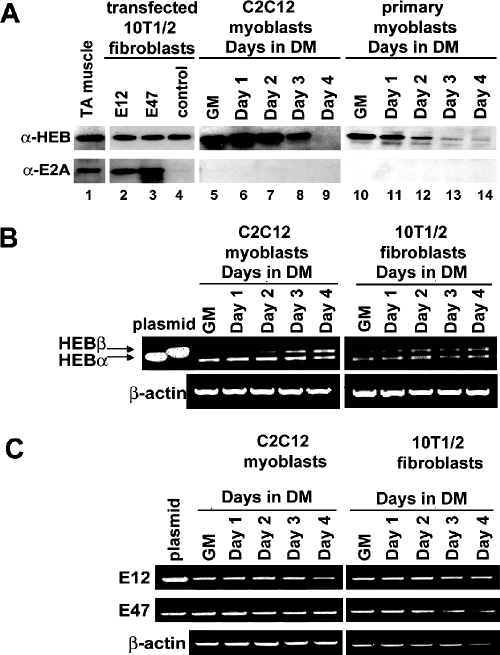
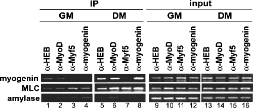
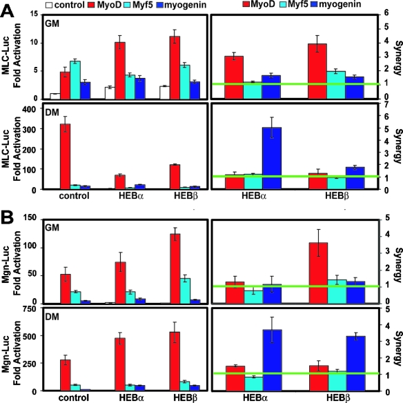
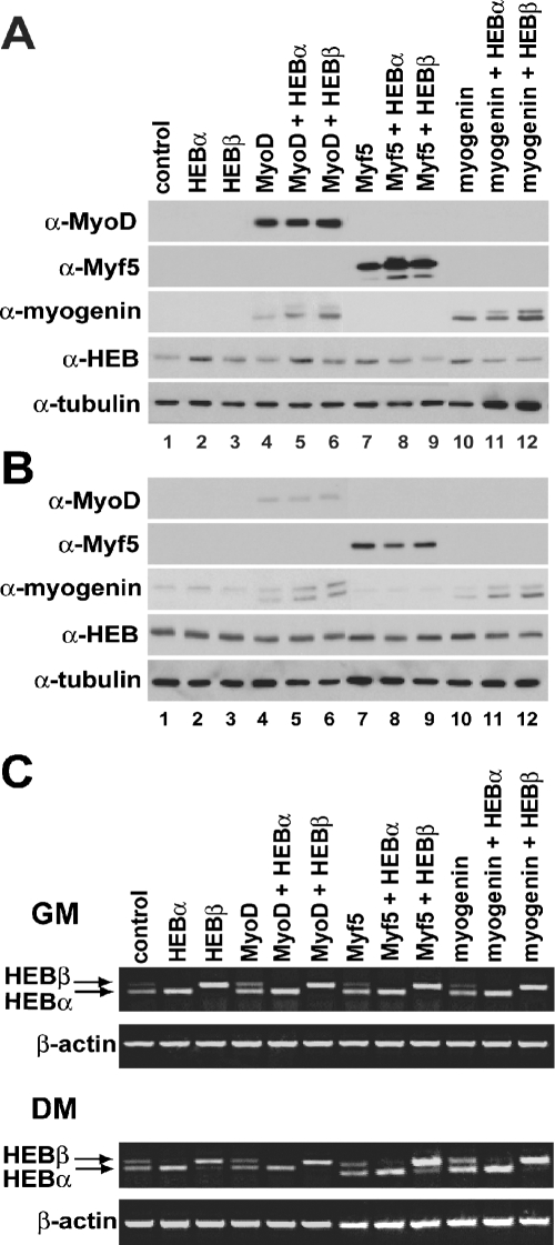
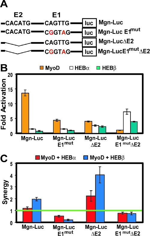
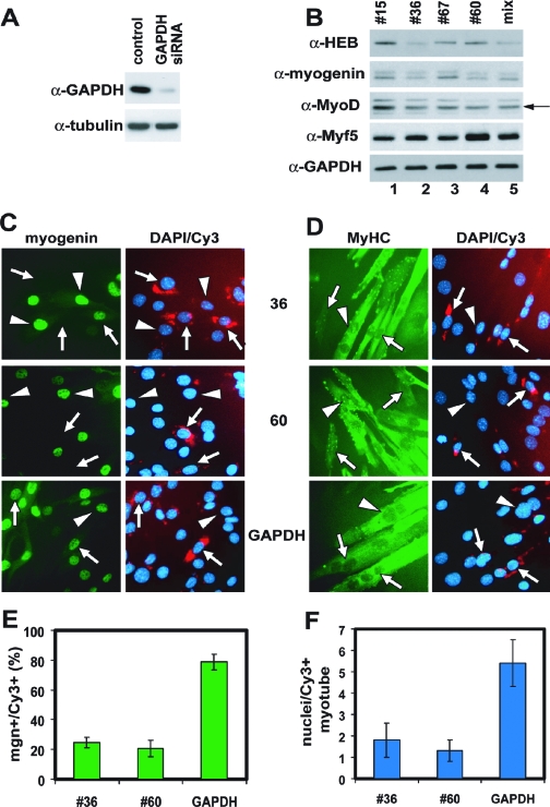
The MyoD family of basic helix-loop-helix transcription factors function as heterodimers with members of the E-protein family to induce myogenic gene activation. The E-protein HEB is alternatively spliced to generate alpha and beta isoforms. While the function of these molecules has been studied in other cell types, questions persist regarding the molecular functions of HEB proteins in skeletal muscle. Our data demonstrate that HEB alpha expression remains unchanged in both myoblasts and myotubes, whereas HEB beta is upregulated during the early phases of terminal differentiation. Upon induction of differentiation, a MyoD-HEB beta complex bound the E1 E-box of the myogenin promoter leading to transcriptional activation. Importantly, forced expression of HEB beta with MyoD synergistically lead to precocious myogenin expression in proliferating myoblasts. However, after differentiation, HEB alpha and HEB beta synergized with myogenin, but not MyoD, to activate the myogenin promoter. Specific knockdown of HEB beta by small interfering RNA in myoblasts blocked differentiation and inhibited induction of myogenin transcription. Therefore, HEB alpha and HEB beta play novel and central roles in orchestrating the regulation of myogenic factor activity through myogenic differentiation.


Figures

FIG. 1.
FIG. 1.
HEB expression is highly regulated in proliferating and differentiating myoblasts. (A) Western analysis of HEB and E2A expression reveals abundant HEB protein but no detectable E2A protein in myoblasts and fibroblasts. (B) RNA was isolated from a differentiation time course of C2C12 myoblasts and 10T1/2 fibroblasts and analyzed by RT-PCR. HEBα was expressed under both growth and differentiation conditions in C2C12 cells, whereas HEBβ was induced upon differentiation. (C) RT-PCR detection of E12 and E47 mRNAs in C2C12 and 10T1/2 cells. α-HEB, anti-HEB; α-E2A, anti-E2A; TA, tibialis anterior muscle.
FIG. 2.
FIG. 2.
HEB binds muscle-specific promoters. ChIP revealed that binding of HEB to the myogenin promoter and MLC enhancer increased markedly after differentiation. Amylase was amplified as a control for specificity, and input DNA was amplified with each primer set to ensure proper DNA quantitation. α-HEB, anti-HEB; α-MyoD, anti-MyoD; α-Myf5, anti-Myf5; α-myogenin, antimyogenin.
FIG. 3.
FIG. 3.
HEB-dependent increase in MRF activity is synergistic and multifaceted. (A) 10T1/2 fibroblasts were transfected with the MLC-Luc reporter plasmid, plasmids expressing MyoD, Myf5, or myogenin, and either HEBα or HEBβ expression plasmids. The panels on the left represent relative activation. The activity of the MLC-Luc reporter plasmid alone was arbitrarily set at 1, and the activity in the presence of the effectors was determined relative to this value (fold activation). The bars represent the mean relative activation, and the error bars represent standard deviations (n = 9). The panels on the right represent synergy (see Materials and Methods). The horizontal line represents the value at which the two effectors function independently or additively. The bars represent the mean synergy value, and the error bars represent standard deviations (n = 9). (B) The same transfection as for panel A but with the Mgn-Luc reporter plasmid.
FIG. 4.
FIG. 4.
HEBβ enhances MyoD-mediated expression of endogenous myogenin. (A) 10T1/2 fibroblasts were transfected as indicated and maintained in growth medium. Protein was isolated, and 25 μg was analyzed by Western blotting with the indicated antibodies. The expression of tubulin was monitored as a control for protein quantitation. (B) The same transfection and Western blots as for panel A, but protein was isolated from cells induced to differentiate for 2 days after transfection. (C) The same transfection as for panels A and B, but RNA was isolated and analyzed by RT-PCR. Transfection of HEBα specifically increases levels of HEBα, and transfection of HEBβ specifically increases levels of HEBβ. α-MyoD, anti-MyoD; α-Myf5, anti-Myf5; α-myogenin, antimyogenin; α-HEB, anti-HEB; α-tubulin, antitubulin.
FIG. 5.
FIG. 5.
MyoD activity on the myogenin promoter is E1 E-box specific. (A) Schematic diagram of the E-boxes in the Mgn-Luc reporter vector and the mutant reporter vectors in which the E1 E-box has been mutated (Mgn-LucE1mut), the E2 E-box has been deleted (Mgn-LucDE2), or both (Mgn-LucE1mutDE2). (B) Activity of Mgn-Luc in transfected 10T1/2 fibroblasts. Cells were transfected with either EMSV control, MyoD, HEBα, or HEBβ in addition to each of the reporter plasmids indicated. The cells were maintained in growth medium, harvested, and assayed for luciferase activity. The bars represent the mean activation relative to the control, and the error bars represent standard deviations (n = 3). (C) Synergy between MyoD and HEB isoforms in the activation of Mgn-Luc in transfected 10T1/2 fibroblasts. Cells were transfected with combinations of MyoD and HEBα or MyoD and HEBβ. The horizontal line represents the value at which the two effectors function in an additive manner. The bars represent the mean synergy values, and the error bars represent standard deviations (n = 3).
FIG. 6.
FIG. 6.
MEK1 specifically inhibits the MyoD-HEBα complex. (A) 10T1/2 fibroblasts were transfected with the Mgn-Luc reporter vector, plasmids expressing either full-length (FL) MyoD or deletion mutants of MyoD in addition to HEBα or HEBβ. The bars represent the mean synergy values, and the error bars represent standard errors of the means (n = 8).The green horizontal line represents the value at which the two effectors function independently or additively. (B) The same transfection as for panel A but including either empty vector or plasmid expressing activated MEK1. The bars represent the mean synergy values, and the error bars represent standard errors of the means (n = 8). The green horizontal line represents the value at which MyoD and HEBα function independently or additively. (C) The same transfection as for panel B but with HEBβ instead of HEBα.
FIG. 7.
FIG. 7.
Knockdown of HEB expression inhibits induction of myogenin and differentiation. (A) Western analysis of C2C12 myoblasts transfected with GAPDH siRNA. Note the reduced levels of GAPDH protein only in cells transfected with siRNA specific for GAPDH. The expression of tubulin was monitored as a control for protein quantitation. (B) Western analysis of C2C12 cells transfected with HEB-specific siRNAs and then induced to differentiate for 48 h. Note the reduced level of HEB and myogenin protein present in cells transfected with siRNAs specific for HEBα/β (no. 35, mix) and HEBβ (no. 60). HEB siRNA no. 15 did not reduce HEB expression. (C and D) Immunofluorescent detection of myogenin and MyHC in siRNA-transfected C2C12 cells. Cells were fixed either after 48 h of differentiation for detection of myogenin (C) or after 96 h for detection of MyHC (D). Nuclei were counterstained with DAPI. Note the inhibition in myogenin induction (C) and delayed formation of multinucleated myotubes (D) after HEBα/β (no. 36) and HEBβ (no. 60) induced knockdown of HEB expression. (E) Quantitation of the results in panel C. The bars represent the average percentages of cells positive for both myogenin and siRNA (Cy3) per field of view (magnification, ×10). The error bars represent standard deviations (n = 4 fields). (F) Quantitation of the results in panel D. The bars represent the average number of nuclei per siRNA-positive myotubes, and the error bars represent standard deviations (n = 25 myotubes). α-HEB, anti-HEB; α-myogenin, antimyogenin; α-MyoD, anti-MyoD; α-Myf5, anti-Myf5; α-GAPDH, anti-GAPDH.

Similar articles

Cited by

References

    1. Asakura, A., A. Fujisawa-Sehara, T. Komiya, and Y. Nabeshima. 1993. MyoD and myogenin act on the chicken myosin light-chain 1 gene as distinct transcriptional factors. Mol. Cell. Biol. 13:7153-7162. - PMC - PubMed
    1. Ballinger, C. A., P. Connell, Y. Wu, Z. Hu, L. J. Thompson, L. Y. Yin, and C. Patterson. 1999. Identification of CHIP, a novel tetratricopeptide repeat-containing protein that interacts with heat shock proteins and negatively regulates chaperone functions. Mol. Cell. Biol. 19:4535-4545. - PMC - PubMed
    1. Bergstrom, D. A., B. H. Penn, A. Strand, R. L. Perry, M. A. Rudnicki, and S. J. Tapscott. 2002. Promoter-specific regulation of MyoD binding and signal transduction cooperate to pattern gene expression. Mol. Cell 9:587-600. - PubMed
    1. Bergstrom, D. A., and S. J. Tapscott. 2001. Molecular distinction between specification and differentiation in the myogenic basic helix-loop-helix transcription factor family. Mol. Cell. Biol. 21:2404-2412. - PMC - PubMed
    1. Berkes, C. A., D. A. Bergstrom, B. H. Penn, K. J. Seaver, P. S. Knoepfler, and S. J. Tapscott. 2004. Pbx marks genes for activation by MyoD indicating a role for a homeodomain protein in establishing myogenic potential. Mol. Cell 14:465-477. - PubMed

Publication types

MeSH terms

LinkOut - more resources