. 2006 Aug;26(15):5921-31.
doi: 10.1128/MCB.00315-06.

Understanding the molecular basis of the interaction between NDPK-A and AMPK alpha 1

Affiliations

Understanding the molecular basis of the interaction between NDPK-A and AMPK alpha 1

Russell M Crawford et al. Mol Cell Biol. 2006 Aug.

Retraction in

Abstract

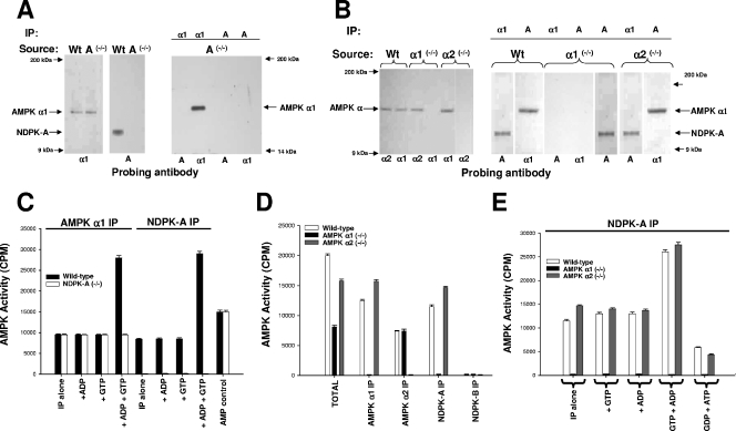
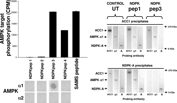
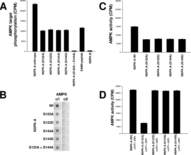
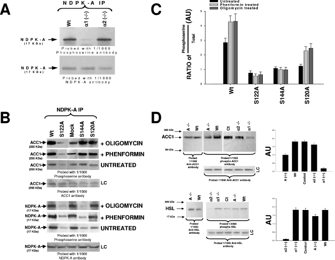
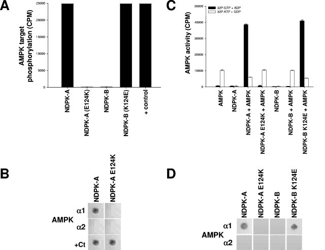
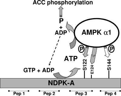
Nucleoside diphosphate kinase (NDPK) (nm23/awd) belongs to a multifunctional family of highly conserved proteins (approximately 16 to 20 kDa) including two well-characterized isoforms (NDPK-A and -B). NDPK catalyzes the conversion of nucleoside diphosphates to nucleoside triphosphates, regulates a diverse array of cellular events, and can act as a protein histidine kinase. AMP-activated protein kinase (AMPK) is a heterotrimeric protein complex that responds to the cellular energy status by switching off ATP-consuming pathways and switching on ATP-generating pathways when ATP is limiting. AMPK was first discovered as an activity that inhibited preparations of acetyl coenzyme A carboxylase 1 (ACC1), a regulator of cellular fatty acid synthesis. We recently reported that NDPK-A (but not NDPK-B) selectively regulates the alpha1 isoform of AMPK independently of the AMP concentration such that the manipulation of NDPK-A nucleotide trans-phosphorylation activity to generate ATP enhanced the activity of AMPK. This regulation occurred irrespective of the surrounding ATP concentration, suggesting that "substrate channeling" was occurring with the shielding of NDPK-generated ATP from the surrounding medium. We speculated that AMPK alpha1 phosphorylated NDPK-A during their interaction, and here, we identify two residues on NDPK-A targeted by AMPK alpha1 in vivo. We find that NDPK-A S122 and S144 are phosphorylated by AMPK alpha1 and that the phosphorylation status of S122, but not S144, determines whether substrate channeling can occur. We report the cellular effects of the S122 mutation on ACC1 phosphorylation and demonstrate that the presence of E124 (absent in NDPK-B) is necessary and sufficient to permit both AMPK alpha1 binding and substrate channeling.


Figures

FIG. 1.
FIG. 1.
Investigation of AMPK α1, AMPK α2, and NDPK-A knockout tissues. The precipitating tissue source is shown above each blot together (where relevant) with the precipitating antibody. The lower part of each blot denotes a 1/1,000 dilution of probing antibody as indicated. (A) Ten micrograms of extracted NDPK-A wild-type (Wt) or NDPK-A-null (−/−) cytosol probed with sheep anti-AMPK α1 or mouse anti-NDPK-A (left and middle panels). The right panel shows anti-AMPK α1 or mouse anti-NDPK-A antibodies used to precipitate (IP) 10 μg of extracted cytosol and probed with either an AMPK α1- or NDPK-A-specific antibody, as indicated. (B) The procedure used was the same as that described above (A), except that the tissue source was extracted liver cytosol from murine wild-type (Wt), AMPK α1 knockout (−/−), or AMPK α2 knockout (−/−) tissue. (C) Ten micrograms of extracted NDPK-A wild-type or NDPK-A-null (−/−) cytosol precipitated using either an AMPK α1- or NDPK-A specific antibody and assayed for AMPK-SAMS activity in the presence/absence of 500 nM ADP, GTP, and ADP plus GTP, as indicated. Error bars (n = 3) on all assay histograms indicate the ranges and not the standard errors. (D) Ten micrograms of extracted AMPK wild-type, AMPK α1-null (−/−), or AMPK α2-null (−/−) cytosol precipitated using either an AMPK α1-, AMPK α2-, NDPK-A-, or NDPK-B-specific antibody and assayed for AMPK-SAMS activity relative to the input (total). (E) Ten micrograms of extracted AMPK wild-type, AMPK α1-null (−/−), or AMPK α2-null (−/−) cytosol precipitated using an NDPK-A-specific antibody and assayed for AMPK-SAMS activity in the presence/absence of 500 nM ADP, GTP, and ADP plus GTP, as indicated; the middle bars show no activity in each triplet.
FIG. 2.
FIG. 2.
NDPK peptides. The left histogram shows pure, active rat liver AMPK used to phosphorylate a selection of peptides as follows. Four peptides (NDPKpep1, 2, 3 and 4) corresponding to regions of NDPK-A (amino acids 49 to 62, 92 to 105, 111 to 125 and 139 to 152, respectively) were used, and the SAMS peptide was used as a positive control, each at 1 mM concentration. For the left overlay, the overlay protocol involves a slight variation of the above-described Western blotting protocol in that each of the NDPK-A peptides was immobilized on a nitrocellulose membrane and then blocked/washed as described above for the Western blot protocol. Furthermore, 10 μg of pure, recombinant AMPK α1 or α2 protein was overlaid onto the peptides, with binding being detected using isoform-specific antibodies as described above for Western blotting. In the right blots, 10 μg of rat liver cytosol either left untreated (UT) or incubated with a molar excess of NDPKpep1 (pep1) or NDPKpep3 (pep3) was precipitated using an ACC1 (upper right) or NDPK-A (lower right) antibody and probed with either ACC1-, AMPK α1 (α1)-, or NDPK-A (A)-specific antibodies as labeled at the bottom of each lane. NDPKpep3 specifically interferes with the ACC1/AMPK α1/NDPK-A complex association.
FIG. 3.
FIG. 3.
Specific NDPK-A amino acids targeted by AMPK and substrate-channeling ability of NDPK-A mutants. (A) Pure, active rat liver AMPK was used to phosphorylate a selection of substrates as follows. Fifteen micrograms (each) of bacterial recombinant purified proteins, wild-type NDPK-A, NDPK-A S122A, NDPK-A S122D, NDPK-A S144A, NDPK-A S144D, NDPK-A S122A-plus-S144A double mutant, and NDPK-B (negative control), was used. SAMS peptide (1 mM) was included as a positive control. (B) Overlay binding analysis where each of the NDPK-A mutants and wild-type recombinant proteins was immobilized on a nitrocellulose membrane and then blocked/washed as described above. Ten micrograms of pure, recombinant AMPK α1 or α2 protein was overlaid onto the membrane, and binding was detected using isoform-specific AMPK antibodies. (C) Histogram measuring the capacity of pure, active rat liver AMPK to phosphorylate as a substrate (10 μg of wild-type NDPK compared to mutant proteins [S122A/D and S144A/D]). (D) Histogram measuring the ability of 10 μg of bacterial recombinant wild-type/mutant NDPK-A protein, as labeled, to channel labeled ATP (generated from labeled GTP plus ADP) to 1 mM SAMS peptide.
FIG. 4.
FIG. 4.
In vivo phosphorylation studies. (A) NDPK-A precipitations (IP) from 10 μg of liver cytosolic protein from wild-type littermate control (Wt), AMPK α1-null (α1 −/−), or AMPK α2-null (α2 −/−) mice run on 4 to 12% SDS-PAGE gels and probed with either a 1/1,000 dilution of anti-phosphoserine antibody or anti-NDPK-A antibody, as labeled. (B) HepG2 human-derived liver cell line transfected with various NDPK-A constructs: wild type, S122A, S144A, S120A, or empty vector control (mock). Transfected cells were either left untreated or incubated with 2 mM phenformin or 5 μg/ml oligomycin for 60 min. Ten micrograms of the cytosolic fraction from each construct was precipitated using an NDPK-A antibody as indicated, run on 4 to 12% SDS-PAGE gels, blotted, and probed using either a 1/1,000 dilution of phosphoserine or NDPK-A or ACC1 antibodies, as labeled. (C) Densitometry (histogram) data from four independent experiments were expressed in arbitrary units (AU) as a ratio of the phosphoserine amount over total protein amount ± standard error of the mean. (D) In the upper left panel, 10 μg of liver cytosolic protein from either wild-type (Wt), NDPK-A-null (A −/−), wild-type littermate control (Ct), AMPK α1-null (α1 −/−), or AMPK α2-null (α2 −/−) mice was run on 4 to 12% SDS-PAGE gels and probed with a 1/1,500 dilution of either anti-ACC1 antibody (leftmost blot) or anti-phospho-ACC1 antibody (center and right blots). In the lower left panel, 10 μg of the cytosolic fraction from mouse skeletal muscle from either wild-type (Wt), NDPK-A-null (A −/−), wild-type littermate control (Ct), AMPK α1-null (α1 −/−), or AMPK α2-null (α2 −/−) mice was run on an SDS-PAGE gel and probed with either a 1/1,500 dilution of anti-HSL antibody or a 1/2,000 dilution of phospho-HSL antibody. In each case, loading controls (LC) are included. Densitometry (D, right histograms) data from four independent experiments were calculated as described above for panel C.
FIG. 5.
FIG. 5.
A single amino acid on NDPK-A determines its association with AMPK α1. (A) Pure, active rat liver AMPK was used to phosphorylate a selection of substrates, 15 μg (each) of NDPK-A, NDPK-A(E124K), NDPK-B, and NDPK-B(K124E). In the final lane, CK2α holoenzyme (+ control) was used to phosphorylate NDPK-A (shown) and NDPK-B (not shown) mutant recombinant proteins, and both were identical. (B) NDPK-A and NDPK-A E124K mutant recombinant proteins were immobilized on nitrocellulose membranes and then blocked/washed as described above for the Western blot protocol. Furthermore, 10 μg of purified, recombinant AMPK α1, AMPK α2, or CK2α protein was overlaid onto the membrane, with binding detected using isoform-specific antibodies for each protein, as described above for the Western blot protocol. (C) Substrate-channeling ability of NDPK mutants was tested by adding, where indicated, active rat liver AMPK to 15 μg of either NDPK-A, NDPK-A E124K, NDPK-B, or NDPK-B K124E recombinant protein. Either [32P]GTP plus ADP (resulting in labeled NDPK-generated ATP) or [32P]ATP plus GDP (resulting in labeled NDPK-generated GTP) was used to demonstrate substrate channeling or substrate stealing, respectively, and also to act as cross-contamination control for each kinase individually. (D) NDPK-A, NDPK-A E124K, NDPK-B, and NDPK-B K124E proteins were immobilized on nitrocellulose membranes and then blocked/washed as described above for the Western blot protocol. Furthermore, 10 μg of purified, recombinant AMPK α1 or AMPK α2 protein was overlaid onto the membrane, with binding detected using isoform-specific antibodies for each protein, as described above for the Western blot protocol.
FIG. 6.
FIG. 6.
Model of interaction between AMPK α1 and NDPK-A. The phosphorylation status of residue S122 on NDPK-A determines whether NDPK-A channels ATP derived from GTP plus ADP to AMPK α1 as the substrate in the phosphorylation of downstream AMPK targets, for example, ACC1. Mutation of residue E124K on NDPK-A eliminates the specific interaction of NDPK-A with AMPK α1 (not shown). Serine 144 was also identified as an AMPK α1 target, but experiments revealed that its phosphorylation status has no bearing on substrate channeling or ACC1 phosphorylation. Peptides (Pep) 1 to 4 refer to surface regions of NDPK-A.

Similar articles

Cited by

References

    1. Arnaud-Dabernat, S., P. M. Bourbon, A. Dierich, M. Le Meur, and J. Y. Daniel. 2003. Knockout mice as model systems for studying nm23/NDP kinase gene functions. Application to the nm23-M1 gene. J. Bioenerg. Biomembr. 35:19-30. - PubMed
    1. Biondi, R. M., M. Engel, M. Sauane, C. Welter, O. G. Issinger, L. Jimenez de Asua, and S. Passeron. 1996. Inhibition of nucleoside diphosphate kinase activity by in vitro phosphorylation by protein kinase CK2. Differential phosphorylation of NDP kinases in HeLa cells in culture. FEBS Lett. 399:183-187. - PubMed
    1. Bosnar, M. H., J. De Gunzburg, R. Bago, L. Brecevic, I. Weber, and J. Pavelic. 2004. Subcellular localization of A and B Nm23/NDPK subunits. Exp. Cell Res. 298:275-284. - PubMed
    1. Bradford, M. M. 1976. A rapid and sensitive method for the quantitation of microgram quantities of protein utilizing the principle of protein-dye binding. Anal. Biochem. 72:248-254. - PubMed
    1. Caligo, M. A., G. Cipollini, L. Fiore, S. Calvo, F. Basolo, P. Collecchi, F. Ciardiello, S. Pepe, M. Petrini, and G. Bevilacqua. 1995. NM23 gene expression correlates with cell growth rate and S-phase. Int. J. Cancer 60:837-842. - PubMed

Publication types

MeSH terms