. 2006 Aug 1;20(15):2067-81.
doi: 10.1101/gad.1430406. Epub 2006 Jul 17.

SUMO modifications control assembly of synaptonemal complex and polycomplex in meiosis of Saccharomyces cerevisiae

Affiliations

SUMO modifications control assembly of synaptonemal complex and polycomplex in meiosis of Saccharomyces cerevisiae

Chung-Hsu Cheng et al. Genes Dev. .

Abstract

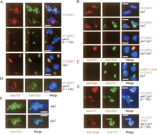
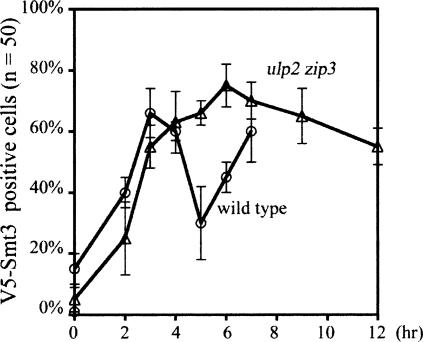
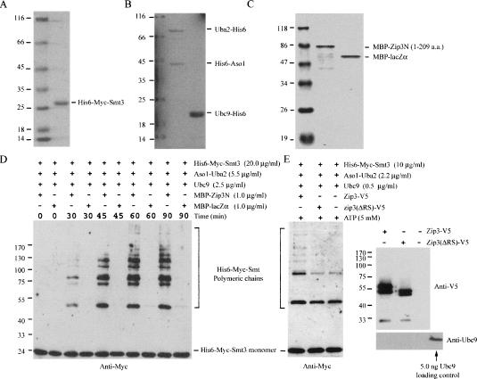
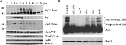
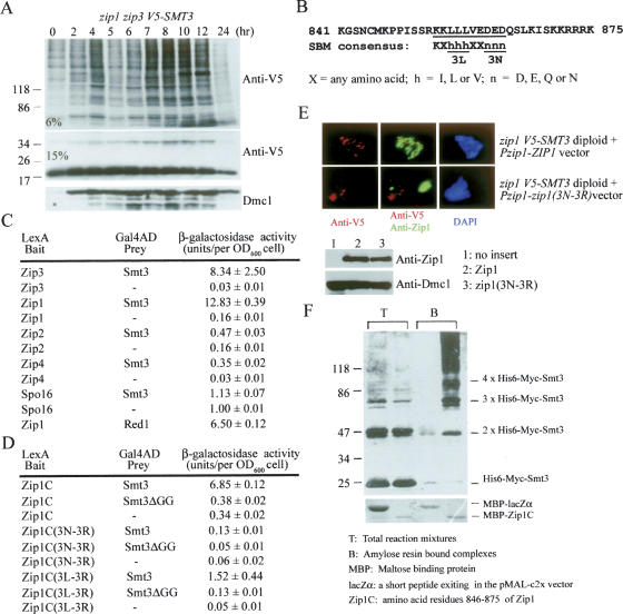
The synaptonemal complex (SC) is a proteinaceous complex that apparently mediates synapsis between homologous chromosomes during meiotic prophase. In Saccharomyces cerevisiae, the Zip1 protein is the integral component of the SC. In the absence of a DNA double-strand break or the SC initiation protein Zip3, Zip1 proteins aggregate to form a polycomplex (PC). In addition, Zip1 is also responsible for DSB-independent nonhomologous centromere coupling at early meiotic prophase. We report here that Zip3 is a SUMO (small ubiquitin-related modifier) E3 ligase and that Zip1 is a binding protein for SUMO-conjugated products. Our results also suggest that at early meiotic prophase, Zip1 interacts with Zip3-independent Smt3 conjugates (e.g., Top2) to promote nonhomologous centromere coupling. At and after mid-prophase, the Zip1 protein begins to associate with Zip3-dependent Smt3 conjugates (e.g., Red1) along meiotic chromosomes in the wild-type cell to form SCs and with Smt3 polymeric chains in the zip3 mutant to form PCs.


Figures

Figure 1.
Figure 1.
Colocalization of Smt3 conjugates but not ubiquitin conjugates with Zip1 at SCs and PCs. Representative images of surface nuclei spreads of various sporulating cells stained with DAPI (in blue), anti-Zip1, anti-V5, anti-Myc, or anti-Top2 antibodies, respectively. All white bars represent 5 μm.
Figure 2.
Figure 2.
Time-course analysis of the Smt3 modification along meiotic chromosomes. Surface spreads of the sporulating wild-type and ulp2 zip3 cells were stained with anti-V5 (for V5-Smt3). Percentages of surface spreads showing positive V5-Smt3 signals along meiotic chromosomes were determined. Representative images of the wild-type surface nuclei spreads are provided in Supplementary Figure S1. In the wild-type cell, loss of early Smt3 foci and appearance of extensive chromosomal Smt3 signals occur between 5 and 6 h after the cells were transferred into sporulating medium. The majority of the wild-type cells (>40%) began to exhibit the first nuclear divisions after 8 h of incubation in the sporulating time point. Our results revealed that Smt3 foci gradually accumulated in the ulp2 zip3 mutant at early prophase and were sustained to at least 12 h. No SC-like structure (i.e., extensive line of Smt3 signal) was observed along meiotic chromosomes in the ulp2 zip3 mutant.
Figure 3.
Figure 3.
Massive accumulation of Smt3 polymeric chains in zip3 during and after mid-prophase. (A) Western time-course analysis of wild-type and zip3 mutant expressing the V5-tagged Smt3 protein. Total cell lysates were prepared by TCA precipitation, and the proteins were separated either by a 6% or 15% SDS-PAGE as indicated. (B) V5-Smt3 covalently links with His6-Myc-Smt3 in the zip3 mutant. Two different zip3 mutants expressing either both V5-Smt3 and His6-Myc-Smt3 (a) or only the V5-Smt3 protein (b) were constructed. Sporulating cells were harvested at the 8-h time point, TCA-precipitated, and dissolved in a denaturing buffer containing 8 M urea. The His6-tagged polypeptides were purified by the Ni2+ chelating resin. Western analysis of total cell extracts (lanes 1,3) and the purified His6-tagged polypeptides (lanes 2,4) was performed using anti-V5 and anti-Myc antibody as indicated. (C) Western time-course analysis of yeast strains expressing the V5-tagged smt3-allR protein. The smt3-allR mutant protein does not form a polymeric chain. Total cell lysates of zip3 mutant expressing the V5-tagged wild-type Smt3 protein were used here as a positive control for showing the Smt3 polymeric chains (the first lane from the left). Zip1 protein was used to monitor progression of the meiotic cell cycle. (D) Immunostaining of Zip1 in the nuclear surface spreads of yeast strains expressing the V5-tagged smt3-allR protein. All white bars represent 5 μm. The ZIP3 V5-smt3allR cell forms aberrant SCs (i.e., Zip1 foci or short lines), and the zip3 V5-smt3allR cell only forms PCs. These PCs were much smaller in size than those observed in the zip3 V5-smt3allR cells (Fig. 1A).
Figure 4.
Figure 4.
Zip3 is a putative Smt3 E3 ligase. (A) Amino acid sequence alignments of the RFM in various E3 ligases. The conserved cysteine (C) and histidine (H) residues are indicated. (B) The putative structure of the C3H2C3-RFM in Zip3. (C) Sequence comparison of the SBMs found in yeast Zip3 and other SUMO enzymes, including the Zip3 homologs in C. elegans (ZHP3) and in Drosophila melanogaster (C331053), and human PIAX, SAE2, and PML proteins. (D) The C3H2C3-RFM and SBM are essential for Zip3’s functions. Yeast CEN/ARS6 vectors harboring the wild-type ZIP3 gene or various zip3 mutants were transformed into a zip3 mutant, respectively. The transformants were plated onto the sporulation medium and incubated for 48–72 h at 30°C. Sporulation frequency was determined microscopically. The SCs and PCs were examined by immunostaining the surface nuclei spreads with anti-Zip1 antibody. (E) The zip3 HIS6-MYC-SMT3 diploid cell was transformed with either the V5-tagged wild-type Zip3 or zip3H80A mutant protein. Representative images of surface nuclei spreads of sporulating cells at the 7-h time point stained with DAPI, anti-Zip1 (in green), and anti-Myc (in red), respectively. (F) Western blot analysis revealed that the V5-tagged wild-type Zip3 and zip3H80A mutant proteins were properly expressed in the sporulating cells in E. Zip1 protein was used as a sample loading control.
Figure 5.
Figure 5.
Zip3 exhibits SUMO E3 ligase activity in vitro. Purified SUMOylating His6-Myc-Smt3(A), E1 and E2 (B), and MBP–Zip3N and MBP–lacZα (C) from E. coli were separated by SDS-PAGE and then visualized by Coomassie blue staining. MBP–Zip3N is a fusion protein (molecular weight 73,830) of MBP and the N-terminal portion of yeast Zip3 protein (amino acid residues 1–209). The latter contains both RFM and SBM. (D) Zip3N exhibits SUMO E3 ligase activity in vitro. Purified MBP–Zip3N or MBP–lacZα (1.0 μg/mL) was mixed with SUMOylating His6-Myc-Smt3, E1, and E2 in the presence of ATP (5 mM). The reaction mixtures were taken out at the indicated time points, separated by SDS-PAGE, and visualized by Western blot using anti-Myc antibody. A MBP–lacZα fusion protein (molecular weight 50,840) was used here as a negative control. (E) RFM and SBM are essential for Zip3’s Smt3 E3 ligase activity. The wild-type and mutant Zip3 proteins were separately expressed in a JEL1 yeast strain transformed using either pYC2-PGAL1-Zip3-V5 or pYC2-PGAL1-Zip3-ΔRS-V5 expression vector, respectively. The Zip3-ΔRS-V5, lacking both RFM and SBM (amino acid resides 52–99), is an internal deletion mutant. Both proteins were immunoaffinity-purified using anti-V5-antibody-conjugated agarose (Sigma). A yeast cell harboring only the pYC2 vector was used as a negative control for immunoaffinity purification. The purified V5-tagged proteins were mixed with SUMOylating His6-Myc-Smt3, E1, and E2 (total 10 ng) in the presence of ATP (5 mM). The reaction products were separated by SDS-PAGE and then visualized by Western blot analysis using anti-Myc antibody. The purified V5-tagged proteins were also examined by Western analysis using anti-V5 and anti-Ubc9 antibodies. A portion of Zip3 proteins migrated slower in SDS-PAGE, and these proteins were shown to be phosphorylated by dephosphorylation assay using calf intestinal alkaline phosphatase (data not shown). The wild-type Zip3 protein itself could not carry out autonomous Smt3 modification because no Smt3-conjugated Zip3-V5 product was detected by anti-V5 antibody. Purified E2 or Ubc9 protein (∼5 ng) was loaded in a separate lane (as indicated) to be used as a positive control for anti-Ubc9 Western blot analysis. No Ubc9 protein was detected in all three immunoaffinity-purified products. Because the Smt3 chain formation reactions were carried out in the presence of 5.0 ng of recombinant Ubc9 protein, it is unlikely that the E3 ligase activity of purified Zip3-V5 protein is due to copurification of Ubc9 or other E3 enzymes from host cells.
Figure 6.
Figure 6.
Zip3 and Ulp2 are differentially regulated at and after meiotic mid-prophase. (A) Western time-course analysis of Zip3-13myc, Ulp2-13myc, Mms21-3HA, Ubc9-YFP, Rad51, and Zip1 proteins. (B) Post-translational modifications of Zip3 are regulated by Cdc28/Clb5, Clb5 kinase activity, and Spo11 protein (or formation of DSBs in DNA). Western analysis of the total cell lysates from wild-type and various mutants at the 6-h sporulation time point. The unmodified, Smt3-modified, and phosphorylated Zip3 proteins were marked on the right, respectively. Rad51 and tubulin were both used here as protein loading controls.
Figure 7.
Figure 7.
Zip1 is a binding protein for Smt3-conjugated products. (A) Protection of polymeric Smt3 chains by Zip1. Western time-course analysis of the V5-Smt3 conjugates in the zip1 zip3 mutant. The Smt3 monomer (apparent molecular weight ∼20,000) and its conjugated products were detected by anti-V5 antibody. Meiotic cell cycle progression was monitored by induction of the meiosis-specific Dmc1 protein. (B) Zip1 contains an SBM. Sequence comparison of the C-terminal of Zip1 protein with a consensus SBM was recently performed (Hannich et al. 2005). The conserved amino acid residues are indicated. (C) Zip1 interacts with Smt3 and Red1. A quantitative yeast two-hybrid analysis was carried out as described in Table 1. (D) The C-terminal portion of Zip1 preferentially interacts with Smt3-conjugated products but not the Smt3 monomer. Zip1C contains the amino acid residues 846–875 of Zip1. A conjugation-incompetent Smt3 mutant, smt3ΔGG, lacks the terminal pair of glycines of the wild-type Smt3 protein. Zip1C(3L–3R) and Zip1C(3N–3R) are constructed by mutating the 3L or 3N into three arginines. (E) The zip1 V5-SMT3 diploid cell was transformed with either the wild-type ZIP1 or the zip13N–3R mutant gene. Representative images of surface nuclei spreads of sporulating cells at the 7-h time point stained with DAPI, anti-Zip1 (in green), and anti-V5 (in red), respectively. Wild-type Zip1 proteins colocalized with staining signals of V5-Smt3 along meiotic chromosomes. In contrast, the zip13N–3R mutant proteins aggregate together to form PC-like structures. The V5-Smt3 signals were detected along meiotic chromosomes but not with these PC-like structures. (F) In vitro binding analysis of Zip1C with Smt3 polymeric chains. Smt3 polymeric chains were synthesized in vitro using purified His6-Myc-Smt3, E1, and E2 in the presence of ATP. The reaction mixtures were mixed either with a MBP–lacZα or MBP–Zip1C fusion protein in the presence of the amylose resins that specifically recognize MBP. The amylose-resin-bound proteins were separated by SDS-PAGE. MBP–lacZα and MBP–Zip1C were visualized by Coomassie blue staining. Smt3 monomers and polymeric chains were detected by Western blots using the anti-Myc antibody.

References

    1. Agarwal S., Roeder G.S. Zip3 provides a link between recombination enzymes and synaptonemal complex proteins. Cell. 2000;102:245–255. - PubMed
    1. Aguilar C., Davidson C., Dix M., Stead K., Zheng K., Hartman T., Guacci V. Topoisomerase II suppresses the temperature sensitivity of Saccharomyces cerevisiae pds5 mutants, but not the defect in sister chromatid cohesion. Cell Cycle. 2005;4:1294–1304. - PubMed
    1. Arora C., Kee K., Maleki S., Keeney S. Antiviral protein Ski8 is a direct partner of Spo11 in meiotic DNA break formation, independent of its cytoplasmic role in RNA metabolism. Mol. Cell. 2004;13:549–559. - PubMed
    1. Aylon Y., Liefshitz B., Bitan-Banin G., Kupiec M. Molecular dissection of mitotic recombination in the yeast Saccharomyces cerevisiae. Mol. Cell. Biol. 2003;23:1403–1417. - PMC - PubMed
    1. Bachant J., Alcasabas A., Blat Y., Kleckner N., Elledge S.J. The SUMO-1 isopeptidase Smt4 is linked to centromeric cohesion through SUMO-1 modification of DNA topoisomerase II. Mol. Cell. 2002;9:1169–1182. - PubMed

Publication types

MeSH terms

Substances