. 1991 Dec 20;67(6):1111-20.
doi: 10.1016/0092-8674(91)90288-a.

Molecular nature of Spemann's organizer: the role of the Xenopus homeobox gene goosecoid

Affiliations

Molecular nature of Spemann's organizer: the role of the Xenopus homeobox gene goosecoid

K W Cho et al. Cell. .

Abstract

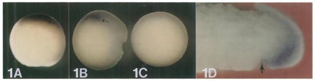
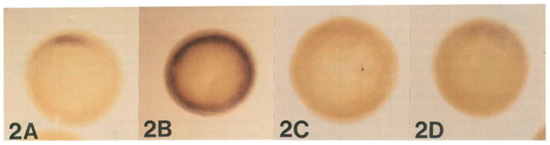
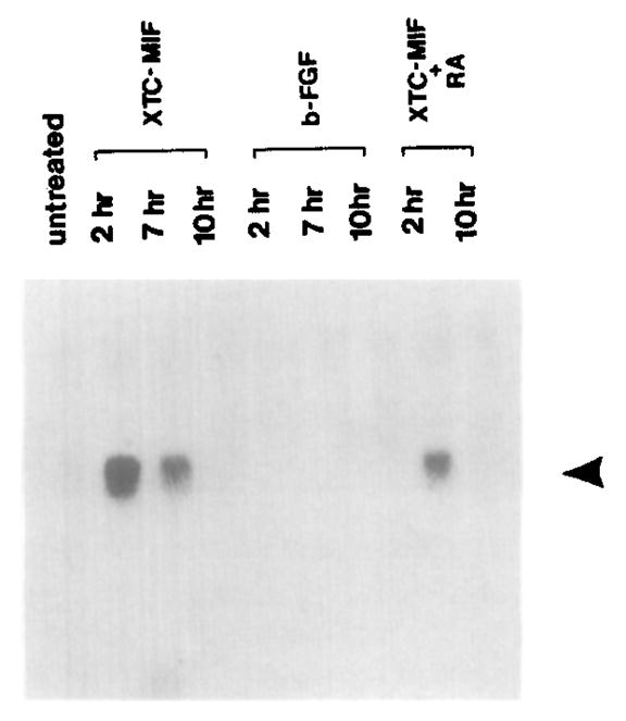
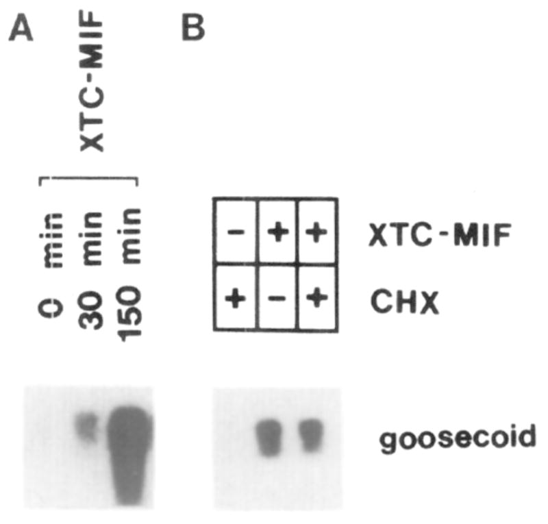
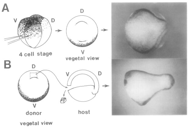
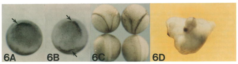
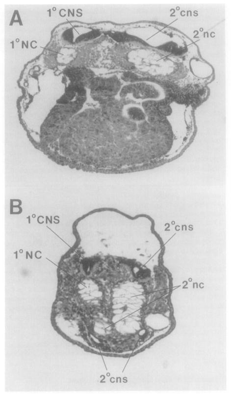
This study analyzes the function of the homeobox gene goosecoid in Xenopus development. First, we find that goosecoid mRNA distribution closely mimics the expected localization of organizer tissue in normal embryos as well as in those treated with LiCl and UV light. Second, goosecoid mRNA accumulation is induced by activin, even in the absence of protein synthesis. It is not affected by bFGF and is repressed by retinoic acid. Lastly, microinjection of goosecoid mRNA into the ventral side of Xenopus embryos, where goosecoid is normally absent, leads to the formation of an additional complete body axis, including head structures and abundant notochordal tissue. The results suggest that the goosecoid homeodomain protein plays a central role in executing Spemann's organizer phenomenon.


Figures

Figure 1
Figure 1. Whole-Mount In Situ Hybridization of goosecoid Expression in Stage 10½ Gastrulae
(A) antisense probe, vegetal view; (B) antisense probe, side view; (C) control sense probe, vegetal view; (D) sagittal section through the dorsal lip. Note that goosecoid hybridization is located in the deep layer of the upper lip of the blastopore. The fate of this region is to become head and notochord mesoderm. The arrow indicates the dorsal blastopore lip.
Figure 2
Figure 2. goosecoid Expression Follows the Expected Behavior of the Organizer Field in Experimentally Treated Embryos
These embryos have been rendered transparent with Murray’s solution (see Experimental Procedures). (A) untreated stage 10½ gastrula; note that the goosecoid field encompasses about 60° of arc of the marginal zone. (B) LiCI-treated gastrula (0.12 M for 40 min at the 32-cell stage); goosecoid expression has become radially symmetric. (C) UV-treated gastrula (60 s, see Experimental Procedures); note that goosecoid expression is abolished. (D) RA-treated embryo (10−6 M, starting at the 2-cell stage); goosecoid expression is inhibited but still weakly detectable.
Figure 3
Figure 3. goosecoid Expression in Animal Cap Fragments Treated with Peptide Growth Factors and RA
Note that goosecoid mRNA (arrowhead) is induced transiently by XTC-MIF (activin), that it is not induced at all by bFGF, and that XTC-MIF induction is inhibited by RA (compare lanes incubated with growth factor for 2 hr). XTC-MIF was used at 1:3 dilution. FGF was used at 200 ng/ml and RA at 10−6 M. Total RNA extracted from 20 animal caps was loaded in each lane and processed as described (Blumberg et al., 1991).
Figure 4
Figure 4. Time Course and Protein Synthesis Independence of the Induction of goosecoid mRNA by XTC-MIF in Animal Caps
(A) Groups of 20 animal caps isolated from stage 8 blastulae were incubated with XTC-MIF for the indicated times and analyzed by Northern blot. Note that goosecoid induction is detectable after 30 min. (B) Inhibition of protein synthesis by cycloheximide (CHX) does not prevent goosecoid expression. Animal caps were preincubated for 30 min with or without cycloheximide and then induced for 90 min with XTC-MIF, following the protocol of Rosa (1989).
Figure 5
Figure 5. Comparable Results Are Obtained by goosecoid mRNA Microinjection and by Dorsal Lip Transplantation
Experimental diagram and embryos resulting from (A) microinjection of goosecoid mRNA into the two ventral blastomeres (as close as possible to the first cleavage plane) and (B) a traditional Spemann organizer transplantation experiment. Note that the resulting embryos resemble each other and have extensive secondary neural tubes (dark lines) at the late neurula stage. In both embryos the two axes originate independently from each other in the posterior region, i.e., two sites of dorsal invagination were present during gastrulation.
Figure 6
Figure 6. Phenotypic Effect of Microinjecting goosecoid or Δgsc mRNA into the Two Ventral Blastomeres at the 4-Cell Stage
(A) Δgsc control, only one dorsal lip is present at early gastrula. (B) goosecoid mRNA injection, two dorsal lip–like structures are present (arrows). (C) Top, two embryos that received goosecoid mRNA; secondary neural axes are visible. The two bottom embryos were injected with Δgsc mRNA, and no secondary axis is present. (D) Twinned embryo produced by goosecoid mRNA injection; note that a complete head structure containing eyes, hatching gland, and cement gland has been induced.
Figure 7
Figure 7. The Additional Axis Induced by goosecoid mRNA Contains Massive Notochord Structures
Two transverse sections from the same animal are shown. Note that the notochord is much larger in the secondary axis (2°nc) than in the primary axis (1 °NC). Note that in more posterior regions (B), two small additional neural tubes (cns) have formed in close proximity to the ectopic notochordal tissue in the ventral side of the embryo.

Similar articles

Cited by

References

    1. Amaya E, Musci TJ, Kirschner MW. Expression of a dominant negative mutant of the FGF receptor disrupts mesoderm formation in Xenopus embryos. Cell. 1991;66:257–270. - PubMed
    1. Blumberg B, Wright CVE, De Robertis EM, Cho KWY. Organizer-specific homeobox genes in Xenopus laevis embryos. Science. 1991;253:194–196. - PubMed
    1. Busa WB, Gimlich RL. Lithium-induced teratogenesis in frog embryos prevented by a polyphosphoinositide cycle intermediate or a diacylglycerol analog. Dev Biol. 1989;132:315–324. - PubMed
    1. Cho KWY, De Robertis EM. Differential activation of Xenopus homeobox genes by mesoderm inducing growth factors and retinoic acid. Genes Dev. 1990;4:1910–1916. - PubMed
    1. Cho KWY, Goetz J, Wright CVE, Fritz A, Hardwicke J, De Robertis EM. Differential utilization of the same reading frame in a Xenopus homeobox gene encodes two proteins sharing the same DNA-binding specificity. EMBO J. 1988;7:2139–2149. - PMC - PubMed

Publication types