Vitamin A active metabolite, all-trans retinoic acid, induces spinal cord sensitization. I. Effects after oral administration
- PMID: 16847436
- PMCID: PMC1629405
- DOI: 10.1038/sj.bjp.0706829
Vitamin A active metabolite, all-trans retinoic acid, induces spinal cord sensitization. I. Effects after oral administration
Abstract
Background and purpose: Retinoic acid is an active metabolite of vitamin A involved in the modulation of the inflammatory and nociceptive responses. The aim of the present study was to analyze the properties of spinal cord neuronal responses of male Wistar rats treated with all-trans retinoic acid (ATRA) p.o. in the normal situation and under carrageenan-induced inflammation. We also studied the expression and distribution of cyclooxygenases (COX) in the spinal cord.
Experimental approach: Properties of spinal cord neurons were studied by means of the single motor unit technique. The expression of COX enzymes in the spinal cord was assessed by Western blot analysis and immunohistochemistry.
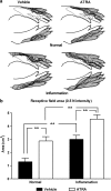
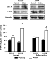
Key results: Intensity thresholds for mechanical and electrical stimulation (C-fibers) were significantly lower in animals treated with ATRA than vehicle, either in normal rats or in rats with inflammation. The size of cutaneous receptive fields was also larger in animals treated with ATRA in the normal and inflammatory conditions. The expression of COX-2 enzyme, but not COX-1, was significantly higher in animals treated with ATRA. COX-2 labeling was observed in dorsal horn cells and in ventral horn motoneurons.
Conclusions and implications: In conclusion, the oral treatment with ATRA in rats induces a sensitization-like effect on spinal cord neuronal responses similar to that observed in animals with inflammation and might explain the enhancement of allodynia and hyperalgesia observed in previously published behavioral experiments. The mechanism of action involves an over-expression of COX-2, but not COX-1, in dorsal and ventral horn areas of the lumbar spinal cord.
Figures


Similar articles
-
All-trans retinoic acid induces COX-2 and prostaglandin E2 synthesis in SH-SY5Y human neuroblastoma cells: involvement of retinoic acid receptors and extracellular-regulated kinase 1/2.J Neuroinflammation. 2007 Jan 4;4:1. doi: 10.1186/1742-2094-4-1. J Neuroinflammation. 2007. PMID: 17204142 Free PMC article.
-
Vitamin A active metabolite, all-trans retinoic acid, induces spinal cord sensitization. II. Effects after intrathecal administration.Br J Pharmacol. 2006 Sep;149(1):65-72. doi: 10.1038/sj.bjp.0706826. Epub 2006 Jul 17. Br J Pharmacol. 2006. PMID: 16847438 Free PMC article.
-
The oral administration of retinoic acid enhances nociceptive withdrawal reflexes in rats with soft-tissue inflammation.Inflamm Res. 2004 Jul;53(7):297-303. doi: 10.1007/s00011-004-1261-5. Epub 2004 Jun 25. Inflamm Res. 2004. PMID: 15241564
-
Prostanoids synthesized by cyclo-oxygenase isoforms in rat spinal cord and their contribution to the development of neuronal hyperexcitability.Br J Pharmacol. 1997 Dec;122(8):1593-604. doi: 10.1038/sj.bjp.0701548. Br J Pharmacol. 1997. PMID: 9422803 Free PMC article.
-
Enhancement of the analgesic activity of paracetamol and nitroparacetamol by the oral administration of all-trans retinoic acid.Neuropharmacology. 2006 Sep;51(4):858-65. doi: 10.1016/j.neuropharm.2006.05.033. Epub 2006 Jul 25. Neuropharmacology. 2006. PMID: 16870215
Cited by
-
Nitroparacetamol (NCX-701) and pain: first in a series of novel analgesics.CNS Drug Rev. 2007 Fall;13(3):279-95. doi: 10.1111/j.1527-3458.2007.00016.x. CNS Drug Rev. 2007. PMID: 17894645 Free PMC article. Review.
-
Anti-inflammatory and anti-hyperalgesic effect of all-trans retinoic acid in carrageenan-induced paw edema in Wistar rats: involvement of peroxisome proliferator-activated receptor-β/δ receptors.Indian J Pharmacol. 2013 May-Jun;45(3):278-82. doi: 10.4103/0253-7613.111944. Indian J Pharmacol. 2013. PMID: 23833373 Free PMC article.
-
RAR/RXR and PPAR/RXR Signaling in Spinal Cord Injury.PPAR Res. 2007;2007:29275. doi: 10.1155/2007/29275. PPAR Res. 2007. PMID: 18060014 Free PMC article.
-
All-trans retinoic acid induces COX-2 and prostaglandin E2 synthesis in SH-SY5Y human neuroblastoma cells: involvement of retinoic acid receptors and extracellular-regulated kinase 1/2.J Neuroinflammation. 2007 Jan 4;4:1. doi: 10.1186/1742-2094-4-1. J Neuroinflammation. 2007. PMID: 17204142 Free PMC article.
References
-
- Bradford MM. A rapid and sensitive method for the quantitation of microgram quantities of protein utilizing the principle of protein-dye binding. Anal Biochem. 1976;72:248–254. - PubMed
-
- Cocco S, Diaz G, Stancampiano R, Diana A, Carta M, Curreli R, et al. Vitamin a deficiency produces spatial learning and memory impairment in rats. Neuroscience. 2002;115:475–482. - PubMed
-
- Devaux Y, Seguin C, Grosjean S, De Talance N, Schwartz M, Burlet A, et al. Retinoic acid and lipopolysaccharide act synergistically to increase prostanoid concentrations in rats in vivo. J Nutr. 2001;131:2628–2635. - PubMed
-
- Duester G, Mic FA, Molotkov A. Cytosolic retinoid dehydrogenases govern ubiquitous metabolism of retinol to retinaldehyde followed by tissue specific metabolism to retinoic acid. Chemico-Biol Interact. 2003;143:201–210. - PubMed
-
- Dubner R, Ruda MA. Activity-dependent neuronal plasticity following tissue injury and inflammation. Trends Neurosci. 1992;15:96–103. - PubMed
Publication types
MeSH terms
Substances
LinkOut - more resources
Full Text Sources
Other Literature Sources
Research Materials

