

Determining the topology of integral membrane peptides using EPR spectroscopy
- PMID: 16848493
- PMCID: PMC2533427
- DOI: 10.1021/ja0622204
Determining the topology of integral membrane peptides using EPR spectroscopy
Abstract
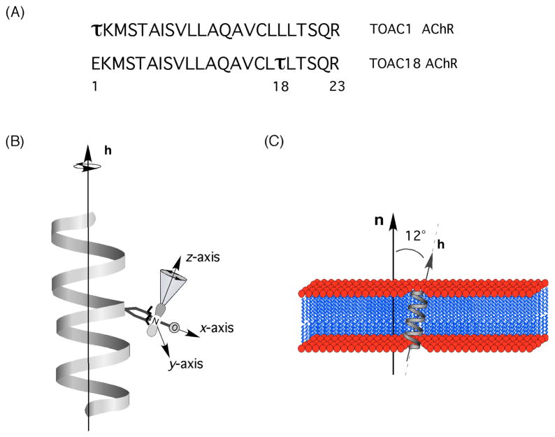
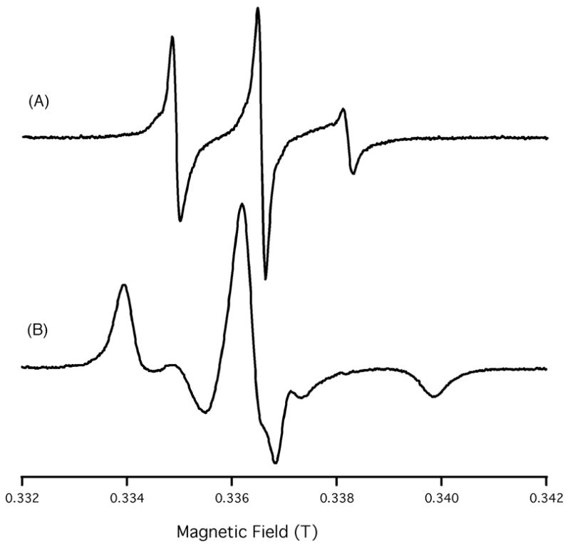
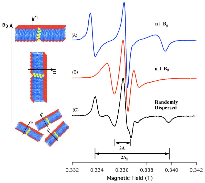
This paper reports on the development of a new structural biology technique for determining the membrane topology of an integral membrane protein inserted into magnetically aligned phospholipid bilayers (bicelles) using EPR spectroscopy. The nitroxide spin probe, 2,2,6,6-tetramethylpiperidine-1-oxyl-4-amino-4-carboxylic acid (TOAC), was attached to the pore-lining transmembrane domain (M2delta) of the nicotinic acetylcholine receptor (AChR) and incorporated into a bicelle. The corresponding EPR spectra revealed hyperfine splittings that were highly dependent on the macroscopic orientation of the bicelles with respect to the static magnetic field. The helical tilt of the peptide can be easily calculated using the hyperfine splittings gleaned from the orientational dependent EPR spectra. A helical tilt of 14 degrees was calculated for the M2delta peptide with respect to the bilayer normal of the membrane, which agrees well with previous 15N solid-state NMR studies. The helical tilt of the peptide was verified by simulating the corresponding EPR spectra using the standardized MOMD approach. This new method is advantageous because: (1) bicelle samples are easy to prepare, (2) the helical tilt can be directly calculated from the orientational-dependent hyperfine splitting in the EPR spectra, and (3) EPR spectroscopy is approximately 1000-fold more sensitive than 15N solid-state NMR spectroscopy; thus, the helical tilt of an integral membrane peptide can be determined with only 100 microg of peptide. The helical tilt can be determined more accurately by placing TOAC spin labels at several positions with this technique.
Figures

Similar articles
-
Probing topology and dynamics of the second transmembrane domain (M2δ) of the acetyl choline receptor using magnetically aligned lipid bilayers (bicelles) and EPR spectroscopy.Chem Phys Lipids. 2017 Aug;206:9-15. doi: 10.1016/j.chemphyslip.2017.05.010. Epub 2017 May 29. Chem Phys Lipids. 2017. PMID: 28571787 Free PMC article.
-
Distance measurements on a dual-labeled TOAC AChR M2δ peptide in mechanically aligned DMPC bilayers via dipolar broadening CW-EPR spectroscopy.J Phys Chem B. 2012 Mar 29;116(12):3866-73. doi: 10.1021/jp212272d. Epub 2012 Mar 19. J Phys Chem B. 2012. PMID: 22379959 Free PMC article.
-
Determining the helical tilt angle and dynamic properties of the transmembrane domains of pinholin S2168 using mechanical alignment EPR spectroscopy.Biochim Biophys Acta Biomembr. 2023 Jun;1865(5):184154. doi: 10.1016/j.bbamem.2023.184154. Epub 2023 Apr 5. Biochim Biophys Acta Biomembr. 2023. PMID: 37023970
-
15N-Labeled 4-oxo-2,2,6,6-tetramethyl-piperidine-1-oxyl.2008 Apr 30 [updated 2008 Jun 9]. In: Molecular Imaging and Contrast Agent Database (MICAD) [Internet]. Bethesda (MD): National Center for Biotechnology Information (US); 2004–2013. 2008 Apr 30 [updated 2008 Jun 9]. In: Molecular Imaging and Contrast Agent Database (MICAD) [Internet]. Bethesda (MD): National Center for Biotechnology Information (US); 2004–2013. PMID: 20641553 Free Books & Documents. Review.
-
Biophysical investigations of membrane perturbations by polypeptides using solid-state NMR spectroscopy (review).Mol Membr Biol. 2000 Jul-Sep;17(3):135-42. doi: 10.1080/09687680050197365. Mol Membr Biol. 2000. PMID: 11128972 Review.
Cited by
-
Biophysical approaches for exploring lipopeptide-lipid interactions.Biochimie. 2020 Mar;170:173-202. doi: 10.1016/j.biochi.2020.01.009. Epub 2020 Jan 21. Biochimie. 2020. PMID: 31978418 Free PMC article. Review.
-
Determining the helical tilt of membrane peptides using electron paramagnetic resonance spectroscopy.J Magn Reson. 2009 May;198(1):1-7. doi: 10.1016/j.jmr.2008.12.007. Epub 2008 Dec 14. J Magn Reson. 2009. PMID: 19254856 Free PMC article.
-
Probing topology and dynamics of the second transmembrane domain (M2δ) of the acetyl choline receptor using magnetically aligned lipid bilayers (bicelles) and EPR spectroscopy.Chem Phys Lipids. 2017 Aug;206:9-15. doi: 10.1016/j.chemphyslip.2017.05.010. Epub 2017 May 29. Chem Phys Lipids. 2017. PMID: 28571787 Free PMC article.
-
The rational search for selective anticancer derivatives of the peptide Trichogin GA IV: a multi-technique biophysical approach.Sci Rep. 2016 Apr 4;6:24000. doi: 10.1038/srep24000. Sci Rep. 2016. PMID: 27039838 Free PMC article.
-
Pulse EPR Measurements of Intramolecular Distances in a TOPP-Labeled Transmembrane Peptide in Lipids.Biophys J. 2016 Dec 6;111(11):2345-2348. doi: 10.1016/j.bpj.2016.10.022. Epub 2016 Nov 9. Biophys J. 2016. PMID: 27836102 Free PMC article.
References
Publication types
MeSH terms
Substances
Grants and funding
LinkOut - more resources
Full Text Sources
