. 2006 Jul;2(7):e71.
doi: 10.1371/journal.ppat.0020071.

Macrophage pro-inflammatory response to Francisella novicida infection is regulated by SHIP

Affiliations

Macrophage pro-inflammatory response to Francisella novicida infection is regulated by SHIP

Kishore V L Parsa et al. PLoS Pathog. 2006 Jul.

Abstract

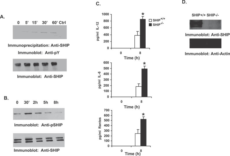
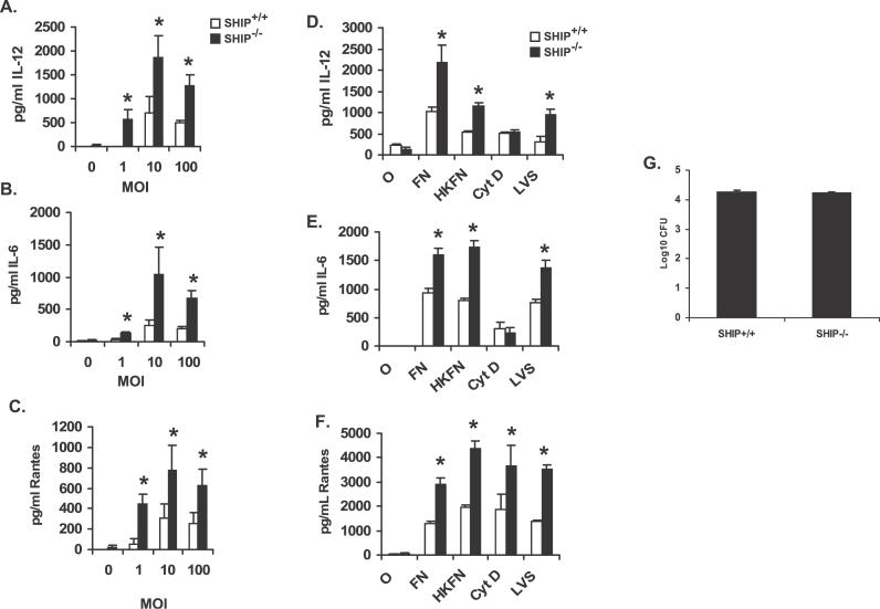
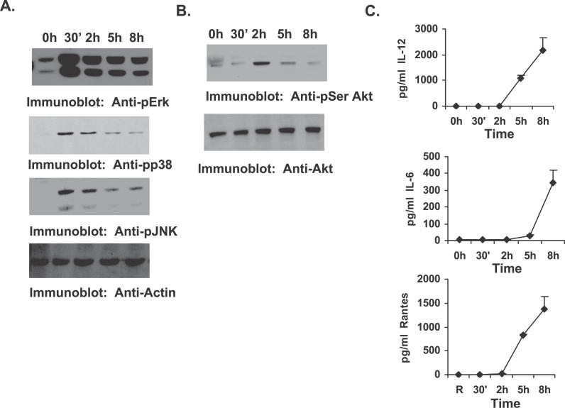
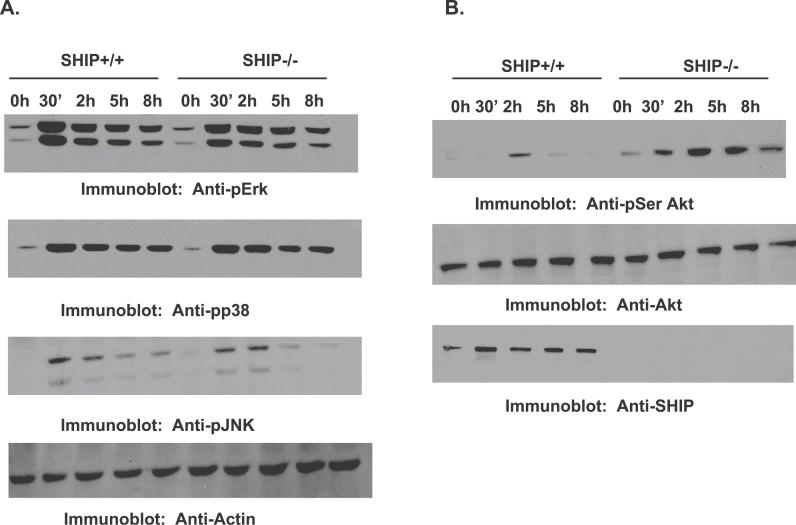
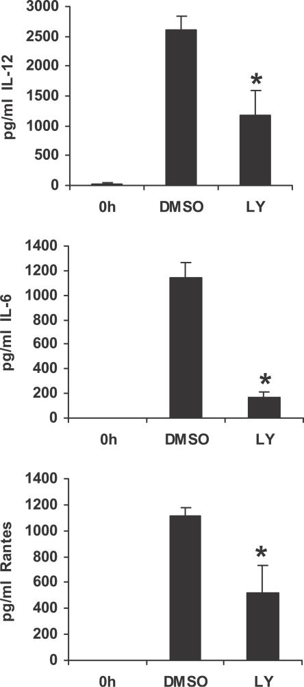
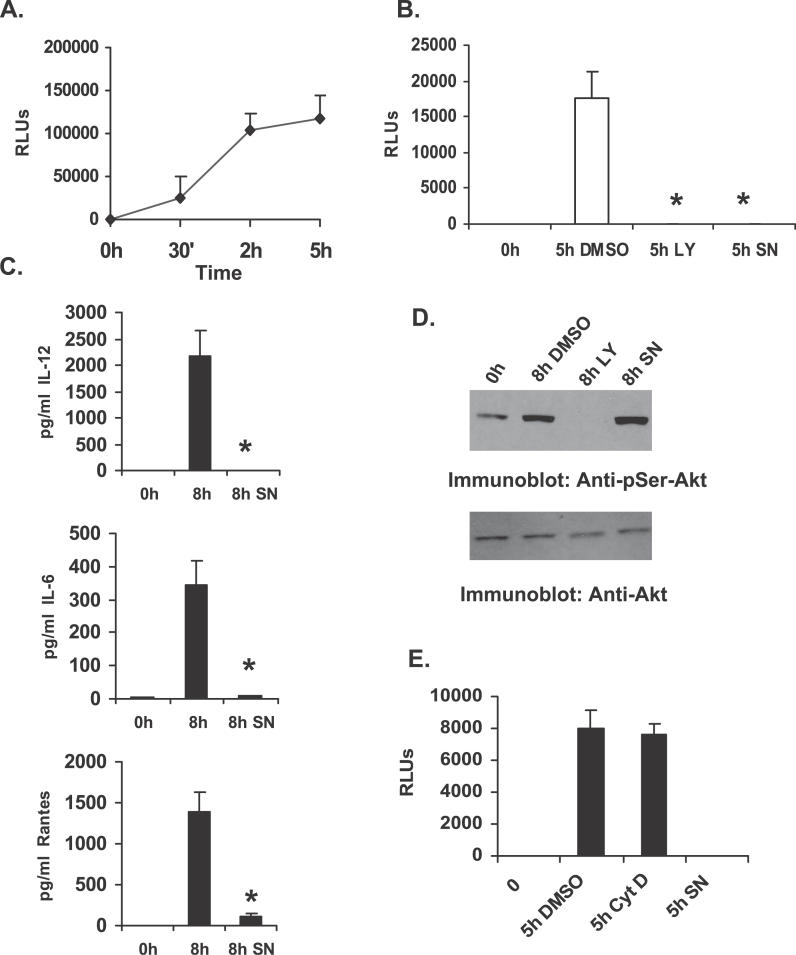
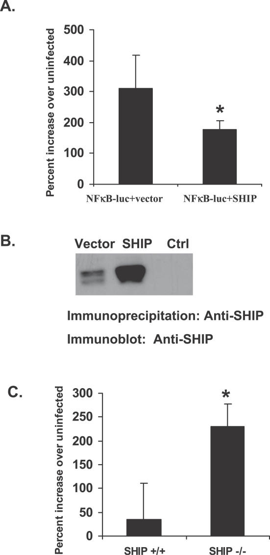
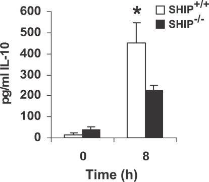
Francisella tularensis, a Gram-negative facultative intracellular pathogen infecting principally macrophages and monocytes, is the etiological agent of tularemia. Macrophage responses to F. tularensis infection include the production of pro-inflammatory cytokines such as interleukin (IL)-12, which is critical for immunity against infection. Molecular mechanisms regulating production of these inflammatory mediators are poorly understood. Herein we report that the SH2 domain-containing inositol phosphatase (SHIP) is phosphorylated upon infection of primary murine macrophages with the genetically related F. novicida, and negatively regulates F. novicida-induced cytokine production. Analyses of the molecular details revealed that in addition to activating the MAP kinases, F. novicida infection also activated the phosphatidylinositol 3-kinase (PI3K)/Akt pathway in these cells. Interestingly, SHIP-deficient macrophages displayed enhanced Akt activation upon F. novicida infection, suggesting elevated PI3K-dependent activation pathways in absence of SHIP. Inhibition of PI3K/Akt resulted in suppression of F. novicida-induced cytokine production through the inhibition of NFkappaB. Consistently, macrophages lacking SHIP displayed enhanced NFkappaB-driven gene transcription, whereas overexpression of SHIP led to decreased NFkappaB activation. Thus, we propose that SHIP negatively regulates F. novicida-induced inflammatory cytokine response by antagonizing the PI3K/Akt pathway and suppressing NFkappaB-mediated gene transcription. A detailed analysis of phosphoinositide signaling may provide valuable clues for better understanding the pathogenesis of tularemia.


Conflict of interest statement

Competing interests. The authors have declared that no competing interests exist.

Figures

Figure 1
Figure 1. SHIP Down-Regulates Macrophage Pro-Inflammatory Response to F. novicida Infection
(A) RAW 264.7 murine macrophage cells were infected with F. novicida (MOI = 100). SHIP immunoprecipitates from uninfected and infected cells were analyzed by Western blotting with anti-phosphotyrosine antibody (Anti-pY; upper panel). The lower panel is a reprobe of the same membrane with anti-SHIP antibody. The lane marked “Ctrl” represents immunoprecipitate with control antibody. (B) BMMs were infected with F. novicida for the times shown in the figure. Protein-matched lysates were separated by SDS/PAGE and analyzed by Western blotting with antibodies specific for phosphorylated SHIP (Anti-pSHIP; upper panel). The lower panel is a reprobe with SHIP antibody (Anti-SHIP). (C) BMMs from SHIP+/+ and SHIP−/− littermate mice were infected for 8 h with F. novicida. Cell supernatants from uninfected and infected cells were analyzed by ELISA for IL-12, IL-6, and RANTES. The graph represents mean and standard deviation (SD) of values obtained from three independent experiments. Data were analyzed by paired Student t test. An asterisk (*) indicates p-value < 0.05. (D) Protein-matched lysates from SHIP+/+ and SHIP−/− BMMs were analyzed by Western blotting with SHIP antibody (upper panel). The same membrane was reprobed with actin antibody (Anti-Actin; lower panel).
Figure 2
Figure 2. Analysis of SHIP Influence on Macrophage Inflammatory Response to F. novicida Infection
(A–C) BMMs from SHIP+/+ and SHIP−/− littermate mice were infected at different MOI (indicated in figure) of F. novicida for 8 h. Cytokine levels were measured in uninfected samples (0 h). Cell supernatants from uninfected and infected cells were assayed for IL-12, IL-6, and RANTES by ELISA. (D–F) BMMs from SHIP+/+ and SHIP−/− littermate mice were infected with 100 MOI of live or heat-killed F. novicida (FN and HKFN, respectively) or with F. tularensis LVS (LVS) for 8 h. Cyt D represents samples that were treated with cytochalasin D (5 μg/ml) before infection with 100 MOI of live F. novicida. Cytokine levels were measured in uninfected samples (0 h). Cell supernatants from uninfected and infected cells were analyzed by ELISA for IL-12, IL-6, and RANTES. (G) BMMs from SHIP +/+ and SHIP−/− littermate mice were infected with 100 MOI of F. novicida for 2 h, then treated with gentamicin (50 μg/ml), lysed in 0.1% SDS and appropriate dilutions of the lysates were plated on Chocolate II agar plates for enumeration of CFUs. All graphs represents mean and SD of values obtained from three independent experiments. Data were analyzed by paired Student t test. An asterisk (*) indicates p-value < 0.05.
Figure 3
Figure 3. PI3K/Akt Pathway Is Activated upon F. novicida Infection
BMMs were infected with F. novicida for the times shown in the figure. (A) Protein-matched lysates were analyzed by Western blotting with phospho-specific antibodies to Erk (Anti-pErk), p38 (Anti-pp38), and JNK (Anti-pJNK). The membranes were reprobed with actin antibody (Anti-Actin) to ensure equal loading of protein in all lanes. (B) Protein-matched lysates were analyzed by Western blotting with phospho-specific antibody to Akt (Anti-pSer Akt; upper panel). The lower panel is a reprobe of the same membrane with total Akt antibody (Anti-Akt). (C) Cell supernatants from the same experiments were assayed by ELISA for IL-12, IL-6, and RANTES. The graph represents mean and SD of values obtained from three independent experiments.
Figure 4
Figure 4. SHIP Negatively Regulates F. novicida–Induced Akt Activation
BMMs from SHIP+/+ and SHIP−/− littermate mice were infected for the times indicated in the figure. (A) Protein-matched lysates were probed with antibodies specific for phosphorylated Erk (Anti-pErk), p38 (Anti-pp38), and JNK (Anti-pJNK). All membranes were reprobed with actin antibody (Anti-Actin). (B) Protein-matched lysates were probed with phospho-specific antibody for Akt (Anti-pSer Akt; upper panel). The membranes were reprobed with total Akt antibody (Anti-Akt; middle panel), and with SHIP antibody (Anti-SHIP; lower panel). These data are representative of three independent experiments.
Figure 5
Figure 5. PI3K Activation Promotes F. novicida–Induced Macrophage Inflammatory Response
BMMs were pretreated for 30 min with PI3K inhibitor LY294002 (LY; 20 μM), or with vehicle control (DMSO), and subsequently infected with F. novicida. Cell supernatants were harvested 8 h post infection and assayed by ELISA for IL-12, IL-6, and RANTES. Cytokine levels were measured in uninfected samples (0 h). The graphs represent mean and SD of values obtained from three independent experiments. Data were analyzed by paired Student t test. An asterisk (*) indicates p-value < 0.05.
Figure 6
Figure 6. PI3K Promotes F. novicida–Induced Inflammatory Response through Its Influence on NFκB
(A) RAW 264.7 cells transfected with plasmid encoding the luciferase gene driven by an NFκB binding element (NFκB-luc). Transfectants were infected with F. novicida for varying times and analyzed for the luciferase activity as a measure of NFκB activation. RLUs, relative light units. (B) RAW 264.7 cells transfected with NFκB-luc plasmid were pretreated with either a PI3K inhibitor LY294002 (LY), a NFκB inhibitor SN50 (SN; 75 μg/ml), or with vehicle control (DMSO), and subsequently infected with F. novicida. At 5 h post infection, cells were lysed and assayed for luciferase activity. RLUs, relative light units. (C) BMMs were pretreated for 30 min with either DMSO (middle bar) or the NFκB inhibitor SN50 (SN), and subsequently infected with F. novicida. Cell supernatants were harvested 8 h post infection and assayed for IL-12, IL-6, and RANTES by ELISA. Cytokine levels were measured in uninfected samples (0 h). The graph represents mean and SD of values obtained from three experiments. Data were analyzed by paired Student t test. An asterisk (*) indicates p-value < 0.05. (D) Protein-matched lysates from BMMs pretreated with either DMSO or with inhibitors of PI3K (LY) or NFκB (SN) and infected with F. novicida were analyzed by Western blotting with phospho-specific antibody to Akt (Anti-pSer-Akt; upper panel). The lower panel is a reprobe of the same membrane with Akt antibody (Anti-Akt). (E) RAW 264.7 cells transfected with NFκB-luc plasmid were pretreated with either cytochalasin D (Cyt D), SN50 (SN), or with vehicle control (DMSO), and subsequently infected with F. novicida. At 5 h post infection, cells were lysed and assayed for luciferase activity. The data are representative of three independent experiments.
Figure 7
Figure 7. SHIP Negatively Regulates F. novicida–Induced NFκB Activation
(A) RAW 264.7 cells were transfected with NFκB-luc plasmid along with either empty vector or plasmid encoding SHIP. Transfectants were infected with F. novicida and assayed for luciferase activity 5 h post infection. Data were analyzed by Student t test. An asterisk (*) indicates p-value < 0.05. (B) SHIP immunoprecipitates from the RAW 264.7 transfectants described in Figure 7A were analyzed by Western blotting for overexpression of SHIP. Ctrl indicates immunoprecipitation with normal rabbit serum. (C) SHIP+/+ and SHIP−/− BMMs were transfected with NFκB-luc plasmid. Transfectants were infected with F. novicida and assayed for luciferase activity. Graphs represent values from three independent experiments. Data were analyzed by Student t test. An asterisk (*) indicates p-value < 0.05.
Figure 8
Figure 8. SHIP Influence on Macrophage IL-10 Response to F. novicida
BMMs from SHIP+/+ and SHIP−/− littermate mice were infected for 8 h with F. novicida. Cell supernatants from uninfected and infected cells were analyzed by ELISA for IL-10. The graph represents mean and SD of values obtained from three independent experiments. Data were analyzed by paired Student t test. An asterisk (*) indicates p-value < 0.05.

Similar articles

Cited by

References

    1. Golovliov I, Baranov V, Krocova Z, Kovarova H, Sjostedt A. An attenuated strain of the facultative intracellular bacterium Francisella tularensis can escape the phagosome of monocytic cells. Infect Immun. 2003;71:5940–5950. - PMC - PubMed
    1. Clemens DL, Lee BY, Horwitz MA. Virulent and avirulent strains of Francisella tularensis prevent acidification and maturation of their phagosomes and escape into the cytoplasm in human macrophages. Infect Immun. 2004;72:3204–3217. - PMC - PubMed
    1. Santic M, Molmeret M, Klose KE, Jones S, Kwaik YA. The Francisella tularensis pathogenicity island protein IglC and its regulator MglA are essential for modulating phagosome biogenesis and subsequent bacterial escape into the cytoplasm. Cell Microbiol. 2005;7:969–979. - PubMed
    1. Nano FE, Zhang N, Cowley SC, Klose KE, Cheung KK, et al. A Francisella tularensis pathogenicity island required for intramacrophage growth. J Bacteriol. 2004;186:6430–6436. - PMC - PubMed
    1. Lindgren H, Golovliov I, Baranov V, Ernst RK, Telepnev M, et al. Factors affecting the escape of Francisella tularensis from the phagolysosome. J Med Microbiol. 2004;53:953–958. - PubMed

Publication types

MeSH terms