

Review
doi: 10.1098/rsif.2006.0115.
Targeting protein-protein interactions by rational design: mimicry of protein surfaces
Affiliations
- PMID: 16849232
- PMCID: PMC1578744
- DOI: 10.1098/rsif.2006.0115
Item in Clipboard
Review
Targeting protein-protein interactions by rational design: mimicry of protein surfaces
J R Soc Interface.
.
Abstract
Protein-protein interactions play key roles in a range of biological processes, and are therefore important targets for the design of novel therapeutics. Unlike in the design of enzyme active site inhibitors, the disruption of protein-protein interactions is far more challenging, due to such factors as the large interfacial areas involved and the relatively flat and featureless topologies of these surfaces. Nevertheless, in spite of such challenges, there has been considerable progress in recent years. In this review, we discuss this progress in the context of mimicry of protein surfaces: targeting protein-protein interactions by rational design.
Figures
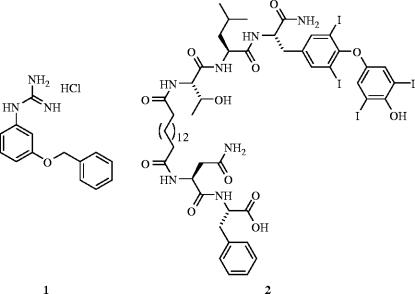
Anti-HIV-1 agents: (1) gp120–CD4 inhibitor (IC50=131 μM); (2) HIV-1 protease dimerization inhibitor (Ki=71 nM).
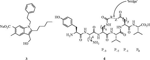
Antagonists of PDZ domains: (3) MAGI3–PTEN inhibitor; (4) PSD-95–NMDA inhibitor.
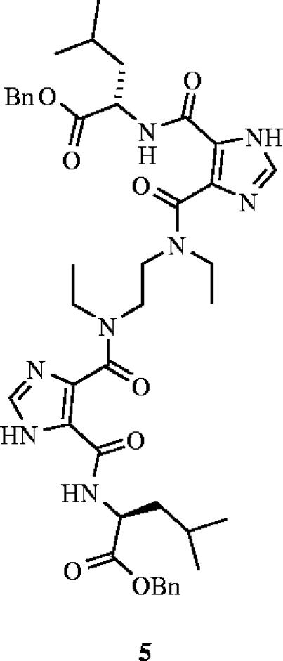
A novel bis-imidazole inhibitor (5) that prevents hepatitis C virus (HCV) cell entry through the disruption of the HCV-E2–CD81 interaction (EC50=38 μM).
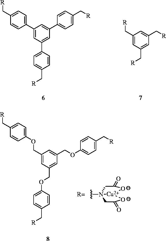
Cu2+-iminodiacetate derivatives that target the surface of carbonic anhydrase: 6, Ka=2.99×10⁵ M−1; 7, Ka=7.5×10⁴ M−1; 8, Ka=3.3×10⁴ M−1.
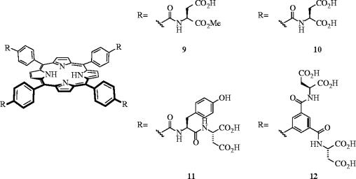
Synthetic porphyrins that recognize the surface of cytochrome c: Kd's=860 nM (9), 160 nM (10), 20 nM (11) and 0.67 nM (12).
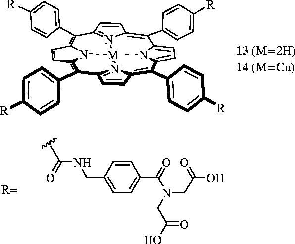
Synthetic porphyrins that denature cytochrome c.
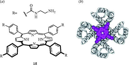
(a) A cationic porphyrin (15) that binds the human Kv1.3 channel with a Ki of 20 nM; (b) overlay of the likely interaction of porphyrin 15 with the human Kv1.3 channel. Part (b) is reproduced with permission from Gradl et al. (2003).
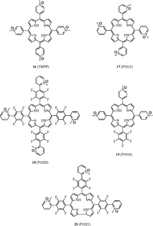
Cationic porphyrins as anti-FGF and anti-VEGF agents.
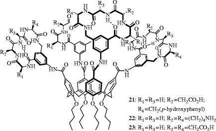
Calix[4]arene derivatives that inhibit the (i) PDGF–PDGFR (21: IC50=250 nM) and (ii) cyt c–cyt c peroxidase (23: Kd=30 nM) interactions.
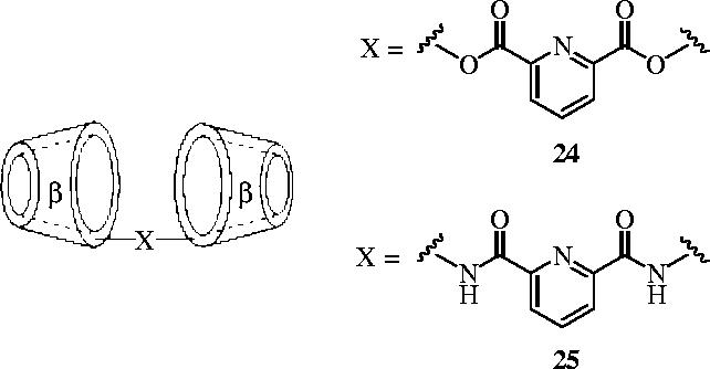
β-Cyclodextrin dimers that inhibit the self-assembly of dimeric citrate synthase (CS) and tetrameric lactate dehydrogenase (LDH).
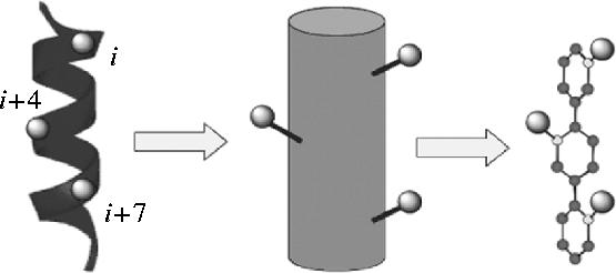
Analogy between the α-helix and the terphenyl scaffold.
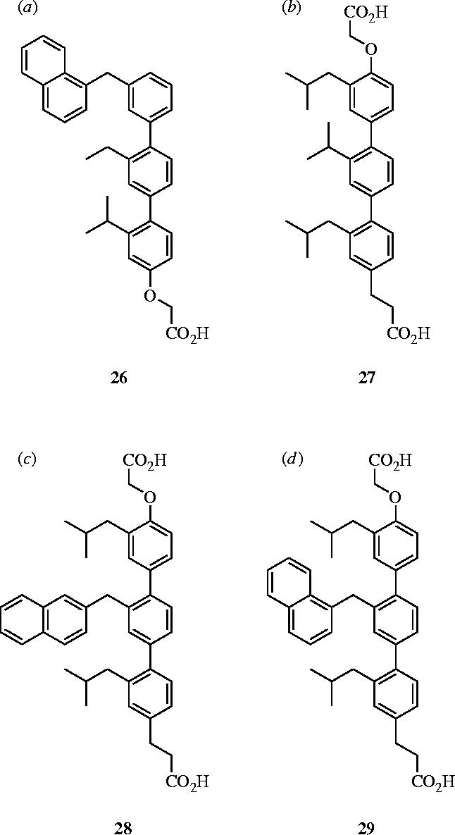
Terphenyl-based α-helical mimetics: (a) antagonist of calmodulin, 26: IC50=9 nM; (b) inhibitor of hexameric gp41 self-assembly, 27: IC50=13.2 μg ml−1; (c) antagonist of HDM2, 28: Ki=182 nM; (d) antagonist of Bcl-xL, 29: Ki=114 nM.
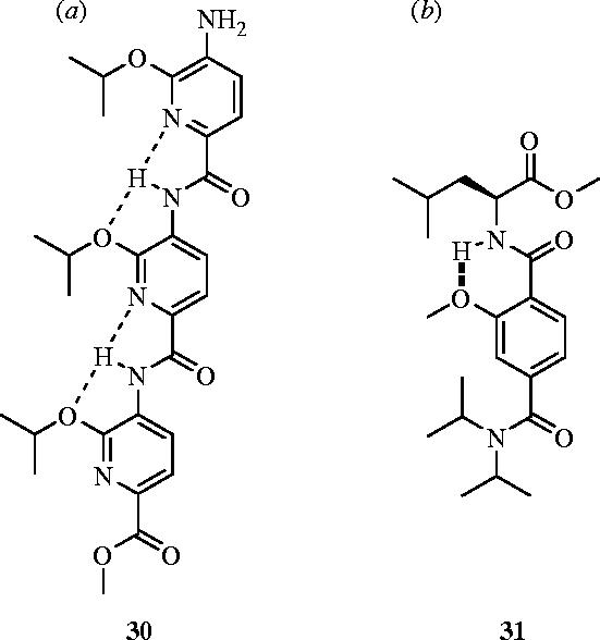
Alternative α-helical mimetics that antagonize Bcl-xL: (a) trispyridylamide 30 (Ki=2.3 μM) and (b) terephthalamide 31 (IC50=35.0 μM).
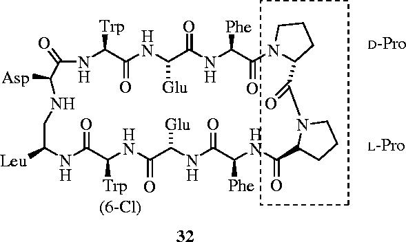
Inhibition of the p53–HDM2 interaction through mimicry of the α-helical region of p53 with β-hairpin 32 (IC50=140 nM).
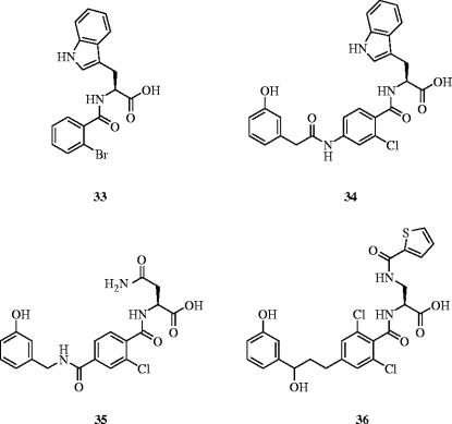
LFA-1–ICAM-1 inhibitors: IC50s=1.4 μM (33), 47 nM (34), 3.7 nM (35) and 1.4 nM (36).
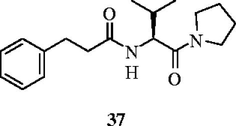
A small molecule, β-turn mimetic that disrupts the interaction between IL-1RI and MyD88.
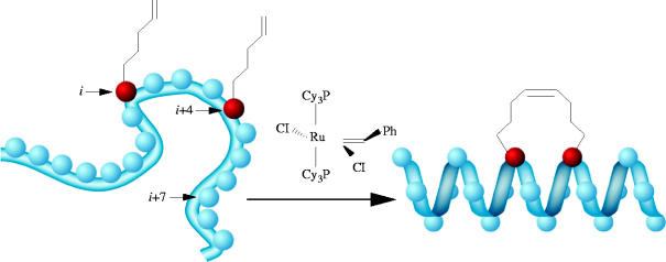
Ring-closing metathesis of olefin-modified i and (i+4) residues to generate hydrocarbon-stapled helices. Reproduced with permission from Walensky et al. (2004).
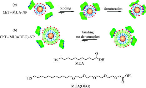
Ligands used for Au and CdSe nanoparticles, and schematic depiction of ChT-nanoparticle leading to: (a) two-step denaturation of ChT and (b) reversible binding with retention of ChT structure. Reproduced and edited with permission from Hong et al. (2004).
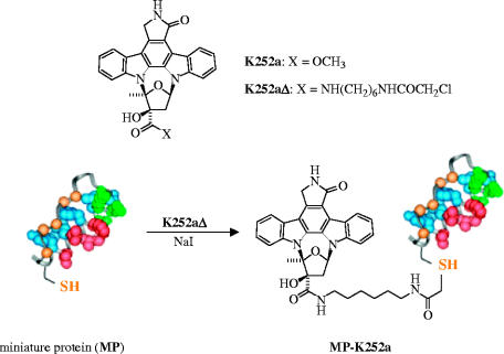
Increasing the kinase specificity of the kinase inhibitor K252a through conjugation of K252a to a miniature protein that features the cAMP-dependent protein kinase (PKA) recognition epitope. Reproduced and edited with permission from Schneider et al. (2005).
References
-
- Adler M, Carter P, Lazarus R.A, Wagner G. Cysteine pairing in the glycoprotein IIbIIIa antagonist kistrin using NMR, chemical analysis, and structure calculations. Biochemistry. 1993;32:282–289. doi:10.1021/bi00052a036 - DOI - PubMed
-
- Anderson K.V. Toll signaling pathways in the innate immune response. Curr. Opin. Immunol. 2000;12:13–19. doi:10.1016/S0952-7915(99)00045-X - DOI - PubMed
-
- Anderson M.E, Yakovleva T, Hu Y, Siahaan T.J. Inhibition of ICAM-1/LFA-1-mediated heterotypic T-cell adhesion to epithelial cells: design of ICAM-1 cyclic peptides. Bioorg. Med. Chem. Lett. 2004;14:1399–1402. doi:10.1016/j.bmcl.2003.09.100 - DOI - PubMed
-
- Arkin M.R, Wells J.A. Small-molecule inhibitors of protein–protein interactions: progressing towards the dream. Nat. Rev. Drug Discov. 2004;3:301–317. doi:10.1038/nrd1343 - DOI - PubMed
-
- Aviezer D, Cotton S, David M, Segev A, Khaselev N, Galili N, Gross Z, Yayon A. Porphyrin analogues as novel antagonists of fibroblast growth factor and vascular endothelial growth factor receptor binding that inhibit endothelial cell proliferation, tumor progression, and metastasis. Cancer Res. 2000;60:2973–2980. - PubMed
Publication types
MeSH terms
Substances
LinkOut - more resources
Full Text Sources
Other Literature Sources