. 2006 Jul 25;103(30):11411-6.
doi: 10.1073/pnas.0601631103. Epub 2006 Jul 18.

Barttin modulates trafficking and function of ClC-K channels

Affiliations

Barttin modulates trafficking and function of ClC-K channels

Ute Scholl et al. Proc Natl Acad Sci U S A. .

Abstract

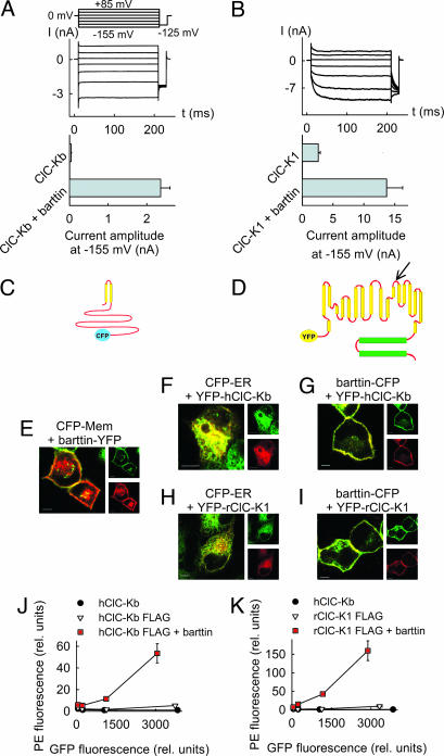
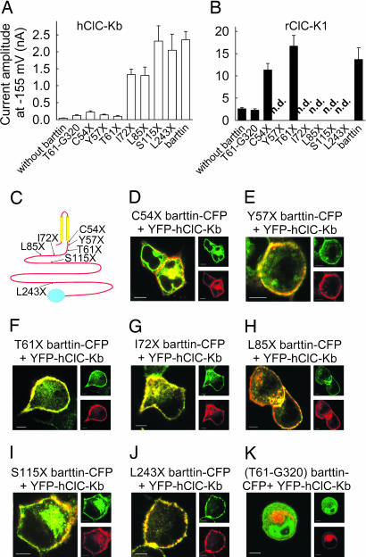
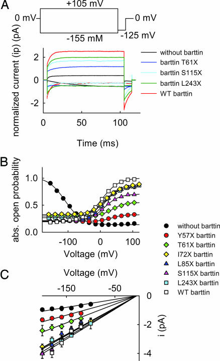
Barttin is an accessory subunit of a subgroup of ClC-type chloride channels expressed in renal and inner ear epithelia. In this study, we examined the effects of barttin on two ClC-K channel isoforms, rat ClC-K1 and human ClC-Kb, using heterologous expression, patch clamping, confocal imaging, and flow cytometry. In the absence of barttin, only a small percentage of rClC-K1 and hClC-Kb channels are inserted into the plasma membrane. Coexpression of barttin enhances surface membrane insertion and furthermore modifies permeation and gating of ClC-K channels. hClC-Kb channels are nonfunctional without barttin and require the coexpressed accessory subunit to become anion conducting. In contrast, rClC-K1 channels are active without barttin, but at the cost of reduced unitary conductance as well as altered voltage dependence of activation. We mapped the separate functions of barttin to structural domains by a deletion analysis. Whereas the transmembrane core is necessary and sufficient to promote ClC-K channel exit from the endoplasmic reticulum, a short cytoplasmic segment following the second transmembrane helix modifies the unitary conductance. The entire cytoplasmic carboxyl terminus affects the open probability of ClC-K channels. The multiple functions of barttin might be necessary for a tight adjustment of epithelial Cl(-) conductances to ensure a precise regulation of body salt content and endocochlear potential.


Conflict of interest statement

Conflict of interest statement: No conflicts declared.

Figures

Fig. 1.
Fig. 1.
Barttin promotes surface membrane insertion of ClC-K channels. (A and B) Representative currents from cells coexpressing hClC-Kb (A) or rClC-K1 (B) with barttin and mean isochronal current amplitudes determined 2 ms after a voltage step to −155 mV (n > 8) from cells expressing channels with and without barttin. (C and D) Topology model of barttin (C) and ClC-K channels (D). The position of the FLAG epitope used for FACS analysis is marked by an arrow. (E) Confocal images of live MDCK cells coexpressing a barttin-YFP fusion protein and a fluorescent marker for the membrane surface (CFP-Mem; Clontech). CFP is shown in green, and YFP is shown in red. This color code results in an orange coloring of regions where both proteins overlap. (F and H) Confocal images of live MDCK cells coexpressing a fluorescent marker for the endoplasmic reticulum (CFP-ER; Clontech) together with YFP-hClC-Kb (F) and YFP-rClC-K1 (H). (G and I) Confocal images of live MDCK cells coexpressing a barttin-CFP fusion protein and YFP-hClC-Kb (G) or YFP-rClC-K1 (I). (Scale bars: 5 μm.) (J and K) Surface expression of hClC-Kb (J) and rClC-K1 (K) with (■) and without (▿) barttin in transiently transfected tsA201 cells determined by flow cytometry. Shown are plots of fluorescence levels of an anti-Flag antibody versus the GFP fluorescence. Data were binned in four groups where means and SEMs were obtained. GFP-hClC-Kb without FLAG (●) was used as a control. For both channels, antibody fluorescence is significantly different from control level (P < 0.05), and barttin significantly increases the fluorescence levels (P < 0.05).
Fig. 2.
Fig. 2.
hClC-Kb protopores are functional only in the presence of barttin. (A) Surface expression of hClC-Kb in transiently transfected tsA201 cells determined by flow cytometry, when expressed alone (▿), together with hClC-1 (■), or with barttin (◇). GFP-hC1C-Kb without FLAG (●) was used as a control. (B and C) Representative current recordings from a cell expressing the hClC-1-hClC-Kb concatamer alone (B) and from another cell expressing it together with barttin (C).
Fig. 3.
Fig. 3.
Barttin modifies the gating of WT and mutant rClC-K1. (A, B, D, and E) Representative current recordings from cells expressing WT rClC-K1 (A and B) or V166E rClC-K1 (D and E) channels, with or without barttin, respectively. (C and F) Voltage dependence of relative open probabilities of WT (C) and absolute open probabilities of V166E (F) rClC-K1 channels, expressed either alone (●) or together with barttin (▿).
Fig. 4.
Fig. 4.
A deletion analysis defines functional domains within barttin. (A and B) Mean isochronal current amplitudes determined 2 ms after a voltage step to −155 mV (n > 8) on cells coexpressing ClC-Kb (A) and ClC-K1 (B) and WT barttin or various truncated versions, respectively. (C) Localization of the tested carboxyl-terminal truncations in a transmembrane topology model of barttin. (DK) Confocal images of live MDCK cells coexpressing hClC-Kb with various barttin mutants. (Scale bars: 5 μm.) CFP is shown in green, and YFP is shown in red.
Fig. 5.
Fig. 5.
Role of the carboxyl terminus of barttin in modifying the unitary conductance and the absolute open probability of V166E rClC-K1. (A) Representative normalized current amplitudes from cells coexpressing V166E rClC-K1 and truncated barttins. To normalize our data, currents were divided by the number of channels per cell determined by noise analysis. (B and C) Voltage dependences of the absolute open probability (B) and the unitary current amplitudes (C) of V166E rClC-K1/mutant barttin channels.

Similar articles

Cited by

References

    1. Hebert S. C. Curr. Opin. Nephrol. Hypertens. 2003;12:527–532. - PubMed
    1. Birkenhager R., Otto E., Schurmann M. J., Vollmer M., Ruf E. M., Maier-Lutz I., Beekmann F., Fekete A., Omran H., Feldmann D., et al. Nat. Genet. 2001;29:310–314. - PubMed
    1. Estevez R., Boettger T., Stein V., Birkenhager R., Otto E., Hildebrandt F., Jentsch T. J. Nature. 2001;414:558–561. - PubMed
    1. Waldegger S., Jeck N., Barth P., Peters M., Vitzthum H., Wolf K., Kurtz A., Konrad M., Seyberth H. W. Pflügers Arch. 2002;444:411–418. - PubMed
    1. Uchida S., Sasaki S., Nitta K., Uchida K., Horita S., Nihei H., Marumo F. J. Clin. Invest. 1995;95:104–113. - PMC - PubMed

Publication types