. 2006 Jul 25;103(30):11405-10.
doi: 10.1073/pnas.0506112103. Epub 2006 Jul 18.

Nonvascular VEGF receptor 3 expression by corneal epithelium maintains avascularity and vision

Affiliations

Nonvascular VEGF receptor 3 expression by corneal epithelium maintains avascularity and vision

Claus Cursiefen et al. Proc Natl Acad Sci U S A. .

Abstract

Transparency of the cornea, the window of the eye, is a prerequisite for vision. Angiogenesis into the normally avascular cornea is incompatible with good vision and, therefore, the cornea is one of the few tissues in the human body where avascularity is actively maintained. Here, we provide evidence for a critical mechanism contributing to corneal avascularity. VEGF receptor 3, normally present on lymphatic and proliferating blood vascular endothelium, is strongly constitutively expressed by corneal epithelium and is mechanistically responsible for suppressing inflammatory corneal angiogenesis.


Conflict of interest statement

Conflict of interest statement: No conflicts declared.

Figures
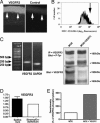
Fig. 1.
Fig. 1.
Ectopic VEGFR3 expression in corneal epithelium. (AD) Strong ectopic expression of both VEGFR3 protein (A and B) and mRNA (C and D) in normal corneal epithelium. (A) Immunofluorescence at ×200. (Left) Anti-VEGFR3 antibody. (Right) Control. Arrows indicate corneal epithelium. (B) Two-color FACS staining against epithelial marker K-12 (phycoerythrin) and VEGFR3 (FITC). Gray indicates isotype; black indicates VEGFR3 staining. Analysis was gated on K-12-phycoerythrin. Arrow indicates VEGFR3 and K12 costained corneal epithelium. (C) RT-PCR for VEGFR3 in normal corneal epithelium (expected sizes: VEGFR3, 290 bp; GAPDH, 245 bp). (D) Quantitative real-time PCR demonstrates higher levels of VEGFR3 in corneal epithelium (Left) compared with stroma and corneal endothelium (Right). (E) VEGF-C binds to corneal epithelial VEGFR3 and leads to VEGFR3 activation. Serum-starved cultured MCE were treated with VEGF-C and VEGFR3 immunoprecipitated from cell lysates by using a polyclonal anti-VEGFR3 antibody (M20) and protein A-Sepharose. Immunoprecipitated proteins were resolved on an 8% SDS/polyacrylamide gel and transferred to nitrocellulose membrane. Phosphotyrosine residues were detected by immunoblotting using PY20 and AG10 antibodies. After stripping the membrane, total VEGFR3 was detected by using M20 antibody. The phosphorylation level of VEGFR3 was quantified by densitometry and corrected to the amount of VEGFR3. A significant increase in epithelial VEGFR3 phosphorylation after exposition to VEGF-C was shown.
Fig. 2.
Fig. 2.
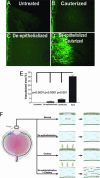
Antiangiogenic effect of corneal epithelium I. (A) Normal corneal–conjunctival border. (B and D) A normally nonangiogenic inflammatory stimulus (cautery; B) causes corneal neovascularization in the absence of epithelium (D). (C) De-epithelialized cornea. Representative segments from corneal flat mounts at the border between normally vascularized conjunctiva (Left) and normally avascular cornea (Right) immunostained with CD31. (E) Morphometry. (F) Diagram of experimental design. (Left) Cross section of normal eye. (Right) Enlargement of cornea. Cells in corneal stroma are keratocytes; red lines are invading blood vessels. (Magnification: AD, ×100.)
Fig. 3.
Fig. 3.
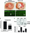
Angiosuppressive effect of corneal epithelium II. (AD) Absence of corneal epithelium (A) significantly enhances the neovascular response (arrowhead: blood vessel) in the model of suture-induced inflammatory angiogenesis [in parallel with increased influx of CD45+ inflammatory cells (C and D; CD45 immunostaining, arrows)] (B). (E) Results of morphometry. (F) Up-regulation of VEGFR3 ligands in inflammatory corneal angiogenesis. The ligands of VEGFR3, VEGF-C, and VEGF–D are significantly up-regulated in conditions associated with inflammatory corneal angiogenesis (suture model from B; P < 0.05 for both ligands; lane 1, control; lane 2, suture-induced inflammatory angiogenesis; expected sizes: VEGF-C, 531 bp; VEGF-D, 307 bp; GAPDH, 245 bp). (Magnification: C and D, ×200.)
Fig. 4.
Fig. 4.
Antiangiogenic effect of a VEGFR3 chimeric protein. A VEGFR3 chimeric protein, ligating VEGF-C and VEGF-D, can substitute for the antiangiogenic effect of VEGFR3-expressing corneal epithelium. The neovascular response after cautery of de-epithelialized corneas (representative segment from CD31-stained corneal flat mount) (A) is significantly diminished (C) when a VEGFR3 chimeric protein is administered locally (B). (Magnification: A and B, ×100.)
Fig. 5.
Fig. 5.
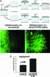
Antiangiogenic effect of corneal epithelium critically depends on epithelial VEGFR3. Direct inhibition of epithelial VEGFR3 using ex vivo treatment with neutralizing antibodies (19) diminishes the epithelium’s ability to dampen angiogenesis. (A) Experimental design. (B and C) The neovascular response to an inflammatory stimulus is significantly increased after ex vivo treatment with a neutralizing anti-VEGFR3 antibody. Representative segments from CD31-stained corneal flat mounts (C) are compared with corneas receiving epithelial transplants that received ex vivo treatment with control IgG (B). (D) Morphometry. (Magnification: B and C, ×100.)

References

    1. Streilein J. W. Nat. Rev. Immunol. 2003;3:879–889. - PubMed
    1. Cursiefen C., Masli S., Ng T. F., Dana M. R., Bornstein P., Lawler J., Streilein J. W. Invest. Ophthalmol. Visual Sci. 2004;45:1117–1124. - PubMed
    1. Cursiefen C., Chen L., Dana M. R., Streilein J. W. Cornea. 2003;22:273–281. - PubMed
    1. Gimbrone M. A., Cotran R. S., Leapman S. B., Folkman J. J. Natl. Cancer Inst. 1974;52:413–427. - PubMed
    1. Kenyon B. M., Voest E. E., Chen C. C., Flynn E., Folkman J., D’Amato R. J. Invest. Ophthalmol. Visual Sci. 1996;37:1625–1632. - PubMed

Publication types

MeSH terms

Substances

LinkOut - more resources