

doi: 10.1534/genetics.106.061705.
Epub 2006 Jul 18.
An efficient genetic screen in Drosophila to identify nuclear-encoded genes with mitochondrial function
Affiliations
- PMID: 16849596
- PMCID: PMC1569793
- DOI: 10.1534/genetics.106.061705
Item in Clipboard
An efficient genetic screen in Drosophila to identify nuclear-encoded genes with mitochondrial function
Genetics.
2006 Sep.
Abstract
We conducted a screen for glossy-eye flies that fail to incorporate BrdU in the third larval instar eye disc but exhibit normal neuronal differentiation and isolated 23 complementation groups of mutants. These same phenotypes were previously seen in mutants for cytochrome c oxidase subunit Va. We have molecularly characterized six complementation groups and, surprisingly, each encodes a mitochondrial protein. Therefore, we believe our screen to be an efficient method for identifying genes with mitochondrial function.
Figures
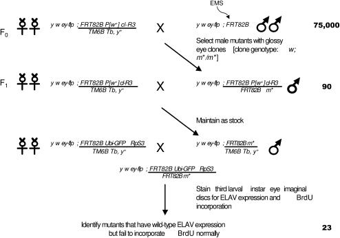
Crossing scheme for genetic screen. A total of 75,000 chromosomes were mutagenized and screened for the adult glossy-eye phenotype. A total of 90 glossy-eye mutants were isolated, and larval eye-disc clones were induced to study their BrdU incorporation and ELAV expression. Twenty-three failed to incorporate BrdU but expressed ELAV normally and were studied further. m* represents the EMS-induced mutation. The cl-R3 mutation is cell lethal and is used to eliminate cells that are homozygous for the chromosome that does not contain m*. Similarly, for larval clones, cells homozygous for the RpS3 (Minute) mutation are eliminated. Cells heterozygous for the cl-R3 and RpS3 mutations grow more slowly, allowing m*/m* cells to form large clones.
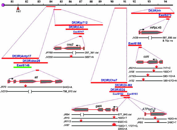
Map positions and molecular lesions of identified mutations. Deficiency stock names and their approximate endpoints, as annotated by FlyBase, are indicated. Solid black lines, alleles identified from the screen; red stars, point mutations; brackets, deletions; red bars, deficiencies that fail to complement the mutant; green bars, deficiencies that complement the mutant.
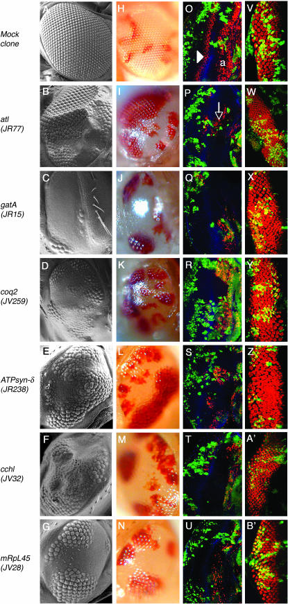
Representative adult and larval phenotypes of mutants from mapped complementation groups. (A–N) Eyes of adult flies examined by scanning electron microscopy (A–G) and bright-field microscopy (H–N). An eye containing mock clones is mosaic in color (H), but the entire eye is faceted as in wild-type tissue (A). In eyes mutant for the indicated mitochondrial genes, cells heterozygous for the mutation are faceted (B–G) and red (I–N) as in mock clones, while homozygous clones are glossy and white. In all images, the representative allele is indicated in parentheses and posterior is to the left. (O–U) 30-min BrdU incorporation (red) in third instar larval eye discs with mock (O) and mutant (P–U) clones. Armadillo (blue) marks the morphogenetic furrow. In an eye disc with mock clones (O), both green and nongreen tissue are wild type. BrdU is randomly incorporated anterior to the furrow (“a”); posterior to the furrow, a single band of BrdU incorporation (arrowhead) marks the SMW. In mutant eye discs, BrdU incorporation is lost in homozygous mutant tissue, marked by the lack of GFP, both anterior and posterior to the furrow. Incorporation remains normal in clones heterozygous for the mutation (green). For comments on the apparent local nonautonomy of this phenotype (arrow), see Mandal et al. (2005). (V–B′) ELAV antibody stainings of eye discs with mock (V) and mutant (W–B′) clones. Expression of ELAV is largely normal in all mutant clones.
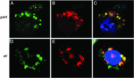
GatA and ATL localize to the mitochondrion. Co-immunostaining of GatA–GFP and ATL–GFP fusion proteins (A and D, green), respectively, with MitoTracker Orange CM-H2TMRos (Molecular Probes, Eugene, OR) (B and E) shows mitochondrial localization of the fusion proteins (C and F, yellow). Full-length cDNAs were cloned using the Gateway cloning system (Invitrogen, San Diego) into the pAWG vector containing the actin promoter and sequence coding for a C-terminal GFP protein. Blue, DAPI staining (C) and TOPRO3 (F) staining.
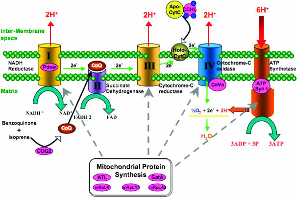
Schematic of complexes involved in oxidative phosphorylation in the mitochondrion. Proteins highlighted in pink are encoded by mutant genes identified from this screen or described in Mandal et al. (2005).
Similar articles
-
Mutations in the Drosophila mitochondrial tRNA amidotransferase, bene/gatA, cause growth defects in mitotic and endoreplicating tissues.Genetics. 2008 Feb;178(2):979-87. doi: 10.1534/genetics.107.084376. Epub 2008 Feb 1. Genetics. 2008. PMID: 18245325 Free PMC article.
-
Comparison of the oxidative phosphorylation (OXPHOS) nuclear genes in the genomes of Drosophila melanogaster, Drosophila pseudoobscura and Anopheles gambiae.Genome Biol. 2005;6(2):R11. doi: 10.1186/gb-2005-6-2-r11. Epub 2005 Jan 31. Genome Biol. 2005. PMID: 15693940 Free PMC article.
-
A screen for dominant modifiers of the irreC-rst cell death phenotype in the developing Drosophila retina.Genetics. 2000 Sep;156(1):205-17. doi: 10.1093/genetics/156.1.205. Genetics. 2000. PMID: 10978286 Free PMC article.
-
The art and design of genetic screens: Drosophila melanogaster.Nat Rev Genet. 2002 Mar;3(3):176-88. doi: 10.1038/nrg751. Nat Rev Genet. 2002. PMID: 11972155 Review.
-
The Heidelberg Screen for Pattern Mutants of Drosophila: A Personal Account.Annu Rev Cell Dev Biol. 2016 Oct 6;32:1-46. doi: 10.1146/annurev-cellbio-113015-023138. Epub 2016 Aug 3. Annu Rev Cell Dev Biol. 2016. PMID: 27501451 Review.
Cited by
-
Genetic analysis of fibroblast growth factor signaling in the Drosophila eye.G3 (Bethesda). 2012 Jan;2(1):23-8. doi: 10.1534/g3.111.001495. Epub 2012 Jan 1. G3 (Bethesda). 2012. PMID: 22384378 Free PMC article.
-
A Genetic Screen Using the Drosophila melanogaster TRiP RNAi Collection To Identify Metabolic Enzymes Required for Eye Development.G3 (Bethesda). 2019 Jul 9;9(7):2061-2070. doi: 10.1534/g3.119.400193. G3 (Bethesda). 2019. PMID: 31036678 Free PMC article.
-
Glycolytic disruption restricts Drosophila melanogaster larval growth via the cytokine Upd3.PLoS Genet. 2025 May 2;21(5):e1011690. doi: 10.1371/journal.pgen.1011690. eCollection 2025 May. PLoS Genet. 2025. PMID: 40315265 Free PMC article.
-
Drosophila follicle stem cells are regulated by proliferation and niche adhesion as well as mitochondria and ROS.Nat Commun. 2012 Apr 3;3:769. doi: 10.1038/ncomms1765. Nat Commun. 2012. PMID: 22473013 Free PMC article.
-
Expression profiling of attenuated mitochondrial function identifies retrograde signals in Drosophila.G3 (Bethesda). 2012 Aug;2(8):843-51. doi: 10.1534/g3.112.002584. Epub 2012 Aug 1. G3 (Bethesda). 2012. PMID: 22908033 Free PMC article.
References
-
- Ackerman, S. H., and A. Tzagoloff, 2005. Function, structure, and biogenesis of mitochondrial ATP synthase. Prog. Nucleic Acid Res. Mol. Biol. 80: 95–133. - PubMed
-
- Altschul, S. F., W. Gish, W. Miller, E. W. Myers and D. J. Lipman, 1990. Basic local alignment search tool. J. Mol. Biol. 215: 403–410. - PubMed
-
- Ashby, M. N., S. Y. Kutsunai, S. Ackerman, A. Tzagoloff and P. A. Edwards, 1992. COQ2 is a candidate for the structural gene encoding para-hydroxybenzoate:polyprenyltransferase. J. Biol. Chem. 267: 4128–4136. - PubMed
-
- Boyer, P. D., 1997. The ATP synthase: a splendid molecular machine. Annu. Rev. Biochem. 66: 717–749. - PubMed
-
- Claros, M. G., and P. Vincens, 1996. Computational method to predict mitochondrially imported proteins and their targeting sequences. Eur. J. Biochem. 241: 779–786. - PubMed
Publication types
MeSH terms
Substances
Grants and funding
LinkOut - more resources
Full Text Sources
Medical
Molecular Biology Databases
