. 2006 Sep;174(1):317-29.
doi: 10.1534/genetics.106.059709. Epub 2006 Jul 18.

Maternal gametophytic baseless1 is required for development of the central cell and early endosperm patterning in maize (Zea mays)

Affiliations

Maternal gametophytic baseless1 is required for development of the central cell and early endosperm patterning in maize (Zea mays)

José F Gutiérrez-Marcos et al. Genetics. 2006 Sep.

Abstract

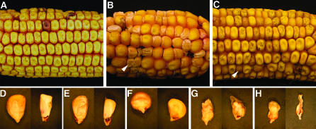
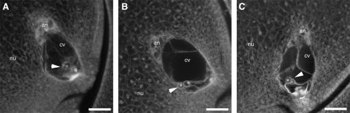
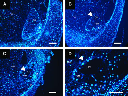
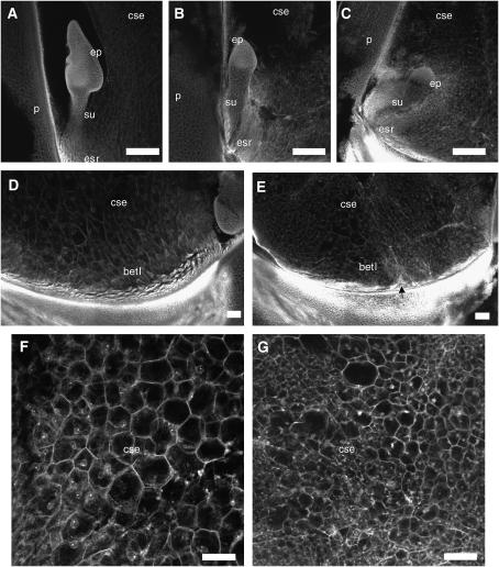
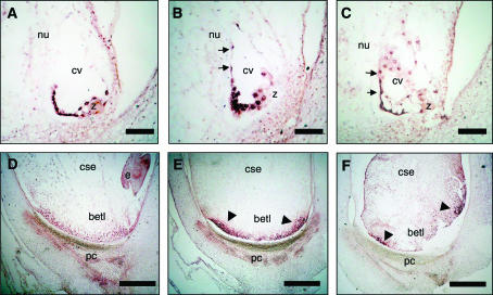
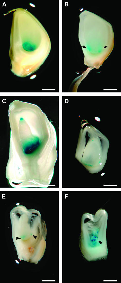
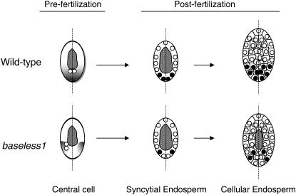
In angiosperms, double fertilization of an egg cell and a central cell with two sperm cells results in the formation of a seed containing a diploid embryo and a triploid endosperm. The extent to which the embryo sac controls postfertilization events in the seed is unknown. The novel gametophytic maternal-effect maize mutation, baseless1 (bsl1) affects central cell development within the embryo sac, frequently by altering the position of the two polar nuclei. Despite this irregularity, fertilization is as efficient as in wild type. The spatial expression of basal endosperm-specific transcripts is altered in free-nuclear and cellular mutant endosperms. At later stages of seed development, bsl1 predominantly affects development of the basal endosperm transfer layer (BETL). When bsl1/+ diploid plants were pollinated by wild-type tetraploid plants, the BETL abnormalities observed in bsl1/bsl1/+/+ tetraploid endosperms were diverse and of variable severity. Moreover, the frequency of kernels with severely perturbed BETL development correlated with the percentage of severely affected bsl1 central cells. Therefore, BSL1 is likely required in the central cell before fertilization for correct BETL patterning to occur. These findings provide new genetic evidence that a maternal gametophytic component is necessary for correct endosperm patterning.


Figures

F<sc>igure</sc> 1.—
Figure 1.—
The bsl1 seed phenotype. (A) Ear from a homozygous wild-type plant pollinated with pollen from a bsl1/+ heterozygous plant, exhibiting normal kernels. (B) Ear from a bsl1/+ heterozygous female pollinated with homozygous wild-type pollen, showing a strong bsl1 maternal effect. Approximately half of the kernels have reduced or aborted endosperms with loose pericarps (arrowhead). (C) Ear from a bsl1/+ heterozygous female pollinated with homozygous wild-type pollen, showing a mild bsl1 maternal effect. Approximately half of the kernels have slightly reduced endosperms (arrowhead). (D–H) Phenotypic classes of kernels produced in test crosses and self-crosses of bsl1/+ plants (germinal face of kernels on the left and median longitudinal sections of kernels on the right). The germinal face of sectioned kernels is oriented to the left for all kernels. (D) Wild type. (E–H) bsl1 phenotypic classes. (E) Reduced endosperm. (F) Germless. (G) Loose pericarp. (H) Empty pericarp.
F<sc>igure</sc> 2.—
Figure 2.—
Embryo sac morphologies in bsl1/+ plants. (A) Wild-type embryo sac with the two central cell polar nuclei centrally located above the egg cell. (B) bsl1 mutant embryo sac with the polar nuclei located adjacent to the abgerminal wall of the central cell. (C) bsl1 mutant embryo sac showing polar nuclei situated off center, abgerminal to the central longitudinal axis. Arrowheads point to the polar nuclei. an, antipodals; cv, central vacuole; e, egg cell; nu, nucellus. Bar, 25 μm.
F<sc>igure</sc> 3.—
Figure 3.—
Developing 3-dap endosperms segregating from bsl1/+ plants pollinated with wild-type pollen. (A) Wild-type fully cellular endosperm. (B–D) Segregating bsl1 mutant endosperms showing delayed cellularization—as indicated by the prolonged presence of the central vacuole (arrowheads), occurring irregularly, in a noncentripetal fashion. Endosperm sections were stained with DAPI and viewed under fluorescence microscopy. cv, central vacuole; en, endosperm; nu, nucellus; p, pericarp; z, zygote. Bar, 50 μm.
F<sc>igure</sc> 4.—
Figure 4.—
Confocal microscopy analysis of 9-dap embryos and endosperms from bsl1/+ plants pollinated with wild-type pollen. (A, D, and F) Wild type. (B, C, E, and G) bsl1. (A) Wild-type coleoptile stage embryo with prominent shoot apical meristem and (B and C) sibling mutant embryos lagging at the transition stage. (D and E) BETL endosperm morphology. (D) Wild-type endosperm with BETL consisting of approximately three layers of slightly elongated cells (autofluorescent) located above the pedicel (intense autofluorescent signal). (E) Typical bsl1 endosperm possessing an irregular BETL containing fewer cell layers. Arrow points to region of basal endosperm devoid of BETL. (F and G) Starchy endosperm morphology. Cells in the central endosperm are large and regularly spaced in wild-type endosperms (F), but appear smaller and of variable size in bsl1 endosperms (G). betl, basal endosperm transfer layer; cse, central starchy endosperm; ep, embryo proper; esr, embryo surrounding region; p, pericarp; su, suspensor. Bar, 100 μm.
F<sc>igure</sc> 5.—
Figure 5.—
mRNA in situ hybridization of basal endosperm-specific transcripts in sibling wild-type and mutant kernels. (A–C) 2-dap syncytial endosperms. (D–F) 9-dap cellular endosperms. (A) ZmEND1 transcript is confined to the basal portion of the wild-type free-nuclear endosperm. (B and C) In sibling mutant endosperms, ZmEND1 expression is displaced from the basal portion of the syncytial endosperm to more lateral regions (arrows). (D) Wild-type meg1 expression is restricted to the BETL, whereas in bsl1 endosperms (E and F) meg1 transcript is localized in discrete areas of the endosperm (arrowheads). betl, basal endosperm transfer layer; cse, central starchy endosperm; e, embryo; pc, placento-chalazal region; cv, central vacuole; nu, nucellus; z, zygote. Bar, 100 μm.
F<sc>igure</sc> 6.—
Figure 6.—
Effects of bsl1 on BETL-specific transgenic reporter expression in triploid and tetraploid endosperms. (A and B) 10-dap sibling kernels taken from a bsl1/+, ProMeg1:GUS/ProMeg1:GUS plant pollinated by a wild-type diploid plant. (A) Wild-type triploid endosperm with uniform ProMeg1:GUS expression (blue) distributed along the basal endosperm. (B) Typical mirror-image GUS expression confined to discrete basal areas of a typical bsl1/bsl1/+ endosperm (indicated by arrows). (C) 10-dap tetraploid endosperm expressing ProMeg1:GUS in the basal and apical endosperm (denoted “top–bottom”). Tetraploid endosperms were generated from crosses between wild-type ProMeg1:GUS/ProMeg1:GUS diploid plants and wild-type tetraploid plants. (D–F) Three distinct classes of 10-dap sibling kernels segregating in an ear from a bsl1/+, ProMeg1:GUS/ProMeg1:GUS plant pollinated by a wild-type tetraploid plant. (D) Typical kernel of the first class showing top–bottom ProMeg1:GUS expression patterns similar to those found in tetraploid endosperms shown in C. (E) Abnormal kernel of the second class exhibiting irregular mirror-image GUS staining patterns in the basal endosperm. (F) Kernel typical of the third class showing highly scattered ProMeg1:GUS staining throughout the endosperm (arrowheads). Bar, 200 μm.
F<sc>igure</sc> 7.—
Figure 7.—
Proposed model for the maternal regulation of BETL development in wild type and bsl1. In wild type, maternal factor(s) required for correct BETL patterning (shading) are putatively distributed along the proximal–distal axis (dashed line) of the central cell before fertilization. Mutation in bsl1 affects development of the central cell, as indicated by the displacement of the polar nuclei (open circles) away from the central longitudinal (proximal–distal) axis. We propose that these maternal factor(s) are also displaced in bsl1 central cells, thus altering BETL patterning. Following fertilization in wild type, BETL specification of basal nuclei (solid circles) takes place in the syncytial endosperm, with subsequent daughter nuclei and BETL cells developing in a lineage-dependent fashion (Costa et al. 2003). Despite fertilization of bsl1/+ plants with wild-type pollen, BETL specification occurs abnormally in bsl1 syncytial endosperms, such that some basal nuclei remain undifferentiated. This leads directly to decreased BETL cell distribution and to subsequent poor nutrient uptake. As a consequence, cellularization is delayed and is indicated by the prolonged presence of the central vacuole (stippled area) in mutant endosperms, at a time when sibling wild-type endosperms are fully cellular. The future germinal side of the kernel is oriented to the right.

Similar articles

Cited by

References

    1. Arthur, K. M., Z. Vejlupkova, R. B. Meeley and J. E. Fowler, 2003. Maize ROP2 GTPase provides a competitive advantage to the male gametophyte. Genetics 165: 2137–2151. - PMC - PubMed
    1. Birchler, J. A., 1993. Dosage analysis of maize endosperm development. Annu. Rev. Genet. 27: 181–204. - PubMed
    1. Boisnard-Lorig, C., A. Colon-Carmona, M. Bauch, S. Hodge, P. Doerner et al., 2001. Dynamic analyses of the expression of the HISTONE∷YFP fusion protein in Arabidopsis show that syncytial endosperm is divided in mitotic domains. Plant Cell 13: 495–509. - PMC - PubMed
    1. Brown, R. C., B. E. Lemmon and H. Nguyen, 2003. Events during the first four rounds of mitosis establish three developmental domains in the syncytial endosperm of Arabidopsis thaliana. Protoplasma 222: 167–174. - PubMed
    1. Chang, M. T., and M. G. Neuffer, 1994. Endosperm-embryo interactions in maize. Maydica 39: 9–18.