

The Tribolium castaneum ortholog of Sex combs reduced controls dorsal ridge development
- PMID: 16849608
- PMCID: PMC1569817
- DOI: 10.1534/genetics.106.058610
The Tribolium castaneum ortholog of Sex combs reduced controls dorsal ridge development
Abstract
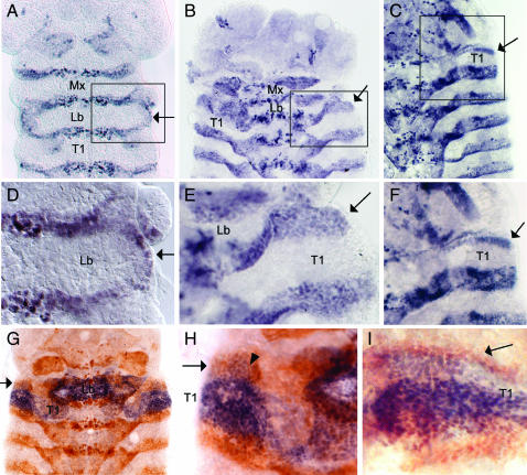
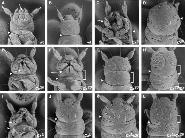
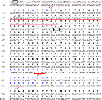
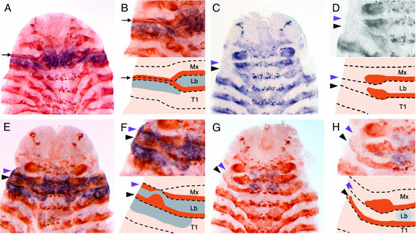
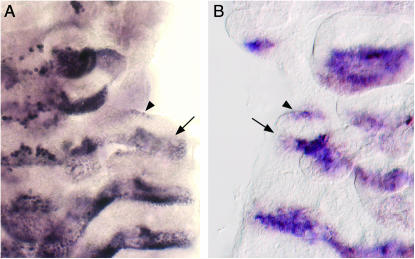
In insects, the boundary between the embryonic head and thorax is formed by the dorsal ridge, a fused structure composed of portions of the maxillary and labial segments. However, the mechanisms that promote development of this unusual structure remain a mystery. In Drosophila, mutations in the Hox genes Sex combs reduced and Deformed have been reported to cause abnormal dorsal ridge formation, but the significance of these abnormalities is not clear. We have identified three mutant allele classes of Cephalothorax, the Tribolium castaneum (red flour beetle) ortholog of Sex combs reduced, each of which has a different effect on dorsal ridge development. By using Engrailed expression to monitor dorsal ridge development in these mutants, we demonstrate that Cephalothorax promotes the fusion and subsequent dorsolateral extension of the maxillary and labial Engrailed stripes (posterior compartments) during dorsal ridge formation. Molecular and genetic analysis of these alleles indicates that the N terminus of Cephalothorax is important for the fusion step, but is dispensable for Engrailed stripe extension. Thus, we find that specific regions of Cephalothorax are required for discrete steps in dorsal ridge formation.
Figures
Similar articles
-
Proboscipedia represses distal signaling in the embryonic gnathal limb fields of Tribolium castaneum.Dev Genes Evol. 2003 Mar;213(2):55-64. doi: 10.1007/s00427-002-0291-7. Epub 2003 Jan 21. Dev Genes Evol. 2003. PMID: 12632174
-
Molecular characterization of Cephalothorax, the Tribolium ortholog of Sex combs reduced.Genesis. 2001 May;30(1):12-20. doi: 10.1002/gene.1027. Genesis. 2001. PMID: 11353513
-
Tribolium castaneum twist: gastrulation and mesoderm formation in a short-germ beetle.Dev Genes Evol. 2005 Jan;215(1):13-31. doi: 10.1007/s00427-004-0446-9. Epub 2004 Nov 30. Dev Genes Evol. 2005. PMID: 15645317
-
Interactions of the Tribolium Sex combs reduced and proboscipedia orthologs in embryonic labial development.Genetics. 2001 Dec;159(4):1643-8. doi: 10.1093/genetics/159.4.1643. Genetics. 2001. PMID: 11779803 Free PMC article.
-
Insect serosa: a head line in comparative developmental genetics.Curr Biol. 2005 Apr 12;15(7):R245-7. doi: 10.1016/j.cub.2005.03.022. Curr Biol. 2005. PMID: 15823522 Review.
Cited by
-
Insights into insect wing origin provided by functional analysis of vestigial in the red flour beetle, Tribolium castaneum.Proc Natl Acad Sci U S A. 2013 Oct 15;110(42):16951-6. doi: 10.1073/pnas.1304332110. Epub 2013 Oct 1. Proc Natl Acad Sci U S A. 2013. PMID: 24085843 Free PMC article.
-
Functional analysis of Scr during embryonic and post-embryonic development in the cockroach, Periplaneta americana.Dev Biol. 2010 May 1;341(1):324-34. doi: 10.1016/j.ydbio.2010.02.018. Epub 2010 Feb 19. Dev Biol. 2010. PMID: 20171962 Free PMC article.
-
Onychophoran Hox genes and the evolution of arthropod Hox gene expression.Front Zool. 2014 Mar 5;11(1):22. doi: 10.1186/1742-9994-11-22. Front Zool. 2014. PMID: 24594097 Free PMC article.
-
Analysis of the Tribolium homeotic complex: insights into mechanisms constraining insect Hox clusters.Dev Genes Evol. 2008 Apr;218(3-4):127-39. doi: 10.1007/s00427-008-0213-4. Epub 2008 Apr 8. Dev Genes Evol. 2008. PMID: 18392875 Free PMC article.
-
Rhinoceros beetle horn development reveals deep parallels with dung beetles.PLoS Genet. 2018 Oct 4;14(10):e1007651. doi: 10.1371/journal.pgen.1007651. eCollection 2018 Oct. PLoS Genet. 2018. PMID: 30286074 Free PMC article.
References
-
- Beeman, R. W., J. J. Stuart, M. S. Haas and R. E. Denell, 1989. Genetic analysis of the homeotic gene complex (HOM-C) in the beetle Tribolium castaneum. Dev. Biol. 133: 196–209. - PubMed
-
- Beeman, R. W., J. J. Stuart, S. J. Brown and R. E. Denell, 1993. Structure and function of the homeotic gene complex (HOM-C) in the beetle, Tribolium castaneum. BioEssays 15: 439–444. - PubMed
-
- Blair, S. S., 1992. Engrailed expression in the anterior lineage compartment of the developing wing blade of Drosophila. Development 115: 21–33. - PubMed
-
- Brown, S. J., J. K. Henry, W. C. Black and R. E. Denell, 1990. Molecular genetic manipulation of the red flour beetle: genome organization and cloning of a ribosomal protein gene. Insect Biochem. 20: 185–193.
-
- Brown, S. J., N. H. Patel and R. E. Denell, 1994. Embryonic expression of the single Tribolium engrailed homolog. Dev. Genet. 15: 7–18. - PubMed
Publication types
MeSH terms
Substances
Associated data
- Actions
- Actions
- Actions
- Actions
- Actions
- Actions
- Actions
- Actions
- Actions
- Actions
Grants and funding
LinkOut - more resources
Full Text Sources
