

Addition of GM-CSF to a peptide/KLH vaccine results in increased frequencies of CXCR3-expressing KLH-specific T cells
- PMID: 16850346
- PMCID: PMC11031059
- DOI: 10.1007/s00262-006-0198-7
Addition of GM-CSF to a peptide/KLH vaccine results in increased frequencies of CXCR3-expressing KLH-specific T cells
Abstract
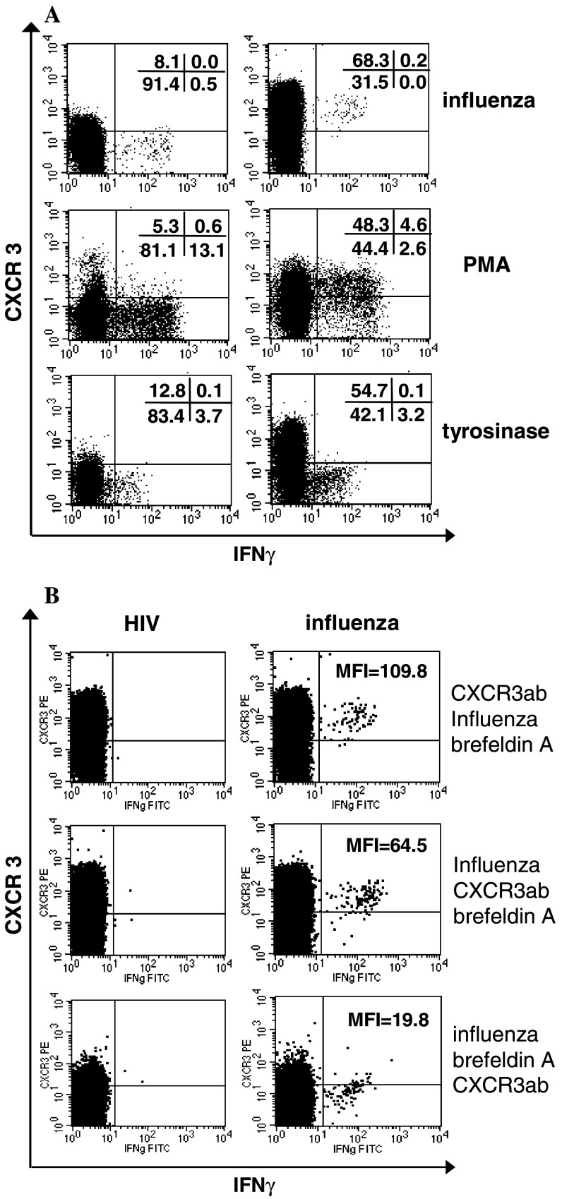
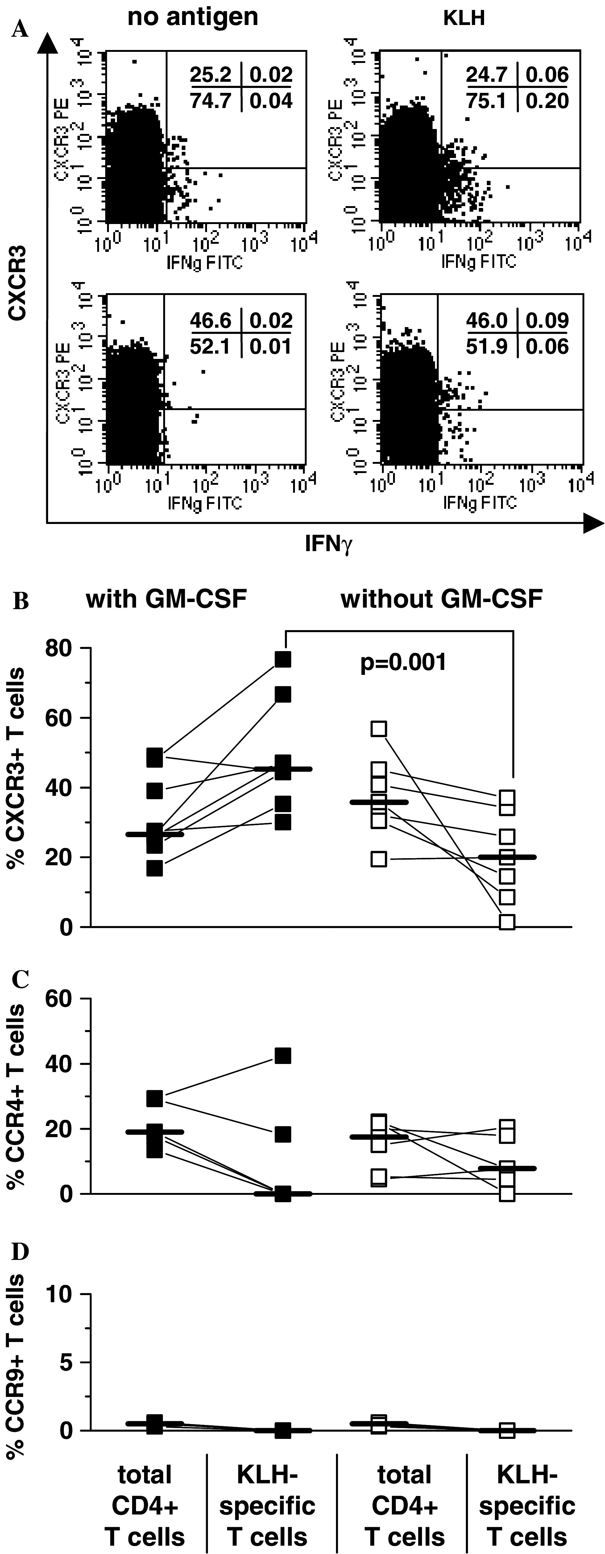
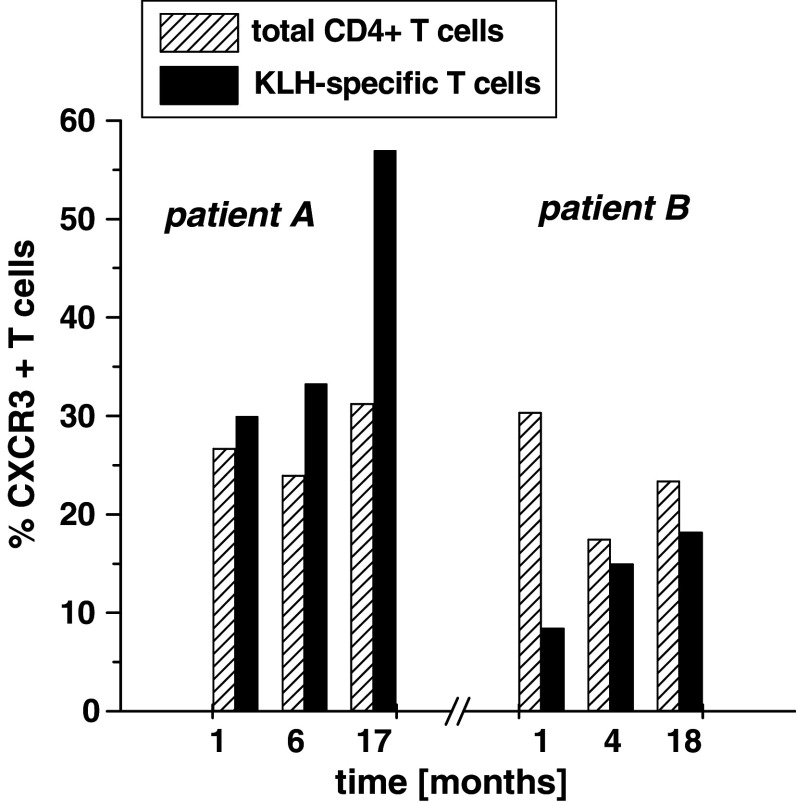
T-cell trafficking is determined by expression patterns of chemokine receptors. The chemokine receptor CXCR3 is expressed on a subpopulation of type 1 T cells and plays an important role for migration of T cells into inflamed and tumor tissues. Here, we studied the chemokine receptor expression on specific T cells generated against the neoantigen keyhole limpet hemocyanin (KLH) in patients who had been immunized in the context of a tumor peptide vaccination trial with or without the adjuvant granulocyte-macrophage colony-stimulating factor (GM-CSF). In patients immunized in the presence of GM-CSF the fraction of CXCR3(+) KLH-specific T cells was significantly higher than in patients immunized in the absence of GM-CSF (median 45 vs. 20%, P = 0.001). In contrast, the chemokine receptor CCR4, associated with migration to the skin was found in both cohorts on less than 10% of KLH-specific T cells. These results show that CXCR3 expression on vaccine-induced T cells can be modulated by modifying the local vaccine milieu.
Figures
Similar articles
-
Peptide vaccination in Montanide adjuvant induces and GM-CSF increases CXCR3 and cutaneous lymphocyte antigen expression by tumor antigen-specific CD8 T cells.Cancer Immunol Res. 2013 Nov;1(5):332-9. doi: 10.1158/2326-6066.CIR-13-0084. Cancer Immunol Res. 2013. PMID: 24377099 Free PMC article. Clinical Trial.
-
Effects of granulocyte-macrophage colony-stimulating factor and foreign helper protein as immunologic adjuvants on the T-cell response to vaccination with tyrosinase peptides.Int J Cancer. 2003 Mar 20;104(2):188-94. doi: 10.1002/ijc.10961. Int J Cancer. 2003. PMID: 12569574 Clinical Trial.
-
Linkage of foreign carrier protein to a self-tumor antigen enhances the immunogenicity of a pulsed dendritic cell vaccine.J Immunol. 2000 May 1;164(9):4797-803. doi: 10.4049/jimmunol.164.9.4797. J Immunol. 2000. PMID: 10779787
-
Use of GM-CSF as an adjuvant with cancer vaccines: beneficial or detrimental?Expert Rev Vaccines. 2010 May;9(5):519-25. doi: 10.1586/erv.10.40. Expert Rev Vaccines. 2010. PMID: 20450326 Review.
-
Novel GM-CSF-based vaccines: One small step in GM-CSF gene optimization, one giant leap for human vaccines.Hum Vaccin Immunother. 2016 Dec;12(12):3020-3028. doi: 10.1080/21645515.2016.1221551. Epub 2016 Aug 25. Hum Vaccin Immunother. 2016. PMID: 27560197 Free PMC article. Review.
Cited by
-
The present and future of peptide vaccines for cancer: single or multiple, long or short, alone or in combination?Cancer J. 2011 Sep-Oct;17(5):343-50. doi: 10.1097/PPO.0b013e318233e5b2. Cancer J. 2011. PMID: 21952285 Free PMC article. Review.
-
Peptide vaccination in Montanide adjuvant induces and GM-CSF increases CXCR3 and cutaneous lymphocyte antigen expression by tumor antigen-specific CD8 T cells.Cancer Immunol Res. 2013 Nov;1(5):332-9. doi: 10.1158/2326-6066.CIR-13-0084. Cancer Immunol Res. 2013. PMID: 24377099 Free PMC article. Clinical Trial.
-
The screening, identification, design and clinical application of tumor-specific neoantigens for TCR-T cells.Mol Cancer. 2023 Aug 30;22(1):141. doi: 10.1186/s12943-023-01844-5. Mol Cancer. 2023. PMID: 37649123 Free PMC article. Review.
-
Update on vaccine development for renal cell cancer.Open Access J Urol. 2010 Aug 4;2:125-41. doi: 10.2147/rru.s7242. Open Access J Urol. 2010. PMID: 24198621 Free PMC article. Review.
-
Activation of GM-CSF and TLR2 signaling synergistically enhances antigen-specific antitumor immunity and modulates the tumor microenvironment.J Immunother Cancer. 2021 Oct;9(10):e002758. doi: 10.1136/jitc-2021-002758. J Immunother Cancer. 2021. PMID: 34599024 Free PMC article.
References
-
- Apodaca G, Aroeti B, Tang K, Mostov K. Brefeldin-A inhibits the delivery of the polymeric immunoglobulin receptor to the basolateral surface of MDCK cells. J Biol Chem. 1993;268:20380–20385. - PubMed
-
- Cole K, Strick C, Paradis T, Ogborne K, Loetscher M, Gladue R, Lin W, Boyd J, Moser B, Wood D, Sahagan B, Neote K. Interferon-inducible T cell alpha chemoattractant (I-TAC), a novel non-ELR CXC chemokine with potent activity on activated T cells through selective high affinity binding to CXCR3. J Exp Med. 1998;187:2009–2021. doi: 10.1084/jem.187.12.2009. - DOI - PMC - PubMed
-
- Disis M, Bernhard H, Shiota F, Hand S, Gralow J, Huseby E, Gillis SMAC. Granulocyte-macrophage colony-stimulating factor: an effective adjuvant for protein and peptide-based vaccines. Blood. 1996;88:202–210. - PubMed
-
- Dranoff G, Jaffee E, Lazenby A, Golumbek P, Levitsky H, Brose K, Jackson V, Hamada H, Pardoll D, Mulligan R. Vaccination with irradiated tumor cells engineered to secrete murine granulocyte-macrophage colony-stimulating factor stimulates potent, specific, and long-lasting anti-tumor immunity. Proc Natl Acad Sci USA. 1993;90:3539–3543. doi: 10.1073/pnas.90.8.3539. - DOI - PMC - PubMed
-
- Foley J, Yu C, Solow R, Yacobucci M, Peden K, Farber J. Roles for CXC chemokine ligands 10 and 11 in recruiting CD4+ T cells to HIV-1-infected monocyte-derived macrophages, dendritic cells, and lymph nodes. J Immunol. 2005;174:4892–4900. - PubMed
Publication types
MeSH terms
Substances
LinkOut - more resources
Full Text Sources
Medical
