

Comparison of the ultrastructure of cortical and retinal terminals in the rat superior colliculus
- PMID: 16850432
- PMCID: PMC2561302
- DOI: 10.1002/ar.a.20359
Comparison of the ultrastructure of cortical and retinal terminals in the rat superior colliculus
Abstract
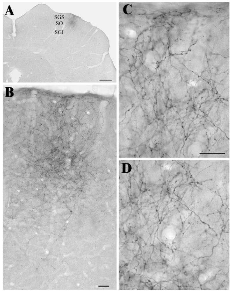
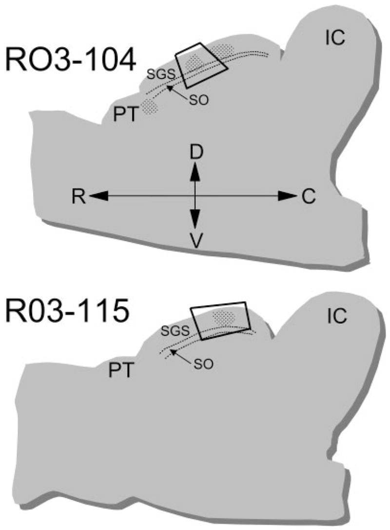
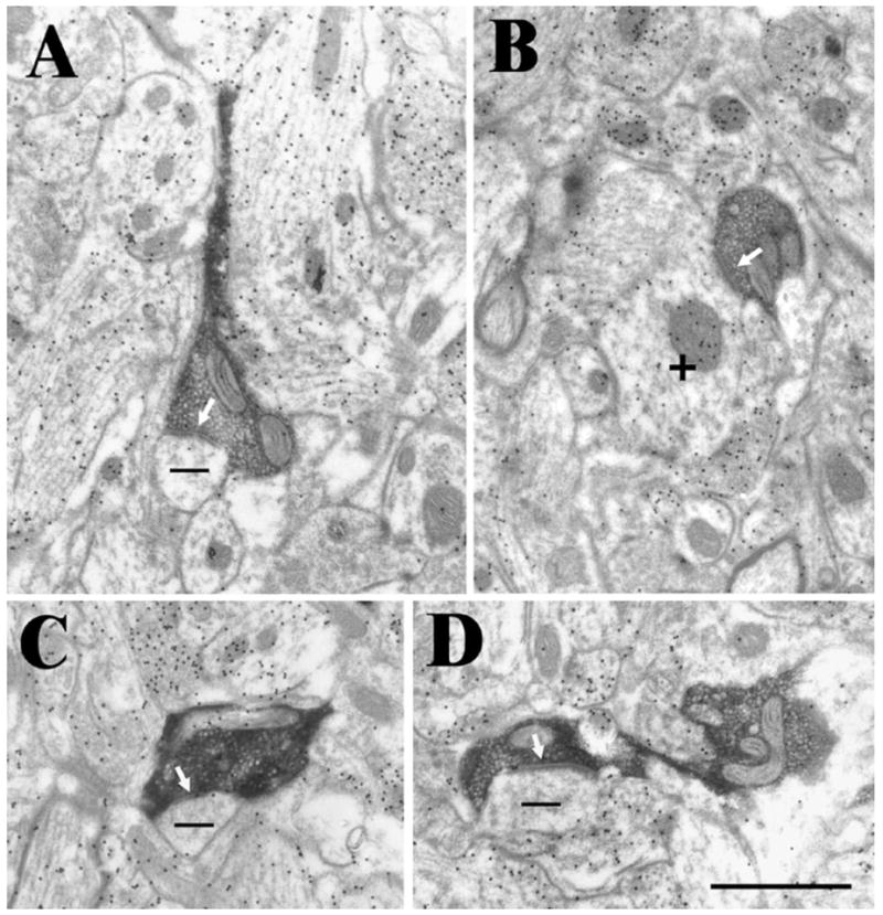
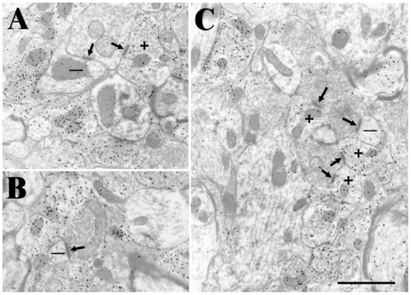
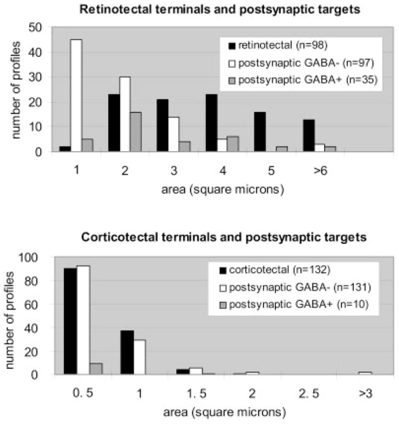
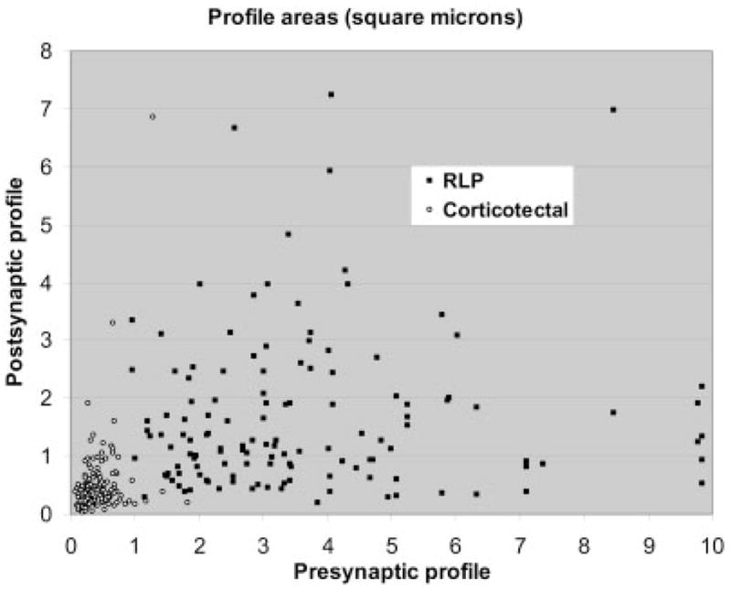
We compared the ultrastructure and synaptic targets of terminals of cortical or retinal origin in the stratum griseum superficiale and stratum opticum of the rat superior colliculus. Following injections of biotinylated dextran amine into cortical area 17, corticotectal axons were labeled by anterograde transport. Corticotectal axons were of relatively small caliber with infrequent small varicosities. At the ultrastructural level, corticotectal terminals were observed to be small profiles (0.44 +/- 0.27 microm(2)) that contained densely packed round vesicles. In tissue stained for gamma amino butyric acid (GABA) using postembedding immunocytochemical techniques, corticotectal terminals were found to contact small (0.51 +/- 0.69 microm(2)) non-GABAergic dendrites and spines (93%) and a few small GABAergic dendrites (7%). In the same tissue, retinotectal terminals, identified by their distinctive pale mitochondria, were observed to be larger than corticotectal terminals (3.34 +/- 1.79 microm(2)). In comparison to corticotectal terminals, retinotectal terminals contacted larger (1.59 +/- 1.70 microm(2)) non-GABAergic dendrites and spines (73%) and a larger proportion of GABAergic profiles (27%) of relatively large size (2.17 +/- 1.49 microm(2)), most of which were vesicle-filled (71%). Our results suggest that cortical and retinal terminals target different dendritic compartments within the neuropil of the superficial layers of the superior colliculus.
Figures
Similar articles
-
Ultrastructural studies of retinal, visual cortical (area 17), and parabigeminal terminals within the superior colliculus of Galago crassicaudatus.J Comp Neurol. 1992 May 1;319(1):85-99. doi: 10.1002/cne.903190109. J Comp Neurol. 1992. PMID: 1592907
-
Electron microscopic study of GABA-immunoreactive neuronal processes in the superficial gray layer of the rat superior colliculus: their relationships with degenerating retinal nerve endings.J Neurocytol. 1991 Apr;20(4):262-76. doi: 10.1007/BF01235544. J Neurocytol. 1991. PMID: 1646864
-
Changes in the pattern of glutamate-like immunoreactivity in rat superior colliculus following retinal and visual cortical lesions.Neuroscience. 1995 Jul;67(1):125-34. doi: 10.1016/0306-4522(95)00057-p. Neuroscience. 1995. PMID: 7477893
-
The NMDAR1 subunit of the N-methyl-D-aspartate receptor is localized at postsynaptic sites opposite both retinal and cortical terminals in the cat superior colliculus.Vis Neurosci. 2000 Jan-Feb;17(1):41-53. doi: 10.1017/s0952523800171044. Vis Neurosci. 2000. PMID: 10750825
-
The organization of GABAergic neurons in the mammalian superior colliculus.Prog Brain Res. 1992;90:219-48. doi: 10.1016/s0079-6123(08)63616-x. Prog Brain Res. 1992. PMID: 1321459 Review.
Cited by
-
The Superior Colliculus: Cell Types, Connectivity, and Behavior.Neurosci Bull. 2022 Dec;38(12):1519-1540. doi: 10.1007/s12264-022-00858-1. Epub 2022 Apr 28. Neurosci Bull. 2022. PMID: 35484472 Free PMC article. Review.
-
The macaque midbrain reticular formation sends side-specific feedback to the superior colliculus.Exp Brain Res. 2010 Apr;201(4):701-17. doi: 10.1007/s00221-009-2090-0. Epub 2009 Nov 26. Exp Brain Res. 2010. PMID: 19940983 Free PMC article.
-
GABAergic cell types in the superficial layers of the mouse superior colliculus.J Comp Neurol. 2020 Feb 1;528(2):308-320. doi: 10.1002/cne.24754. Epub 2019 Aug 19. J Comp Neurol. 2020. PMID: 31396959 Free PMC article.
-
Visual Maps Development: Reconsidering the Role of Retinal Efnas and Basic Principle of Map Alignment.Front Cell Neurosci. 2018 Mar 21;12:77. doi: 10.3389/fncel.2018.00077. eCollection 2018. Front Cell Neurosci. 2018. PMID: 29618973 Free PMC article. No abstract available.
-
Retinal input integration in excitatory and inhibitory neurons in the mouse superior colliculus in vivo.Elife. 2023 Sep 8;12:RP88289. doi: 10.7554/eLife.88289. Elife. 2023. PMID: 37682267 Free PMC article.
References
-
- Appell PP, Behan M. Sources of subcortical GABAergic projections to the superior colliculus in the cat. J Comp Neurol. 1990;302:143–158. - PubMed
-
- Baldauf ZB, Wang XP, Wang S, Bickford ME. Pretectotectal pathway: an ultrastructural quantitative analysis in cats. J Comp Neurol. 2003;464:141–158. - PubMed
-
- Behan M. Identification and distribution of retinocolliculuar terminals in the cat: an electron microscopic autoradiographic analysis. J Comp Neurol. 1981;199:1–15. - PubMed
-
- Behan M. An EM-autoradiographic analysis of the projection from cortical areas 17, 18, and 19 to the superior colliculus in the cat. J Comp Neurol. 1984;225:591–604. - PubMed
Publication types
MeSH terms
Substances
Grants and funding
LinkOut - more resources
Full Text Sources
