

HEX expression and localization in normal mammary gland and breast carcinoma
- PMID: 16854221
- PMCID: PMC1550255
- DOI: 10.1186/1471-2407-6-192
HEX expression and localization in normal mammary gland and breast carcinoma
Abstract
Background: The homeobox gene HEX is expressed in several cell types during different phases of animal development. It encodes for a protein localized in both the nucleus and the cytoplasm. During early mouse development, HEX is expressed in the primitive endoderm of blastocyst. Later, HEX is expressed in developing thyroid, liver, lung, as well as in haematopoietic progenitors and endothelial cells. Absence of nuclear expression has been observed during neoplastic transformation of the thyroid follicular cells. Aim of the present study was to evaluate the localization and the function of the protein HEX in normal and tumoral breast tissues and in breast cancer cell lines.
Methods: HEX expression and nuclear localization were investigated by immunohistochemistry in normal and cancerous breast tissue, as well as in breast cancer cell lines. HEX mRNA levels were evaluated by real-time PCR. Effects of HEX expression on Sodium Iodide Symporter (NIS) gene promoter activity was investigated by HeLa cell transfection.
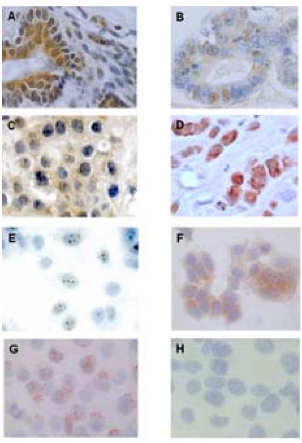
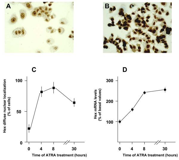
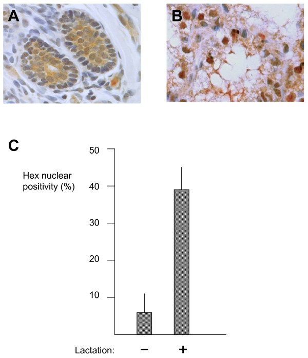
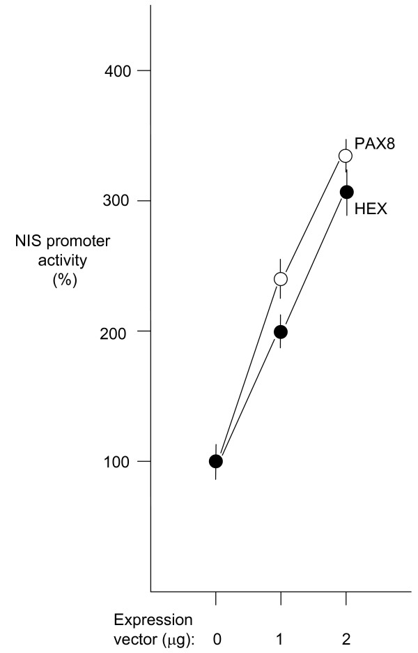
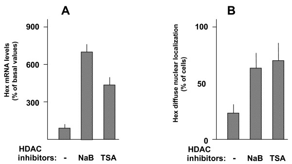
Results: In normal breast HEX was detected both in the nucleus and in the cytoplasm. In both ductal and lobular breast carcinomas, a great reduction of nuclear HEX was observed. In several cells from normal breast tissue as well as in MCF-7 and T47D cell line, HEX was observed in the nucleolus. MCF-7 treatment with all-trans retinoic acid enhanced HEX expression and induced a diffuse nuclear localization. Enhanced HEX expression and diffuse nuclear localization were also obtained when MCF-7 cells were treated with inhibitors of histone deacetylases such as sodium butyrate and trichostatin A. With respect to normal non-lactating breast, the amount of nuclear HEX was greatly increased in lactating tissue. Transfection experiments demonstrated that HEX is able to up-regulate the activity of NIS promoter.
Conclusion: Our data indicate that localization of HEX is regulated in epithelial breast cells. Since modification of localization occurs during lactation and tumorigenesis, we suggest that HEX may play a role in differentiation of the epithelial breast cell.
Figures
Similar articles
-
Effects of histone acetylation on sodium iodide symporter promoter and expression of thyroid-specific transcription factors.Endocrinology. 2005 Sep;146(9):3967-74. doi: 10.1210/en.2005-0128. Epub 2005 May 26. Endocrinology. 2005. PMID: 15919754
-
Transcription factor Nkx-2.5 induces sodium/iodide symporter gene expression and participates in retinoic acid- and lactation-induced transcription in mammary cells.Mol Cell Biol. 2004 Sep;24(18):7863-77. doi: 10.1128/MCB.24.18.7863-7877.2004. Mol Cell Biol. 2004. PMID: 15340050 Free PMC article.
-
Expression and localization of the homeodomain-containing protein HEX in human thyroid tumors.J Clin Endocrinol Metab. 2002 Mar;87(3):1376-83. doi: 10.1210/jcem.87.3.8344. J Clin Endocrinol Metab. 2002. PMID: 11889211
-
Enhancement of sodium/iodide symporter expression in thyroid and breast cancer.Endocr Relat Cancer. 2006 Sep;13(3):797-826. doi: 10.1677/erc.1.01143. Endocr Relat Cancer. 2006. PMID: 16954431 Review.
-
The biology of human breast epithelial progenitors.Semin Cell Dev Biol. 2012 Jul;23(5):606-12. doi: 10.1016/j.semcdb.2012.04.009. Epub 2012 May 17. Semin Cell Dev Biol. 2012. PMID: 22609813 Review.
Cited by
-
Interaction between Hhex and SOX13 modulates Wnt/TCF activity.J Biol Chem. 2010 Feb 19;285(8):5726-37. doi: 10.1074/jbc.M109.046649. Epub 2009 Dec 22. J Biol Chem. 2010. PMID: 20028982 Free PMC article.
-
Genome-wide association studies for milk production traits and persistency of first calving Holstein cattle in Türkiye.Front Vet Sci. 2024 Oct 24;11:1461075. doi: 10.3389/fvets.2024.1461075. eCollection 2024. Front Vet Sci. 2024. PMID: 39512914 Free PMC article.
-
PRH/Hex: an oligomeric transcription factor and multifunctional regulator of cell fate.Biochem J. 2008 Jun 15;412(3):399-413. doi: 10.1042/BJ20080035. Biochem J. 2008. PMID: 18498250 Free PMC article. Review.
-
Regulation of liver growth by glypican 3, CD81, hedgehog, and Hhex.Am J Pathol. 2013 Jul;183(1):153-9. doi: 10.1016/j.ajpath.2013.03.013. Epub 2013 May 8. Am J Pathol. 2013. PMID: 23665349 Free PMC article.
-
A mechanism of nucleocytoplasmic trafficking for the homeodomain protein PRH.Mol Cell Biochem. 2009 Dec;332(1-2):173-81. doi: 10.1007/s11010-009-0188-0. Epub 2009 Jul 9. Mol Cell Biochem. 2009. PMID: 19588232 Free PMC article.
References
-
- Martinez Barbera JP, Clements M, Thomas P, Rodriguez T, Meloy D, Kioussis D, Beddington RS. The homeobox gene Hex is required in definitive endodermal tissues for normal forebrain, liver and thyroid formation. Development. 2000;127:2433–2445. - PubMed
-
- Brickman JM, Jones CM, Clements M, Smith JC, Beddington RS. Hex is a transcriptional repressor that contributes to anterior identity and suppresses Spemann organiser function. Development. 2000;127:2303–2315. - PubMed
-
- Thomas PQ, Brown A, Beddington RS. Hex: a homeobox gene revealing peri-implantation asymmetry in the mouse embryo and an early transient marker of endothelial cell precursors. Development. 1998;125:85–94. - PubMed
-
- Keng VW, Yagi H, Ikawa M, Nagano T, Myint Z, Yamada K, Tanaka T, Sato A. Muramatsu I, Okabe M et al. Homeobox gene Hex is essential for onset of mouse embryonic liver development and differentiation of the monocyte lineage. Biochem Biophys Res Commun. 2000;20:279–739. - PubMed
Publication types
MeSH terms
Substances
LinkOut - more resources
Full Text Sources
Medical
Molecular Biology Databases
Research Materials
