. 2006 Sep 15;281(37):27327-34.
doi: 10.1074/jbc.M605500200. Epub 2006 Jul 19.

Retinoic acid regulates the expression of photoreceptor transcription factor NRL

Affiliations

Retinoic acid regulates the expression of photoreceptor transcription factor NRL

Hemant Khanna et al. J Biol Chem. .

Abstract

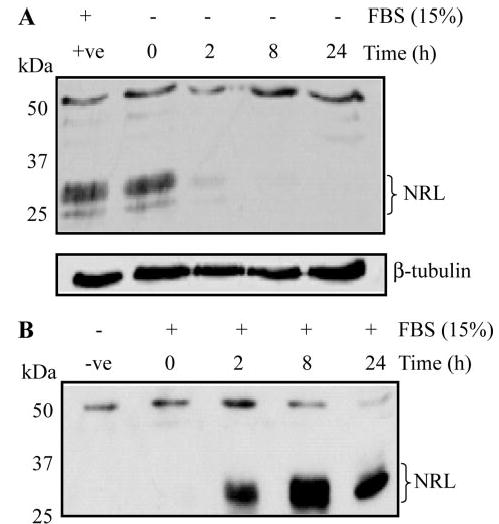
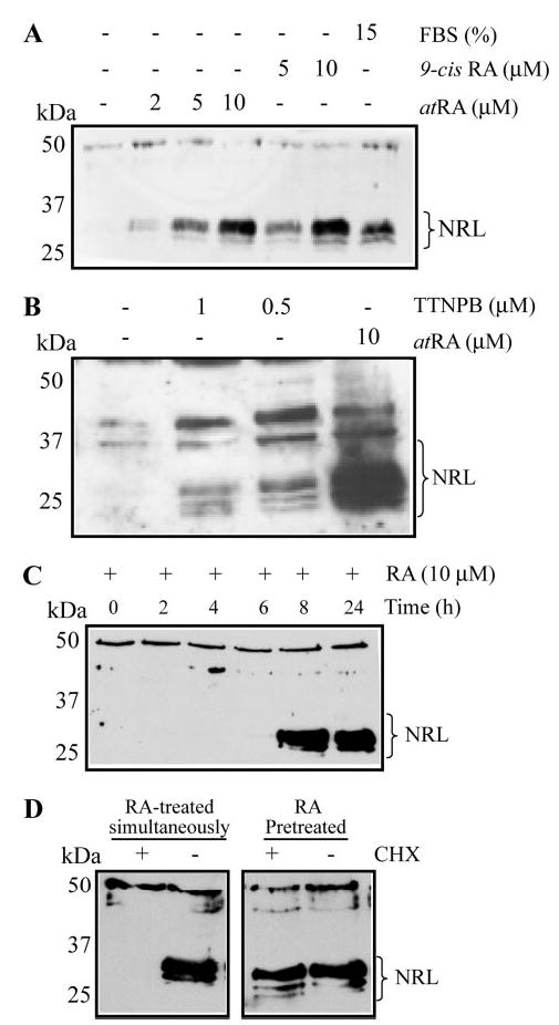
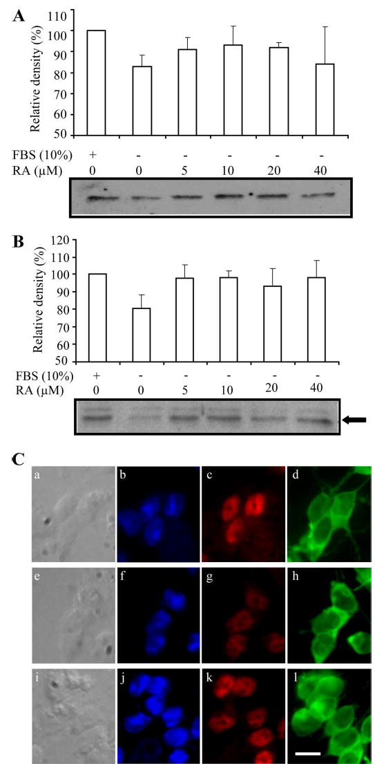
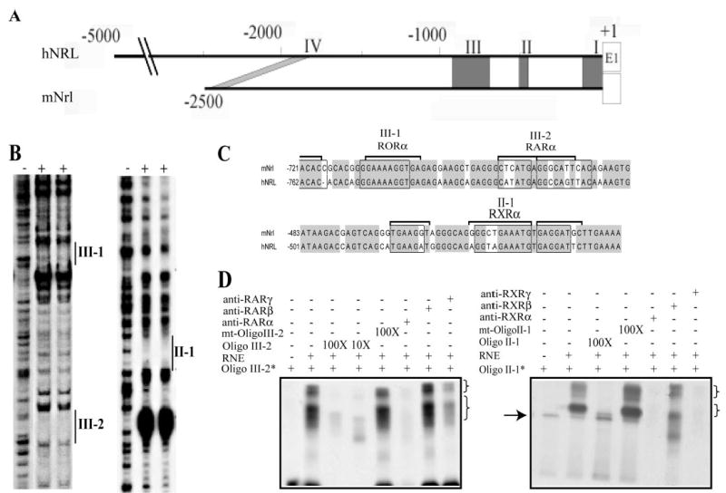
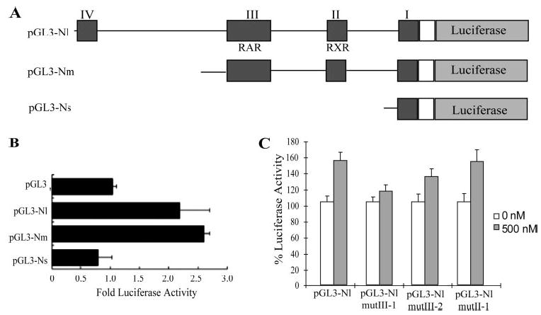
NRL (neural retina leucine zipper) is a key basic motif-leucine zipper (bZIP) transcription factor, which orchestrates rod photoreceptor differentiation by activating the expression of rod-specific genes. The deletion of Nrl in mice results in functional cones that are derived from rod precursors. However, signaling pathways modulating the expression or activity of NRL have not been elucidated. Here, we show that retinoic acid (RA), a diffusible factor implicated in rod development, activates the expression of NRL in serum-deprived Y79 human retinoblastoma cells and in primary cultures of rat and porcine photoreceptors. The effect of RA is mimicked by TTNPB, a RA receptor agonist, and requires new protein synthesis. DNaseI footprinting and electrophoretic mobility shift assays (EMSA) using bovine retinal nuclear extract demonstrate that RA response elements (RAREs) identified within the Nrl promoter bind to RA receptors. Furthermore, in transiently transfected Y79 and HEK293 cells the activity of Nrl-promoter driving a luciferase reporter gene is induced by RA, and this activation is mediated by RAREs. Our data suggest that signaling by RA via RA receptors regulates the expression of NRL, providing a framework for delineating early steps in photoreceptor cell fate determination.


Figures

FIGURE 1
FIGURE 1. Serum induces NRL expression in Y79 cells
Y79 cells were grown in RPMI media without (A) or with (B) FBS (15%) for indicated time intervals, and protein extracts were analyzed by immunoblotting using anti-NRL antibody. Multiple isoforms of NRL are indicated by a bracket. Lanes are as indicated. Lower panel in A shows that the same blot was probed with anti-β-tubulin antibody, which served as a loading control. Molecular masses of markers are shown in kDa. The positive control (+ve) represents Y79 cells grown in 15% FBS.
FIGURE 2
FIGURE 2. RA stimulates expression of NRL protein in Y79 cells
Serum-starved Y79 cells were incubated with indicated concentrations of 9-cis atRA, 15% FBS (A) or TTNPB (B) for 24 h. Cell extracts were analyzed by SDS-PAGE and immunoblotting using anti-NRL antibody. Negative controls included 1% ethanol or Me2SO in lieu of the soluble factors. A bracket indicates multiple phosphorylated NRL isoforms. Lanes are as indicated. Molecular mass markers are indicated on the left. Additional bands in the higher molecular mass range may represent cross-reacting proteins (23). C, time-dependent effect of RA: serum-deprived Y79 cells were incubated with medium containing 10 μM RA for indicated time intervals. At the end of incubation, cells extract was analyzed by SDS-PAGE and immunoblotting using anti-NRL antibody. Lanes are as indicated. D, effect of protein synthesis inhibitor CHX on RA-mediated NRL induction was studied by incubating serum-starved Y79 cells with media containing atRA (10 μM) and CHX (20 μg/ml) (left panel; RA-treated simultaneously). In a similar experiment, cells were pretreated with RA for 24 h followed by addition of CHX (right panel). Cell extracts were analyzed by SDS-PAGE and immunoblotting using the anti-NRL antibody.
FIGURE 3
FIGURE 3. RA increases NRL protein levels in cultured rat and porcine photoreceptors
Analyses of rat (A) and porcine (B) retinal cultures after incubation with indicated concentrations of RA or FBS. Newborn rat retinal cells and adult pig photoreceptors were cultured in vitro, as described under “Experimental Procedures.” Cell extracts were analyzed by SDS-PAGE and immunoblotting using anti-NRL antibody. In both panels, the intensity of the NRL immunoreactive band was reduced in serum-free culture compared with +FBS, and was partially restored by increasing doses of RA. This reduction was significantly different (p <0.05) compared with serum-supplemented controls (*). For rat cultures, this reduction was also significantly different from 20 μM RA, but not for other values. 40 μM RA was toxic for cell survival in newborn rat retina. For pig cultures, the decrease was significantly different compared with all RA concentrations, except 20 μM. The arrow in B indicates the major NRL immunoreactive band used for scanning. Histograms show densitometric scan of representative blots for each culture model. Experiments were performed three times on independent cultures with similar results. C, adult pig photoreceptor cultures were prepared and immuno-stained as described under “Experimental Procedures.” Nomarski differential contrast images of cells are depicted in panels a, e, and i; DAPI staining (blue) of the nuclei in the same fields is shown in panels b, f, and j; NRL immunolabeling (red) of the same fields is shown in panels c, g, and k; and anti-rhodopsin immunolabeling (green) of the same fields is shown in panels d, h, and l. Positive control cultures, maintained in chemically defined medium to which serum-supplemented medium was added for 24 h, revealed strong nuclear NRL immunoreactivity (panel c), as did cells treated with RA (10 μM) for 24 h (panel k); however cells maintained in chemically defined medium demonstrated less intense nuclear staining (panel g). In all cases, rhodopsin staining was not detectably different. Scale bar in panel l is 4 μm for all panels. Experiments were repeated using three independent cultures with similar results.
FIGURE 4
FIGURE 4. Putative RAREs within the Nrl promoter are protected by retinal nuclear proteins
A, schematic representation of the Nrl promoter showing regions of homology (I, II, III, and IV) between human (h) and mouse (m) Nrl. E1 denotes exon 1 of the Nrl gene. B, DNaseI footprinting using bovine RNE was performed as described under “Experimental Procedures.” Footprints corresponding to regions II and III are shown. Vertical lines indicate footprinted regions. (−) denotes footprint in the absence of RNE whereas (+) indicates the experiment in the presence of RNE. Footprints containing the putative RAREs are indicated by III-1, III-2, and II-1. C, sequence of the putative RAREs in the footprints (II and III) of both mouse and human Nrl promoter region. Regions III-1 and III-2 contain putative ROR (orphan receptor) and RAR response elements whereas region II-1 contains a putative RXR binding element. D, EMSA, oligonucleotides corresponding to the regions III-2 (Oligo III-2) and II-1 (Oligo II-1) were radiolabeled using [γ-32P]dATP and incubated with bovine retinal nuclear extract followed by analysis using non-denaturing PAGE, as described under “Experimental Procedures.” Competition experiments were performed with unlabeled oligonucleotides to validate the specificity of the band shift. Experiments in the presence of antibody against various receptor ligands showed the presence or absence of the specific proteins. Arrow indicates a nonspecific band shift. * indicates radiolabeled oligo used in the experiment; mt-Oligo represents mutant oligonucleotide from which the putative RAREs have been deleted. Lanes are as indicated. Brackets indicate specific gel-shifted bands.
FIGURE 5
FIGURE 5. RA receptors bind to and activate Nrl promoter
A, schematic representation of the mouse Nrl promoter-luciferase constructs used to study the response to RA. The deletion fragments were cloned into pGL3-basic plasmid in-frame with the luciferase reporter gene. RAR and RXR response elements in regions III and II, respectively are depicted. These constructs were used in a separate assay to check for intrinsic promoter activity (data not shown). B, Nrl promoter-luciferase constructs were transfected into Y79 cells as described under “Experimental Procedures.” Promoterless vector, pGL3 vector was used as negative control and the value of luciferase activity was set to 1. Results are expressed as a ratio of luciferase values obtained in the presence or absence of RA and represent an average of three independent experiments. C, site-directed mutants of the pGL3-Nl construct (pGL3-Nl-mut III-1, III-2, or II-1), containing deletions of the putative RAREs, were used to transfect HEK293 cells in the presence of indicated concentrations of atRA. The value of the control (transfected with the wild-type pGL3-Nl with no atRA) was set at 100% luciferase activity. Results are expressed as percent luciferase activity as compared with the control.

Similar articles

Cited by

References

    1. Livesey FJ, Cepko CL. Nat Rev Neurosci. 2001;2:109–118. - PubMed
    1. Levine EM, Fuhrmann S, Reh TA. Cell Mol Life Sci. 2000;57:224–234. - PMC - PubMed
    1. Cayouette M, Barres BA, Raff M. Neuron. 2003;40:897–904. - PubMed
    1. Roberts MR, Srinivas M, Forrest D, Morreale de Escobar G, Reh TA. Proc Natl Acad Sci U S A. 2006;103:6218–6223. - PMC - PubMed
    1. Chen Y, Ma JX, Crouch RK. Mol Vis. 2003;9:345–354. - PubMed

Publication types

MeSH terms

LinkOut - more resources