. 2006 Oct;17(10):4364-78.
doi: 10.1091/mbc.e06-02-0143. Epub 2006 Jul 19.

Yeast-to-hyphal transition triggers formin-dependent Golgi localization to the growing tip in Candida albicans

Affiliations

Yeast-to-hyphal transition triggers formin-dependent Golgi localization to the growing tip in Candida albicans

Padmashree C G Rida et al. Mol Biol Cell. 2006 Oct.

Abstract

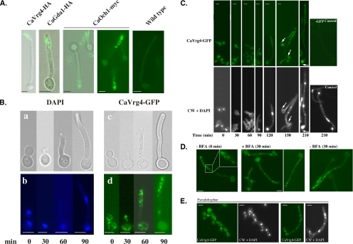
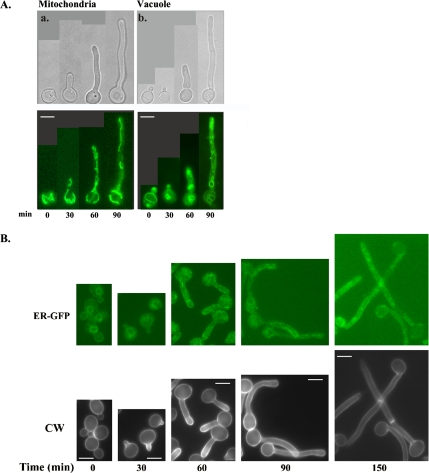
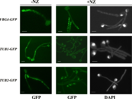
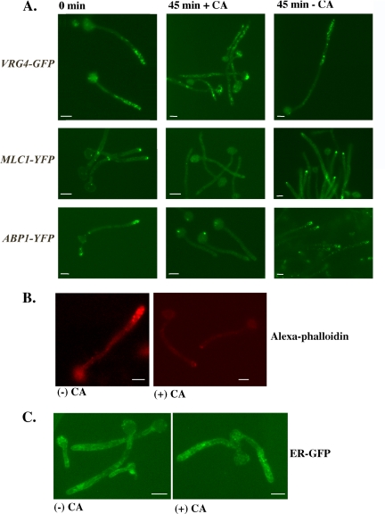
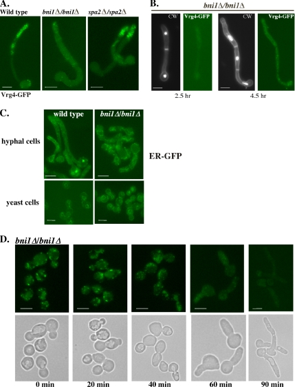
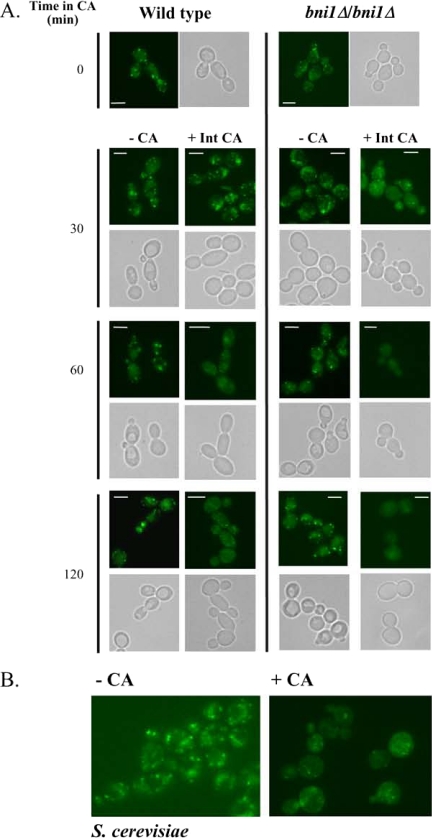
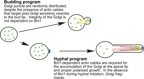
Rapid and long-distance secretion of membrane components is critical for hyphal formation in filamentous fungi, but the mechanisms responsible for polarized trafficking are not well understood. Here, we demonstrate that in Candida albicans, the majority of the Golgi complex is redistributed to the distal region during hyphal formation. Randomly distributed Golgi puncta in yeast cells cluster toward the growing tip during hyphal formation, remain associated with the distal portion of the filament during its extension, and are almost absent from the cell body. This restricted Golgi localization pattern is distinct from other organelles, including the endoplasmic reticulum, vacuole and mitochondria, which remain distributed throughout the cell body and hypha. Hyphal-induced positioning of the Golgi and the maintenance of its structural integrity requires actin cytoskeleton, but not microtubules. Absence of the formin Bni1 causes a hyphal-specific dispersal of the Golgi into a haze of finely dispersed vesicles with a sedimentation density no different from that of normal Golgi. These results demonstrate the existence of a hyphal-specific, Bni1-dependent cue for Golgi integrity and positioning at the distal portion of the hyphal tip, and suggest that filamentous fungi have evolved a novel strategy for polarized secretion, involving a redistribution of the Golgi to the growing tip.


Figures

Figure 1.
Figure 1.
Polarized localization of the Golgi to the apical tip of hyphal cells. (A) Indirect immunofluorescence of Golgi proteins in hyphal cells. Cells (BWP17) expressing Ca-VRG4-HA, CaGDA1-HA, or CaOCH1-myc were induced to form hyphae and processed for indirect immunofluorescence with anti-HA antibodies as described in Materials and Methods. The last panel shows the background signal in an untagged control strain. (B) Localization of CaVrg4-GFP in hyphal cells. Cells expressing CaVRG4-GFP were induced to form hyphae, removed at the indicated times, and viewed directly for CaVrg4-GFP localization or stained with DAPI to visualize nuclear division. (C) Branching is accompanied by accumulation of Vrg4-GFP. Cells expressing CaVRG4-GFP were induced to form hyphae on coverslips. At the indicated time points, cells were stained with DAPI for visualization of nuclei and with calcofluor white for visualization of cell walls and septa. CaVrg4-GFP was visualized by fluorescence microscopy. The last panel shows the background fluorescence in a strain where Ca-VRG4 is not GFP-tagged. (D) Brefeldin A treatment of hyphal cells expressing CaVrg4-GFP. Cells expressing CaVRG4-GFP were induced to form hypha for 1.5 h to allow the redistribution of the Golgi to the distal region of hyphae. An aliquot was removed (−BFA; 0 min), and cells were incubated in the presence or absence BFA for a further 30 min before harvesting to visualize CaVrg4-GFP by fluorescence microscopy. The inset shows some of the cisternae in a wild-type hyphal cell at higher magnification. (E) Random distribution of Golgi puncta in pseudohyphal cells. For induction of pseudohyphae, cells expressing CaVRG4-GFP were incubated at 36°C in YPD buffered at pH 6.0 with citric acid. For each time point, cells seeded on coverslips were stained with DAPI and then with calcofluor white. The coverslips were mounted on slides for fluorescence microscopy. Bars, 5 μm.
Figure 2.
Figure 2.
ER, mitochondria, and vacuoles are randomly distributed in hyphal cells. Cells (BWP17) were induced to form hyphae by growth at 37°C and the addition of serum. Aliquots of cells were removed every 30 min and processed for fluorescence microscopy. (A) Mitochondria (a) were visualized by staining cells with DiOC6. Vacuoles (b) were visualized using MDY-64. (B) ER was visualized in a strain expressing an HDEL-tagged Kar2-GFP fusion protein (ER-GFP; see description in Materials and Methods). Cells expressing ER-GFP were induced to form hyphae, removed at the indicated times, and viewed directly for GFP localization, or stained with CW to visualize cell wall and septa as described in Materials and Methods. Bars, 5 μm.
Figure 3.
Figure 3.
Redistribution of Golgi to the distal region of hyphae does not require microtubules. Cultures of wild-type cells expressing either CaVRG4-GFP, CaTUB1-GFP, or CaTUB2-GFP were induced to form hyphae in the presence (+NZ) or absence (−NZ) of 5 μM NZ. Samples were harvested after 1.5 h of hyphal induction and stained with DAPI for the visualization of CaVrg4-GFP, Tub1-GFP and Tub2-GFP or nuclei, as described in Materials and Methods. Bars, 5 μm.
Figure 4.
Figure 4.
Maintenance of Golgi organization near the hyphal tip requires an intact actin cytoskeleton. (A) Actin inhibition results in the disorganization of Golgi in hyphal cells. Stationary phase cultures of wild-type cells harboring CaVRG4-GFP, MLC1-YFP, or ABP1-YFP were induced to form hyphae for 1 h 45 min. Cells were treated with buffer alone (DMSO + SDS) (−CA) or buffer containing 20 μg/ml CA (+CA) for 45 min before visualization of cells by fluorescence microscopy. (B) CA inhibits actin organization in hyphal cells. Cells grown as described in A were incubated in the presence or absence of CA, fixed, and stained with Alexa 546-phalloidin as described in Materials and Methods. (C) Treatment of hyphal cells with CA has no effect on ER organization. BWP17 cells expressing ER-GFP were induced to form hyphae and then incubated in the presence or absence of 20 μg/ml CA for 45 min as described in A and visualized by fluorescence microscopy. Bars, 5 μm.
Figure 5.
Figure 5.
Bni1 has a hyphal-specific function in the maintenance of Golgi integrity and localization. (A) Golgi disassembly in the bni1Δ/bni1Δ strain. The Golgi was visualized in wild-type (BWP17), bni1Δ/bni1Δ, or spa2Δ/spa2Δ cells harboring CaVRG4-GFP. Overnight cultures of were induced to form hyphae with serum and viewed after 1.5 h of hyphal induction (WT) and 2.5 h for bni1Δ/bni1Δ, spa2Δ/spa2Δ (B) bni1Δ/bni1Δ form true hyphae but are delayed. bni1Δ/bni1Δ cells were induced to form hyphae as in A but harvested after 4.5 h and stained with DAPI and calcofluor white to visualize the nuclei, and cell walls and septa, respectively. (C) Loss of Bni1 has no effect on ER morphology. The ER was visualized in wild-type or bni1Δ/bni1Δ yeast or hyphal cells, harboring pER-GFP. (D) Loss of Golgi puncta in hyphal but not yeast bni1Δ mutant cells. An overnight culture of the bni1Δ/bni1Δ strain harboring VRG4-GFP was diluted and grown into logarithmic phase at 30°C and harvested for microscopy (time, 0 min). These cells were induced to form hyphae and samples were collected at the indicated time points for the visualization of CaVrg4-GFP distribution. Bars, 5 μm.
Figure 6.
Figure 6.
Golgi membranes from bni1Δ/Δ cells have the same density as wild-type Golgi, but differ in size. (A) Western blot of CaVrg4-HA in hyphal cells. Wild-type or bni1Δ/bni1Δ mutant cells, expressing CaVRG4-HA, were induced to form hyphae, and protein extracts prepared from aliquots collected over time of induction. Equal amounts of protein extract were separated by SDS-PAGE and immunoblotted with anti-HA antibody. (B) Sedimentation equilibrium analysis of Golgi membranes. Protein extracts were prepared from wild-type or bni1Δ/bni1Δ mutant hyphal cells expressing VRG4- HA and subjected to differential centrifugation. The Golgi-enriched P100 fraction was layered on to a 22–60% sucrose gradient, centrifuged at 135,000× g for 18 h. Fractions were removed from the top, subjected to SDS-PAGE, and analyzed by immunoblotting with anti-HA antibodies. (C) Velocity sedimentation analysis of Golgi membranes. The Golgi enriched P100 fraction from wild-type or bni1Δ/bni1Δ cells was layered on to a 5–25% sucrose gradient and centrifuged at 135,000× g for 30 min. Fractions were removed from the top, subjected to SDS-PAGE, and analyzed by immunoblotting with anti-HA antibodies.
Figure 7.
Figure 7.
An intact actin cytoskeleton is essential for preserving the structural integrity of the Golgi in yeast cells. (A) Overnight cultures of wild-type and the bni1Δ/bni1Δ strain harboring VRG4-GFP were grown to logarithmic phase and treated with 5 μg/ml CA in DMSO, 0.06% SDS (+Int CA) or with 0.06% SDS and an equivalent volume of DMSO. Aliquots were removed every 30 min and processed for microscopy. Note that the images of bni1Δ/bni1Δ cells were captured at longer exposure times (1.5- to 2-fold) than the wild type due to their decreased signal intensity. Bars, 5 μm. (B) S. cerevisiae cells expressing VRG4-GFP were grown to logarithmic phase and treated with 5 μg/ml CA in DMSO, 0.06% SDS (+CA) or with 0.06% SDS and an equivalent volume of DMSO (−CA). Cells were visualized after 30 min.
Figure 8.
Figure 8.
Model of Golgi organization in C. albicans yeast and hyphal cells. The Golgi consists of puncta (denoted in green) that are randomly distributed throughout the cell during the cell division cycle. On induction of the hyphal program, Bni1-dependent nucleation of actin cables (denoted in red) mediates the redistribution of Golgi cisternae to the distal portion of the extending hyphae. During execution of the hyphal, but not the budding program, Bni1 is also required for the structural integrity of the Golgi (blue denotes Bni1). See text for further discussion.

References

    1. Akashi T., Kanbe T., Tanaka K. The role of the cytoskeleton in the polarized growth of the germ tube in Candida albicans. Microbiology. 1994;140:271–280. - PubMed
    1. Bates S., et al. Outer chain N-glycans are required for cell wall integrity and virulence of Candida albicans. J. Biol. Chem. 2006;281:90–98. - PubMed
    1. Cole L., Orlovich D.A., Ashford A. E. Structure, function and motility of vacuoles in filamentous fungi. Fungal Genet. Biol. 1998;24:86–100. - PubMed
    1. Cooper J. A. Effects of cytochalasin and phalloidin on actin. J. Cell Biol. 1987;105:1473–1478. - PMC - PubMed
    1. Cormack B. P., Bertram G., Egerton M., Gow N. A., Falkow S., Brown A. J. Yeast-enhanced green fluorescent protein (yEGFP)a reporter of gene expression in Candida albicans. Microbiology. 1997;143:303–311. - PubMed

Publication types

MeSH terms

LinkOut - more resources