

Stereological analysis of amygdala neuron number in autism
- PMID: 16855095
- PMCID: PMC6674270
- DOI: 10.1523/JNEUROSCI.1285-06.2006
Stereological analysis of amygdala neuron number in autism
Abstract
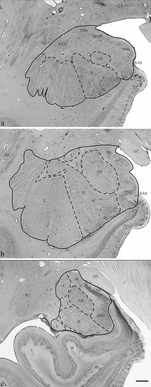
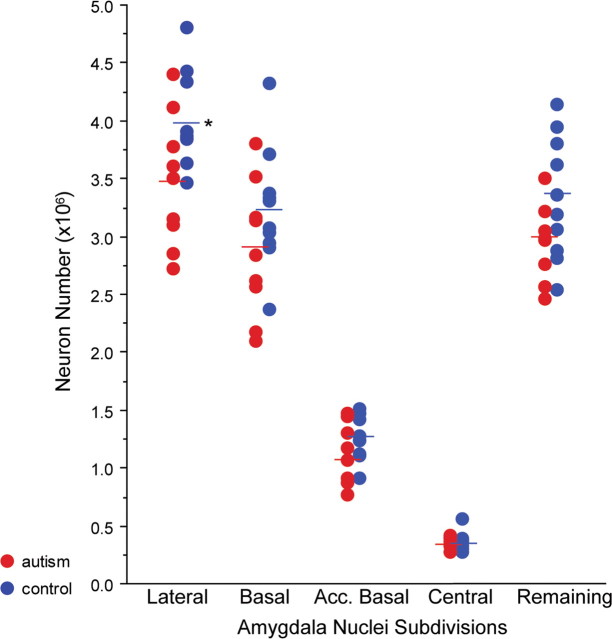
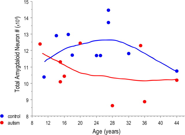
The amygdala is one of several brain regions suspected to be pathological in autism. Previously, we found that young children with autism have a larger amygdala than typically developing children. Past qualitative observations of the autistic brain suggest increased cell density in some nuclei of the postmortem autistic amygdala. In this first, quantitative stereological study of the autistic brain, we counted and measured neurons in several amygdala subdivisions of 9 autism male brains and 10 age-matched male control brains. Cases with comorbid seizure disorder were excluded from the study. The amygdaloid complex was outlined on coronal sections then partitioned into five reliably defined subdivisions: (1) lateral nucleus, (2) basal nucleus, (3) accessory basal nucleus, (4) central nucleus, and (5) remaining nuclei. There is no difference in overall volume of the amygdala or in individual subdivisions. There are also no changes in cell size. However, there are significantly fewer neurons in the autistic amygdala overall and in its lateral nucleus. In conjunction with the findings from previous magnetic resonance imaging studies, the autistic amygdala appears to undergo an abnormal pattern of postnatal development that includes early enlargement and ultimately a reduced number of neurons. It will be important to determine in future studies whether neuron loss in the amygdala is a consistent characteristic of autism and whether cell loss occurs in other brain regions as well.
Figures
References
-
- Adolphs R, Tranel D, Damasio H, Damasio A (1994). Impaired recognition of emotion in facial expressions following bilateral damage to the human amygdala. Nature 372:669–672. - PubMed
-
- Aylward EH, Minshew NJ, Goldstein G, Honeycutt NA, Augustine AM, Yates KO, Barta PE, Pearlson GD (1999). MRI volumes of amygdala and hippocampus in non-mentally retarded autistic adolescents and adults. Neurology 53:2145–2150. - PubMed
-
- Bailey A, Luthert P, Dean A, Harding B, Janota I, Montgomery M, Rutter M, Lantos P (1998). A clinicopathological study of autism. Brain 121:889–905. - PubMed
-
- Baron-Cohen S, Ring HA, Bullmore ET, Wheelwright S, Ashwin C, Williams SC (2000). The amygdala theory of autism. Neurosci Biobehav Rev 24:355–364. - PubMed
Publication types
MeSH terms
Grants and funding
LinkOut - more resources
Full Text Sources
Medical
Miscellaneous