Review
. 2006 Jul 19;34(12):3494-510.
doi: 10.1093/nar/gkl498. Print 2006.

Defective splicing, disease and therapy: searching for master checkpoints in exon definition

Affiliations
Review

Defective splicing, disease and therapy: searching for master checkpoints in exon definition

Emanuele Buratti et al. Nucleic Acids Res. .

Abstract

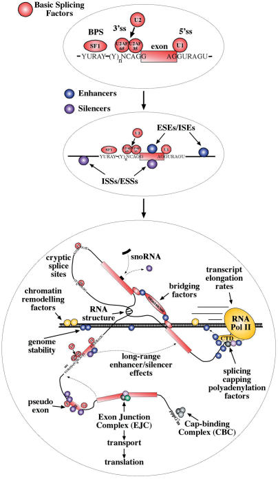
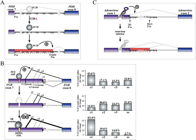
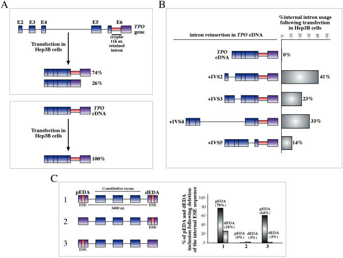
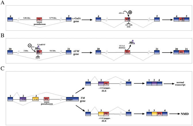
The number of aberrant splicing processes causing human disease is growing exponentially and many recent studies have uncovered some aspects of the unexpectedly complex network of interactions involved in these dysfunctions. As a consequence, our knowledge of the various cis- and trans-acting factors playing a role on both normal and aberrant splicing pathways has been enhanced greatly. However, the resulting information explosion has also uncovered the fact that many splicing systems are not easy to model. In fact we are still unable, with certainty, to predict the outcome of a given genomic variation. Nonetheless, in the midst of all this complexity some hard won lessons have been learned and in this survey we will focus on the importance of the wide sequence context when trying to understand why apparently similar mutations can give rise to different effects. The examples discussed in this summary will highlight the fine 'balance of power' that is often present between all the various regulatory elements that define exon boundaries. In the final part, we shall then discuss possible therapeutic targets and strategies to rescue genetic defects of complex splicing systems.


Figures

Figure 1
Figure 1
The three schematic representations show the increase during the last decade of the number of interactions reported to occur during RNA processing. The upper diagram depicts a classic pre-mRNA molecule in which the exon (closed box) is defined by a small number of relatively conserved sequences that bind a well-defined set of basic splicing factors. The complexity of the system begins to increase, as shown in the middle diagram, when the vast and still growing array of enhancer and silencer factors are to be added to the basic picture. These factors can bind particular enhancer and silencer sequences either within the exon itself (ESE and ESS) or in the nearby flanking regions (ISE and ISS) and help increase/decrease exon recognition. As shown in the bottom diagram, in these recent years the complexity of the regulatory elements that participate in splicing control has taken an enormous leap forward by the discovery of the potential connections between splicing, the specific structural features of pre-RNAs and many of the processes that participate in a their life-cycle such as genome stability, RNA processing speed, transport and translation.
Figure 2
Figure 2
(A) Schematic diagram showing how local context arrangements can influence cryptic splice site activation. For example, in intron 7 of the PDH gene a G26A mutation greatly enhances a preexisting binding site for the SC35 protein (upper and middle panels). The increased binding of this protein causes the shift of donor site usage towards a cryptic donor site in the downstream sequence (lower panel). (B) Another example is represented by a strong SF2/ASF-binding sequence that is not normally needed by the splicing machinery to recognize fibrinogen exon 7 (upper panel). However, if the natural donor site is inactivated by a +1G > U substitution the relative usage of the three cryptic sites which become activated (c1, c2 and c3) and the levels of exon skipping (es) are profoundly influenced by its presence, especially c1 (middle panel). In this case, the importance of local context has also been demonstrated by replacing the natural SF2/ASF-binding sequence with a well-known ESE sequence from the fibronectin gene (fnESE) and observing that the level of c1 usage are substantially increased with respect to the natural sequence (lower panel). (C) Finally, in the case of RNA secondary structure it has been shown that correct utilization of the natural donor site sequence (D1) can be achieved only in the presence of a highly conserved stem-loop structure within Adenovirus type 2 first exon (upper panel). Removal of this sequence has the consequence of activating usage of a cryptic donor site sequence (Dcr1) (lower panel).
Figure 3
Figure 3
(A) This figure shows the effect on internal intron retention in exon 6 of the TPO gene. When the TPO gene is transfected in Hep3B cells only two processed mRNAs are produced, a major form lacking all the intervening introns (74%) and a minor form in which there is the removal of an additional 116 nt long sequence (26%) from the processed mRNA (upper panel, boxed in red). On the other hand, lower panel shows that when the TPO cDNA is transfected in Hep3B cells no processing of this internal intron can be observed. (B) The only way of recovering internal intron processing is that of reintroducing at least one of the normally processed introns of the TPO gene. An analogous situation in which the system has to be ‘primed’ before it can function efficiently is provided by the recent observation that coordinated splicing can occur in correspondence of distant alternatively spliced exons. (C) Shows such an experiment where two alternatively spliced EDA exons (pEDA and dEDA) separated by a 3400 nt long spacer containing three constitutively spliced exons are inserted in a minigene systems in either wild type or mutated form (lacking an important ESE sequence). The graph on the left shows that efficient inclusion of the proximal exon strictly correlates with inclusion efficiencies of the distal exon.
Figure 4
Figure 4
Shows several factors that determine pseudoexon inclusion aside the simple creation of ex novo functional splice sites. In the a-GalA gene (A), a single point mutation G > A observed to occur in position-4 with respect to a cryptic donor site has the effect of creating a novel A/C rich enhancer element that causes inclusion of a 57 bp. pseudoexon in the processed mRNA. In a second example, a 4 nt deletion (GUAA) in the intronic region of the ATM gene between exons 20 and 21 caused the insertion of a 65 nt long pseudoexon in the final mRNA (B). Functional analysis has demonstrated that this deletion abolished binding of an U1snRNP molecule in this position and activated a 3′ss lying 12 nt upstream of this element. Finally, (C) shows the effect of the inclusion of a pseudoexon sequence in the a-tropomyosin pre-mRNA with respect to two mutually exclusive exons 2 and 3. When the pseudoexon sequence (red box) is spliced to exon 2 a 5′ss ZLE sequence is regenerated, thus allowing splicing to occur again giving rise to a normal transcript bearing exon 2 joined to exon 4 (upper panel). On the other hand, when the pseudoexon is joined to exon 3 the ZLE sequence that is regenerated is insufficient to promote joining together of exon 3 to exon 4 and the resulting transcript is thus degraded by NMD (lower panel). This difference in resplicing activity has been proposed to be consistent with the match to consensus of the two ZLE elements (higher for exon 2 than exon 3) and for the higher exon spicing enhancer activity of exon 2.
Figure 5
Figure 5
(A) Shows a schematic diagram (drawn to scale) of the exon-rich genomic region surrounding exon 37 of the neurofibromatosis (NF-1) gene. This exon is normally included constitutively in the mature mRNA but in the presence of a pathological substitution in position 6792 (a C is replaced by a G) the analysis from patient lymphoblasts reveals both the double skipping of exons 36 and 37 and also skipping of only exon 37. The influence of the genomic context has been tested by engineering a series of minigene constructs that gradually included larger portions of this genomic region, and their functionality compared with the in vivo pattern. (B) Wild-type exon 37 alone with a small portion of flanking intronic regions is not sufficient to yield the observed >99% inclusion in normal lymphoblasts. However, this result can be readily achieved when larger portions of this genomic region are included in the genomic construct. In addition to the normal situation, an increased genomic context also provides a closer correspondence with regards to the exon skipping effects of the C6792G substitution (C). In fact, only the construct carrying most of the NF-1 exons 31 to 38 together with a mutated exon 37 yields a percentage of normal transcript, exon 37 skipping and exon 36–37 skipping that reflects the observed percentage in a patient's lymphoblasts that carry the C6792G substitution (the asterisk indicates the levels of normal mRNA from the C6792G-carrying allele after deducting the estimated contribution of the un mutated allele).
Figure 6
Figure 6
Modulation of tissue- and developmental-specific expression of four different exons. (A) Shows that in non-neuronal cells PTB binds to two CU elements which are located both upstream and downstream c-src exon N1 and form a silencing complex that interferes with the interaction between U1snRNP bound at the donor site of this exon and U2AF. This complex does not interfere with binding of U2AF with the U1snRNP present at the donor site of exon 3 and this allows splicing of exon 3 to exon 4, thereby excluding exon N1 from the mature mRNA. On the other hand, in neuronal cells PTB is removed from the CU elements by an unknown factor ‘X’ in the presence of ATP and exon inclusion is promoted by the SF2/ASF protein binding to this exon (which is also bound by its antagonistic molecule, hnRNP A1) and by the DCS complex (which contains hnRNP H/F (H/F), KSRP, plus a neuronal form of PTB known as nPTB or WERI PTB). (B) Shows the Ras signaling pathway-dependent regulation of splicing of the variable v5 exon in the CD44 gene that occurs during T cell activation. In normal situations the enhancer complex in correspondence to a GAA-rich motif near the donor site of this exon is repressed by a strong hnRNP A1-binding sequence. During T cell activation, however, the repression can be overcome by either inhibition or displacement of A1 by phosphorylation of Sam 68 (Src-associated in mitosis) and involvement of powerful splicing coactivators such as SRm160. (C) Represents a model of cTNT exon 5 alternative splicing in the chicken developing heart. In the embryonic cardiomyocite CUG-BP and ETR-3 are expressed at high levels in the nucleus and they bind to the regulatory regions downstream of exon 5 (MSE2 to MSE4) to promote its inclusion. On the other hand, in the adult cardiomyocite CUG-BP and ETR-3 expression is downregulated and skipping is favored by PTB and MBNL proteins that are now free to bind on either side of the exon. Finally, (D) contains a model of HipK3 ‘T’ (testis-specific) differential exon inclusion in both somatic cells and in germ lines. In somatic cells, hnRNP A1 (A1) is principally responsible for the inhibition of the T exon donor site, probably with some help by hnRNP H (H). This is favored by the fact that in these cells the Tra2β protein tends to be in low amounts and is mostly phosphorylated. On the other hand, in germ cells Tra2β expression is upregulated and tends to be in its hypo-phosphorylated form whilst hnRNP A1 is downregulated. This arrangement allows the Tra2β protein to bind in place of A1 and to promote T exon inclusion with the assistance of SF2/ASF and SRp40 which also bind within this exon.
Figure 7
Figure 7
(A) Shows a schematic diagram of the splicing regulatory regions that have been described to affect recognition of SMN1/SMN2 exon 7. Recently, a functional SELEX analysis has been applied to this whole exonic sequence. (B) Shows the minigene system based on the human SMN gene used for this assay in which the entire exon 7 is partially randomized. These sequences can then be selected for inclusion in the final spliced products by subjecting them to rounds of selection in vivo. The borders of this randomized exon are dotted in (B) to indicate the fact that these sequences will not always be used as an exon in this technique. After each round of selection the spliced exon 7 sequences are recovered by RT–PCR and reinserted in the minigene splicing cassette. This selection process has allowed the precise mapping, at the single nucleotide level, of all the major determinants involved in the control of exon 7 splicing. Interestingly, 82% of all the final clones contained the same substitution in correspondence to the last position of this exon, and this change was quite capable of overcoming the effects of other negative regulatory mutations. (C) Shows a comparison of the two donor site strengths (the wild type and the one carrying the A54G substitution) using a panel of splice site prediction programs.
Figure 8
Figure 8
(A) Shows a diagrammatic representation of several positive elements that regulate SMN1/SMN2 exon 7 inclusion. The elements analyzed in this experiment are the first nucleotide of exon 7 (1G), the Tra2β-binding site (Tra2), the conserved tract in the middle of the exon (ConsTract) and the intronic splicing enhancer (element-2) in IVS7. Also shown is the dominant ISS element (ISS-N1). The graphical representation underneath shows the levels of exon 7 inclusion following mutation of all of these elements in a suitable minigene construct. Also included are the basal exon 7 inclusion levels from SMN2 exon 7 that carries a natural C6U substitution. (B) Shows the effect of the same mutations/substitutions when the ISS-N1 sequence has been deleted from the minigene constructs. An analogous situation which regards the CFTR exon 9 sequence is schematically represented in (C) in which the (UG)m(U)n repeated motif near the 3′ss of this exon has been shown to bind a powerful negative regulator of splicing, TDP-43. Several other cis-acting elements are also shown in this diagram: a CERES element within the exon, a positive polypyrimidine sequence near the 5′ss (PCE) that binds TIA-1, and a human-specific ISS in IVS9 that binds members of the SR protein family. The graph below shows that mutations within these elements are strongly inhibitory with respect to the basal inclusion level on a TG11T5 background (lane CFTR exon 9). Nonetheless, also in this case the removal of TDP-43 using siRNA technology rescues the effect of all these negative mutations (D).

Similar articles

Cited by

References

    1. Nissim-Rafinia M., Kerem B. The splicing machinery is a genetic modifier of disease severity. Trends Genet. 2005;21:480–483. - PubMed
    1. Caceres J.F., Kornblihtt A.R. Alternative splicing: multiple control mechanisms and involvement in human disease. Trends Genet. 2002;18:186–193. - PubMed
    1. Garcia-Blanco M.A., Baraniak A.P., Lasda E.L. Alternative splicing in disease and therapy. Nat. Biotechnol. 2004;22:535–546. - PubMed
    1. Cartegni L., Chew S.L., Krainer A.R. Listening to silence and understanding nonsense: exonic mutations that affect splicing. Nature Rev. Genet. 2002;3:285–298. - PubMed
    1. Faustino N.A., Cooper T.A. Pre-mRNA splicing and human disease. Genes Dev. 2003;17:419–437. - PubMed

Publication types

Substances