. 2006 Sep 1;498(1):46-57.
doi: 10.1002/cne.21040.

Synaptic input to OFF parasol ganglion cells in macaque retina

Affiliations

Synaptic input to OFF parasol ganglion cells in macaque retina

Andrea S Bordt et al. J Comp Neurol. .

Abstract

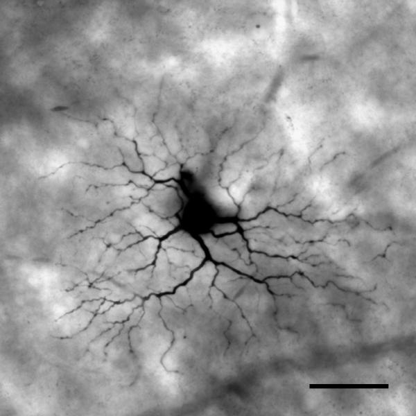
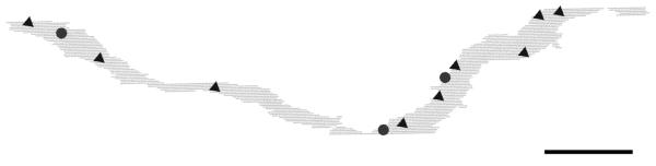
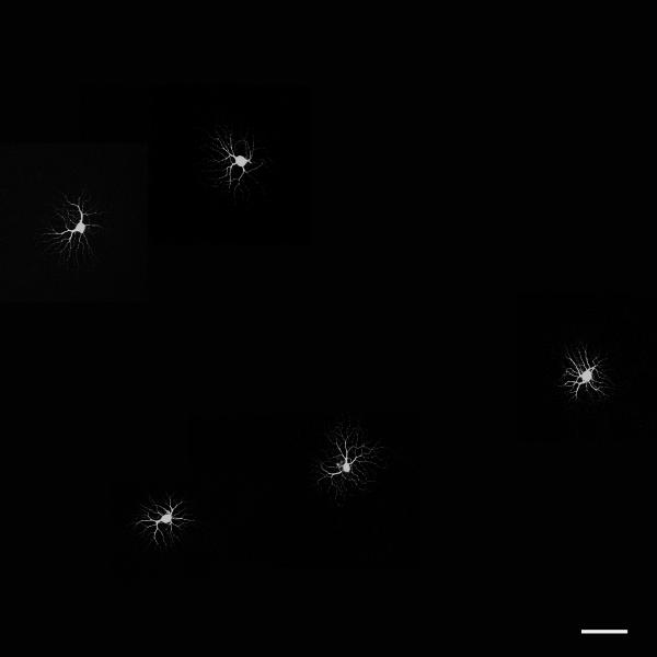
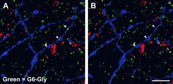
A Neurobiotin-injected OFF parasol cell from midperipheral macaque retina was studied by reconstruction of serial ultrathin sections and compared with ON parasol cells studied previously. In most respects, the synaptic inputs to the two subtypes were similar. Only a few of the amacrine cell processes that provided input to the labeled OFF parasol ganglion cell dendrites made or received inputs within the series, and none of these interactions were with the bipolar cells or other amacrine cells presynaptic to the OFF parasol cell. These findings suggest that the direct inhibitory input to OFF parasol cells originates from other areas of the retina. OFF parasol cells were known to receive inputs from two types of diffuse bipolar cells. To identify candidates for the presynaptic amacrine cells, OFF parasol cells were labeled with Lucifer yellow by using a juxtacellular labeling technique, and amacrine cells known to costratify with them were labeled via immunofluorescent methods. Appositions were observed with amacrine cells containing immunoreactive calretinin, parvalbumin, choline acetylatransferase, and G6-Gly, a cholecystokinin precursor. These findings suggest that the inhibitory input to parasol cells conveys information about several different attributes of visual stimuli and, particularly, about their global properties.


Figures

Figure 1
Figure 1
The labeled OFF parasol ganglion cell was imaged digitally in a whole mount preparation before it was re-embedded and sectioned for electron microscopy. It was identified as a parasol ganglion cell by the size of its dendritic field and the density of its dendritic arbor. Parts of the cell body and some proximal dendrites were obscured due to leakage of Neurobiotin at the injection site. Scale bar = 50 μm.
Figure 2
Figure 2
A low power electron micrograph of the inner plexiform layer (IPL) and parts of the inner nuclear layer (INL) and ganglion cell layer (GCL). Filled triangles denote the borders of the IPL. Labeled OFF parasol ganglion cell dendrites (arrows) ramify at approximately 30% depth, with the lower boundary of the INL being 0% and the upper boundary of the GCL being 100%. Scale bar = 10 μm.
Figure 3
Figure 3
A large, labeled OFF parasol ganglion cell dendrite receives synapses from an amacrine cell (hollow triangles) and a bipolar cell axon at a dyad synapse (arrow). The bipolar cell axon receives a reciprocal synapse (filled triangles) from an electron lucent amacrine cell process. Later in the series, this bipolar cell made another synapse onto the labeled dendrite. Scale bar = 1 μm.
Figure 4
Figure 4
Serial sections demonstrating a monad synapse onto the labeled OFF parasol ganglion cell dendrite. The diffuse bipolar (DB) 3 bipolar cell axon is filled with vesicles and has a prominent synaptic ribbon (arrow). The bipolar cell also makes a gap junction (triangles) with another bipolar axon of the same type. Scale bar = 0.5 μm.
Figure 5
Figure 5
Serial sections through a DB2 bipolar cell axon making a monad type ribbon synapse onto the labeled OFF parasol ganglion cell dendrite. The bipolar cell axon is small in diameter, irregularly shaped and contains microtubules. The ribbon (arrow) first appears in the second section, and the third section shows the synaptic ribbon at the presynaptic membrane and the associated postsynaptic density on the membrane of the labeled OFF parasol ganglion cell dendrite. Scale bar = 1 μm.
Figure 6
Figure 6
Two electron lucent, vesicle-filled amacrine cell processes make conventional synapses (triangles) onto a labeled OFF parasol ganglion cell dendrite. Neither amacrine cell process made other synaptic contacts within the series. Scale bar = 0.5 μm.
Figure 7
Figure 7
The labeled OFF parasol ganglion cell receives a synapse from an electron lucent amacrine cell process (triangles). Three presynaptic dense bars are visible there. The dendrite had three spines (asterisks) within the series, two of which are visible in this section. The amacrine cell process did not make other synaptic contacts within the series. Scale bar = 0.5 μm.
Figure 8
Figure 8
An irregularly-shaped, electron dense amacrine cell dendrite makes a synapse onto the labeled parasol ganglion cell dendrite (triangles). The presynaptic amacrine cell also made and received synapses with other amacrine cells within the series. Scale bar = 0.5 μm.
Figure 9
Figure 9
Parts of a dendrite of the labeled OFF parasol ganglion cell were reconstructed from serial sections; they are viewed here as if in the whole mount preparation. Soon after the beginning of the series, the dendrite bifurcated, and 12 synapses were found on its branches. The circles represent bipolar cell inputs at ribbon synapses; all 3 were found on the side of the dendrite facing the INL. Triangles represent amacrine cell input at a conventional synapse; these were found on both sides of the reconstructed dendrite. Scale bar = 5 μm.
Figure 10
Figure 10
A photomontage of juxtacellularly-labeled ON and OFF parasol ganglion cells. ON parasol cells were more commonly labeled. Scale bar = 100 μm.
Figure 11
Figure 11
(A) Two parvalbumin-IR processes (green) partially surround labeled OFF parasol ganglion cell dendrites (blue) (filled triangles). A parvalbumin-IR process apposes a terminal of a labeled OFF parasol ganglion cell dendrite (blue) (hollow triangle). A 3D reconstruction showing processes from the vitreal orientation. (B) A 3D reconstruction showing processes from the scleral orientation. Twenty-three single optical sections were used in the 3D reconstruction. Scale bar = 5 μm. (C) A calretinin-IR lobuluar appendage (green) apposes a labeled OFF parasol ganglion cell dendrite (blue) at the base of a spine (filled triangle). A ChAT-IR dendrite (red) apposes another dendrite (hollow triangle). A 3D reconstruction showing processes from the vitreal orientation. (D) A 3D reconstruction showing processes from the scleral orientation. Fourteen single optical sections were used in the 3D reconstruction. Scale bar = 5 μm.
Figure 12
Figure 12
(A) Three G6-Gly-IR processes (green) appose a labeled OFF parasol ganglion cell dendrite (blue) (filled triangles). A 3D reconstruction showing processes from the vitreal orientation. (B) A 3D reconstruction showing processes from the scleral orientation. Twenty-one single optical sections were used in the 3D reconstruction. Scale bar = 5 μm.

Similar articles

Cited by

References

    1. Boycott BB, Wässle H. Morphological classification of bipolar cells of the primate retina. Eur J Neurosci. 1991;3:1069–1088. - PubMed
    1. Burr DC, Morrone MC, Ross J. Selective suppression of the magnocellular visual pathway during saccadic eye movements. Nature. 1994;371:511–3. - PubMed
    1. Calkins DJ. Synaptic orgainization of cone pathways in the primate retina. In: Gegenfurtner KR, Sharpe LT, editors. Color Vision: From Genes to Perception. Cambridge University Press; Cambridge: 1999. pp. 163–179.
    1. Calkins DJ, Schein SJ, Tsukamoto Y, Sterling P. M and L cones in macaque fovea connect to midget ganglion cells by different numbers of excitatory synapses. Nature. 1994;371:70–72. - PubMed
    1. Calkins DJ, Sterling P. Absence of spectrally specific lateral inputs to midget ganglion cells in primate retina. Nature. 1996;381:613–615. - PubMed

Publication types

MeSH terms