

Evolution of melanopsin photoreceptors: discovery and characterization of a new melanopsin in nonmammalian vertebrates
- PMID: 16856781
- PMCID: PMC1514791
- DOI: 10.1371/journal.pbio.0040254
Evolution of melanopsin photoreceptors: discovery and characterization of a new melanopsin in nonmammalian vertebrates
Erratum in
- PLoS Biol. 2006 Oct;4(10):e320
Abstract
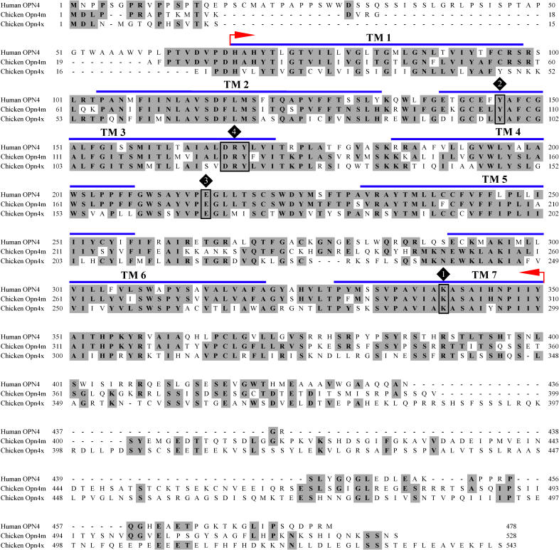
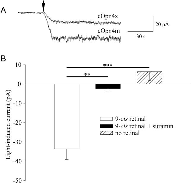
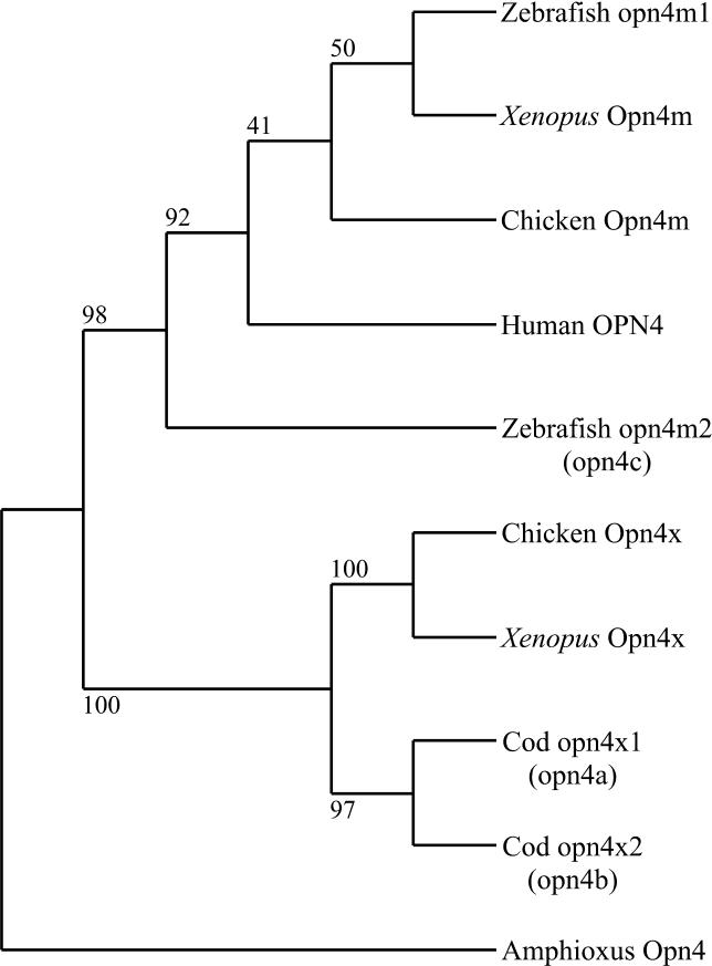
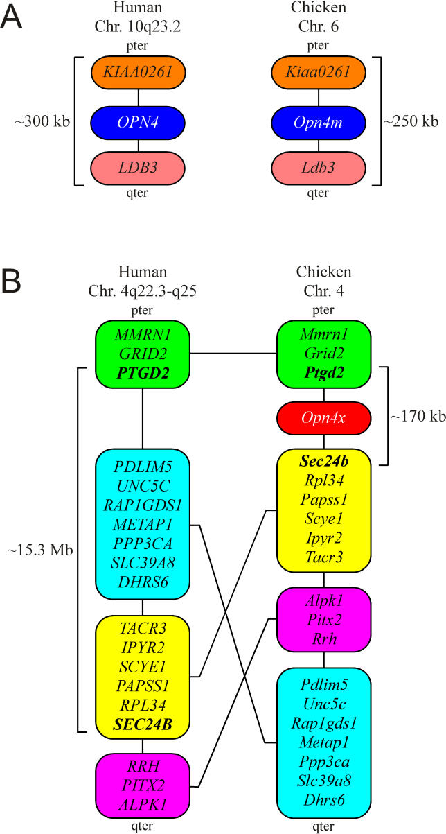
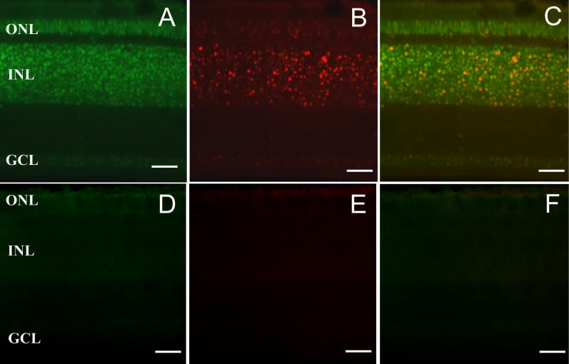
In mammals, the melanopsin gene (Opn4) encodes a sensory photopigment that underpins newly discovered inner retinal photoreceptors. Since its first discovery in Xenopus laevis and subsequent description in humans and mice, melanopsin genes have been described in all vertebrate classes. Until now, all of these sequences have been considered representatives of a single orthologous gene (albeit with duplications in the teleost fish). Here, we describe the discovery and functional characterisation of a new melanopsin gene in fish, bird, and amphibian genomes, demonstrating that, in fact, the vertebrates have evolved two quite separate melanopsins. On the basis of sequence similarity, chromosomal localisation, and phylogeny, we identify our new melanopsins as the true orthologs of the melanopsin gene previously described in mammals and term this grouping Opn4m. By contrast, the previously published melanopsin genes in nonmammalian vertebrates represent a separate branch of the melanopsin family which we term Opn4x. RT-PCR analysis in chicken, zebrafish, and Xenopus identifies expression of both Opn4m and Opn4x genes in tissues known to be photosensitive (eye, brain, and skin). In the day-14 chicken eye, Opn4m mRNA is found in a subset of cells in the outer nuclear, inner nuclear, and ganglion cell layers, the vast majority of which also express Opn4x. Importantly, we show that a representative of the new melanopsins (chicken Opn4m) encodes a photosensory pigment capable of activating G protein signalling cascades in a light- and retinaldehyde-dependent manner under heterologous expression in Neuro-2a cells. A comprehensive in silico analysis of vertebrate genomes indicates that while most vertebrate species have both Opn4m and Opn4x genes, the latter is absent from eutherian and, possibly, marsupial mammals, lost in the course of their evolution as a result of chromosomal reorganisation. Thus, our findings show for the first time that nonmammalian vertebrates retain two quite separate melanopsin genes, while mammals have just one. These data raise important questions regarding the functional differences between Opn4x and Opn4m pigments, the associated adaptive advantages for most vertebrate species in retaining both melanopsins, and the implications for mammalian biology of lacking Opn4x.
Figures
Comment in
-
Melanopsin photopigment comes in two distinct forms.PLoS Biol. 2006 Aug;4(8):e263. doi: 10.1371/journal.pbio.0040263. Epub 2006 Jul 25. PLoS Biol. 2006. PMID: 20076619 Free PMC article. No abstract available.
References
-
- Lucas RJ, Freedman MS, Muñoz M, García-Fernández JM, Foster RG. Regulation of the mammalian pineal by non-rod, non-cone, ocular photoreceptors. Science. 1999;284:505–507. - PubMed
-
- Berson DM, Dunn FA, Takao M. Phototransduction by retinal ganglion cells that set the circadian clock. Science. 2002;295:1070–1073. - PubMed
-
- Lucas RJ, Douglas RH, Foster RG. Characterization of an ocular photopigment capable of driving pupillary constriction in mice. Nat Neurosci. 2001;4:621–626. - PubMed
-
- Panda S, Provencio I, Tu DC, Pires SS, Rollag MD, et al. Melanopsin is required for non-image-forming photic responses in blind mice. Science. 2003;301:525–527. - PubMed
Publication types
MeSH terms
Substances
Associated data
- Actions
- Actions
- Actions
- Actions
- Actions
- Actions
- Actions
- Actions
- Actions
- Actions
- Actions
- Actions
Grants and funding
LinkOut - more resources
Full Text Sources
Molecular Biology Databases
